. 2022 Sep 24;20(1):426.
doi: 10.1186/s12951-022-01634-z.

A physicochemical double-cross-linked gelatin hydrogel with enhanced antibacterial and anti-inflammatory capabilities for improving wound healing

Affiliations

A physicochemical double-cross-linked gelatin hydrogel with enhanced antibacterial and anti-inflammatory capabilities for improving wound healing

Yapeng Lu et al. J Nanobiotechnology. .

Erratum in

Abstract

Background: Skin tissue is vital in protecting the body from injuries and bacterial infections. Wound infection caused by bacterial colonization is one of the main factors hindering wound healing. Wound infection caused by colonization of a large number of bacteria can cause the wound to enter a continuous stage of inflammation, which delays wound healing. Hydrogel wound dressing is composed of natural and synthetic polymers, which can absorb tissue fluid, improve the local microenvironment of wound, and promote wound healing. However, in the preparation process of hydrogel, the complex preparation process and poor biological efficacy limit the application of hydrogel wound dressing in complex wound environment. Therefore, it is particularly important to develop and prepare hydrogel dressings with simple technology, good physical properties and biological effects by using natural polymers.

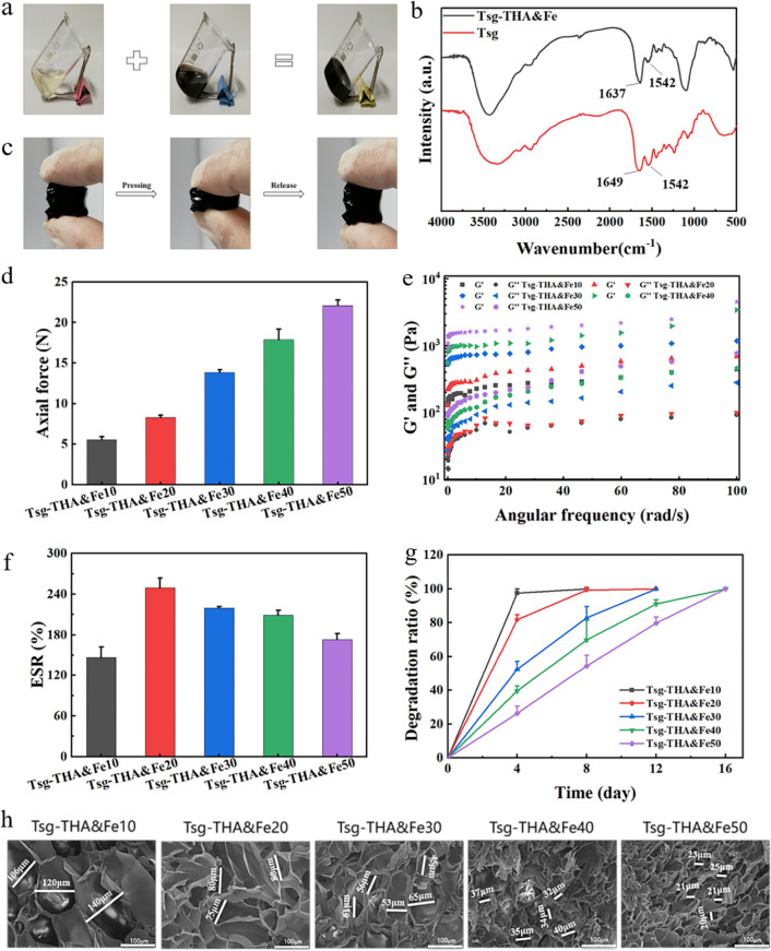
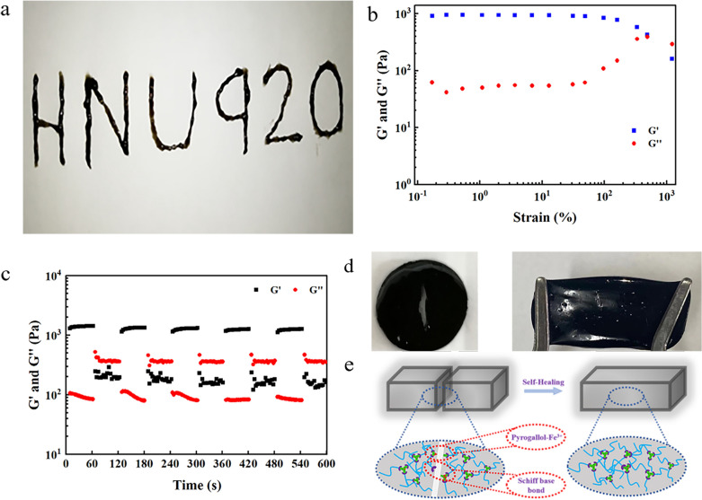
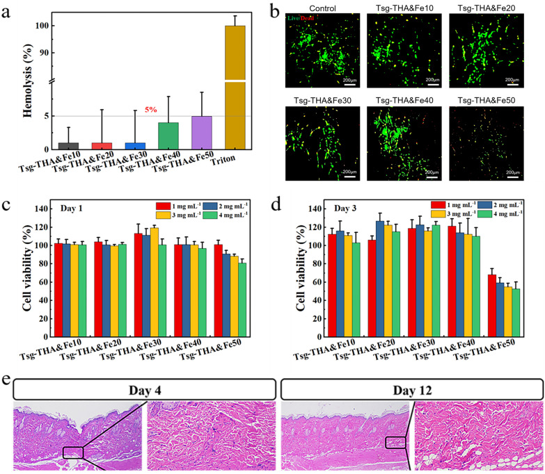
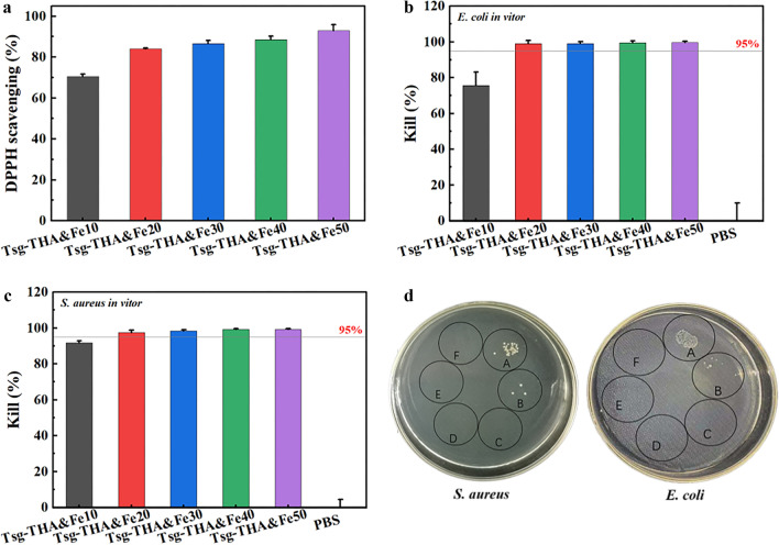
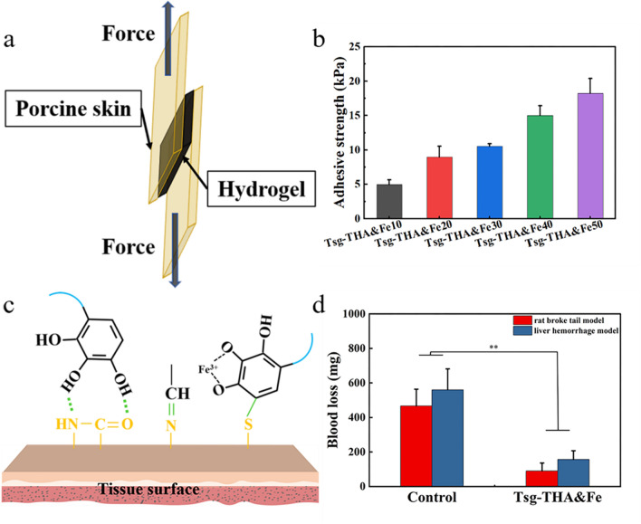
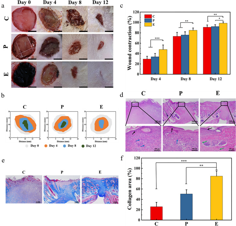
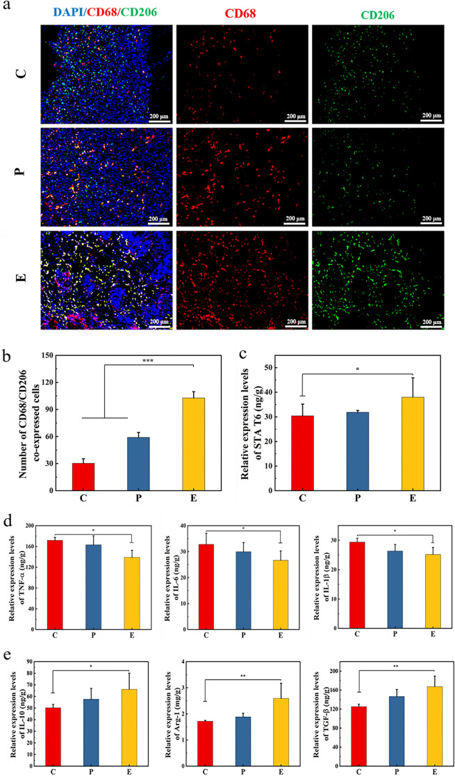
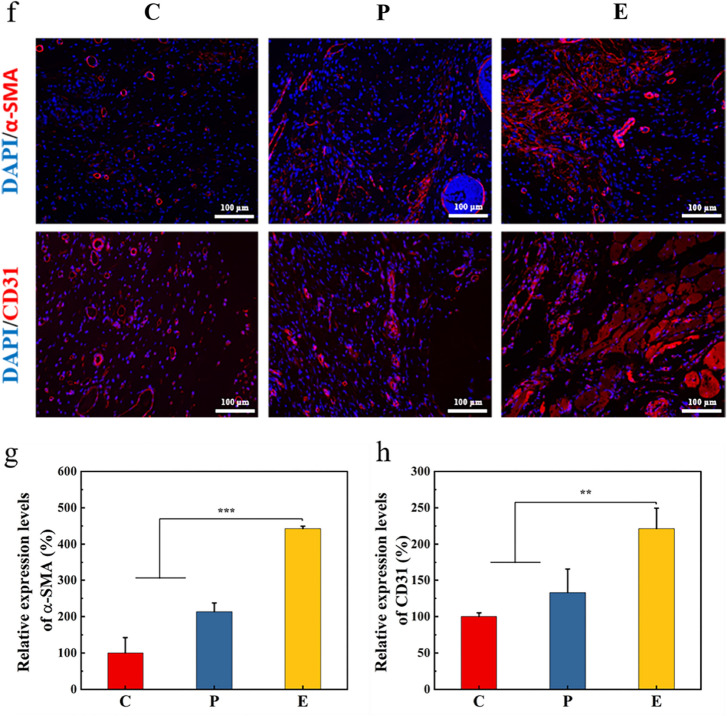
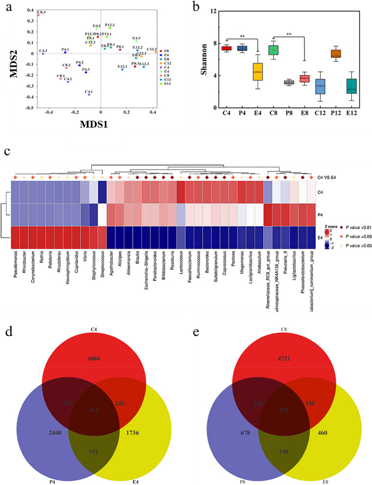
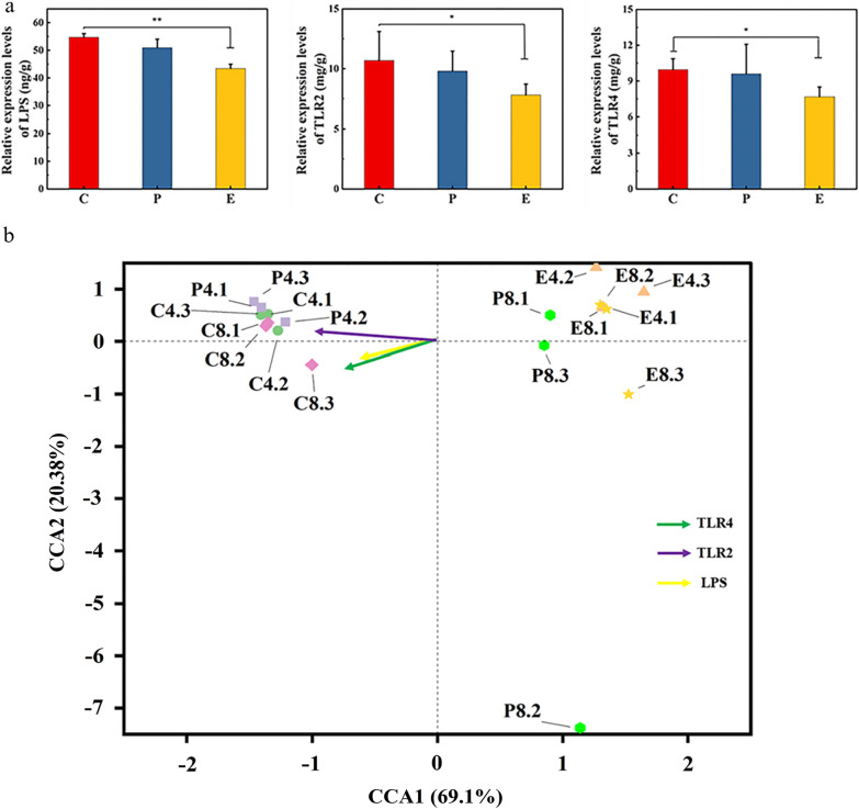
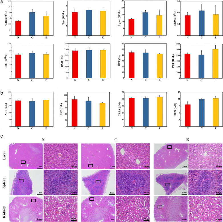
Results: In this study, a gelatin-based (Tsg-THA&Fe) hydrogel was created by mixing trivalent iron (Fe3+) and 2,3,4-trihydroxybenzaldehyde (THA) to form a complex (THA&Fe), followed by a simple Schiff base reaction with tilapia skin gelatin (Tsg). The gel time and rheological properties of the hydrogels were adjusted by controlling the number of complexes. The dynamic cross-linking of the coordination bonds (o-phthalmictriol-Fe3+) and Schiff base bonds allows hydrogels to have good self-healing and injectable properties. In vitro experiments confirmed that the hydrogel had good biocompatibility and biodegradability as well as adhesion, hemostasis, and antibacterial properties. The feasibility of Tsg-THA&Fe hydrogel was studied by treating rat skin trauma model. The results showed that compared with Comfeel® Plus Transparent dressing, the Tsg-THA&Fe hydrogel could obvious reduce the number of microorganisms, prevent bacterial colonization, reduce inflammation and accelerate wound healing. Local distribution of the Tsg-THA&Fe hydrogel in the skin tissue did not cause organ toxicity.

Conclusions: In summary, the preparation process of Tsg-THA&Fe hydrogel is simple, with excellent performance in physical properties and biological efficacy. It can effectively relieve inflammation and control the colonization of wound microbes, and can be used as a multi-functional dressing to improve wound healing.

Keywords: Anti-inflammatory; Hydrogels; Tilapia skin gelatin; Wound healing; Wound microbiology.


Conflict of interest statement

The autors declare no competing interests.

Figures

Fig. 1
Fig. 1
Preparation and characterization of the Tsg-THA&Fe hydrogel. a Photographs of Tsg solution, THA&Fe solution and the formed Tsg-THA&Fe hydrogel. b FT-IR spectra of Tsg and Tsg-THA&Fe hydrogel. c The original compressed and recovered states of the prepared hydrogels. d The compression force of the Tsg-THA&Fe hydrogel at 90% strain (n = 3). e Frequency dependence of the dynamic storage modulus (G′) and loss modulus (G″) of Tsg-THA&Fe hydrogel. f The equilibrium swelling rate (ESR) of Tsg-THA&Fe hydrogels (n = 5). g In vitro degradation properties of Tsg-THA&Fe hydrogel with pH 7.4 at 37 °C (n = 5). h The SEM images of Tsg-THA&Fe hydrogel
Fig. 2
Fig. 2
Injectable and self-healing properties of the hydrogels. a Injectable properties of Tsg-THA&Fe hydrogel under normal environment. b Strain scan of Tsg-THA&Fe40 hydrogel at G′ and G″. c Rheological properties of the Tsg-THA&Fe40 hydrogel at alternating strain switching from 1 to 500%. d Demonstration of self-healing of hydrogels with cracks. e) Self-healing mechanism of Tsg-THA&Fe hydrogel
Fig. 3
Fig. 3
Biocompatibility of the Tsg-THA&Fe hydrogel. a Hemolysis assay of Tsg-THA&Fe hydrogel (n = 3). b Cell staining of NIH-3T3 cells cultured in the Tsg-THA&Fe hydrogel for 3 days. Survival rate of NIH-3T3 cells cultured in each group at different concentrations of hydrogel leachate for 1 c and 3 d days (n = 6). e Hematoxylin–eosin (H&E) staining of skin tissue implanted subcutaneously with Tsg-THA&Fe40 hydrogel, the box shows the approximate location of hydrogel implantation (n = 5)
Fig. 4
Fig. 4
In vitro antioxidant and bacterial inhibition properties of the hydrogels. a DPPH scavenging of Tsg-THA&Fe hydrogel (n = 3). Quantitative results of the inhibition performance of Tsg-THA&Fe hydrogel against b E. coli and c S. aureus (n = 5). d Images of bacterial clones formed by the inhibitory effects of the control group (A), Tsg-THA&Fe10 hydrogel group (B), Tsg-THA&Fe20 hydrogel group (C), Tsg-THA&Fe30 hydrogel group (D), Tsg-THA&Fe40 hydrogel group (E), Tsg-THA&Fe50 hydrogel group (F) on S. aureus and E. coli on agar plates are shown
Fig. 5
Fig. 5
Adhesive and hemostatic properties of the Tsg-THA&Fe. a Schematic diagram of the lap shear test of Tsg-THA&Fe hydrogel by using pigskin. b Adhesion strength test of Tsg-THA&Fe to pigskin (n = 3). c Skin adhesion mechanism of Tsg-THA&Fe. d The hemostatic effect was evaluated in the rat broke tail model and liver hemorrhage model (n = 3, *P < 0.05, **P < 0.01)
Fig. 6
Fig. 6
Evaluation of the effect of the Tsg-THA&Fe hydrogel on wound healing in vivo. a Wounds of untreated wound group (group C), Comfeel® Plus Transparent treatment group (group P) and Tsg-THA&Fe40 hydrogel treatment group (group E) on day 4, 8 and 12. (scale bar: 5 mm). b Schematic diagrams of contraction changes in the groups C, P, and E on days 4, 8, and 12 during wound healing. c Wound contraction rates of each group at days 4, 8, and 12 (n = 6). d H&E staining of skin tissue at the wound on day 12 in each group (Black arrow: cuticula, Green arrow: blood vessels, Blue arrows: inflammatory cells). e Masson staining of skin tissues at the wound on day 12 in each group. f Quantitative analysis of collagen deposition area in Masson staining (n = 4). *P < 0.05, **P < 0.01, ***P < 0.001
Fig. 7
Fig. 7
Hydrogel promotes wound healing by modulating the inflammatory response. a Immunofluorescence staining images of the pan-macrophage marker CD68 (red) and M2 macrophage marker CD206 (green) in the group C, group P and group E on day 8. b The number of CD68 and CD206 co-expressed cells in group C, group P and group E on day 8 (n = 4, ***P < 0.001). c The expression of signal transducer and activators of transcription 6 (STA T6) in wound of group C, P and E on day 8 (n = 4, *P < 0.05). d The expression of tumor necrosis factor-alpha (TNF-α), interleukin-6 (IL-6), and interleukin-1β (IL-1β) in wound of group C, P and E on day 8 (n = 4, *P < 0.05). e The expression of interleukin-10 (IL-10), arginase-1 (Arg-1), and tumor growth factor-beta (TGF-β) in wound of group C, P and E on day 8 (n = 4, *P < 0.05). f Immunofluorescence staining images of α-SMA (red) and CD31 (red) in the wounds on day 12 in each group. Quantitative analysis of relative fluorescence intensity of g α-SMA and h CD31 stained skin tissue (n = 4, **P < 0.01, ***P < 0.001)
Fig. 7
Fig. 7
Hydrogel promotes wound healing by modulating the inflammatory response. a Immunofluorescence staining images of the pan-macrophage marker CD68 (red) and M2 macrophage marker CD206 (green) in the group C, group P and group E on day 8. b The number of CD68 and CD206 co-expressed cells in group C, group P and group E on day 8 (n = 4, ***P < 0.001). c The expression of signal transducer and activators of transcription 6 (STA T6) in wound of group C, P and E on day 8 (n = 4, *P < 0.05). d The expression of tumor necrosis factor-alpha (TNF-α), interleukin-6 (IL-6), and interleukin-1β (IL-1β) in wound of group C, P and E on day 8 (n = 4, *P < 0.05). e The expression of interleukin-10 (IL-10), arginase-1 (Arg-1), and tumor growth factor-beta (TGF-β) in wound of group C, P and E on day 8 (n = 4, *P < 0.05). f Immunofluorescence staining images of α-SMA (red) and CD31 (red) in the wounds on day 12 in each group. Quantitative analysis of relative fluorescence intensity of g α-SMA and h CD31 stained skin tissue (n = 4, **P < 0.01, ***P < 0.001)
Fig. 8
Fig. 8
Bioinformatics analysis of wound microorganisms. In order to facilitate the comparison and analysis of data in each group, the labels of each group were defined as XY.n by Bioinformatics analysis. The X position mainly represented the untreated group (group C), Comfeel® Plus Transparent treatment group (group P) and Tsg-THA&Fe40 hydrogel treatment group (group E). Y positions respectively represent different time points, mainly including day 4, 8 and 12. The n position represents the serial number of different samples in the group. a The results of Non-metric multidimensional scaling (NMDS) analysis based on OTU level using each group of samples are shown. each point in the figure indicates a sample, the distance between points indicates the degree of variation, and the samples of the same group are indicated using the same color. when Stress is less than 0.2, it indicates that NMDS can accurately reflect the degree of variation among samples. b The Alpha Diversity analysis index Shannon was counted for different samples at a 97% consistency threshold, and Shannon is the total number of taxa in the samples and their percentage. The higher the community diversity and the more evenly spread the species, the greater the Shannon index (n = 3, *P < 0.05, **P < 0.01). c On day 4, Metastat heat map analysis for groups C4. The corresponding values in the heat map are the z-values obtained by normalizing the species in each row, and the z-values for samples in each classification are obtained by dividing the difference between the relative abundance of samples in that classification and the average relative abundance of all samples in that classification by the standard deviation of all samples in that classification. d and e The common and unique the operational taxonomic unit (OUT) among different groups are analyzed and plotted as a Venn Graph, where each circle represents a sample (group), and the number of overlapping circles represents the number of common OTU among samples (groups), and the number of numbers without overlapping circles represents the number of unique OTU of samples (groups)
Fig. 9
Fig. 9
Relationship between wound microorganisms and innate immunity. a The expression levels of wound inflammatory factors lipopolysaccharide (LPS), toll-like receptor 2 (TLR2) and toll-like receptor 4 (TLR4) in untreated wound group (group C), Comfeel Plus Transparent treatment group (group P) and Tsg-THA&Fe40 hydrogel treatment group on day 8 (n = 3, *P < 0.05, **P < 0.01). b The canonical correspondence analysis (CCA) was used mainly to reflect the relationship between the groups' colony-based distances and environmental factors. The horizontal coordinate indicates the first principal component, and the percentage indicates the contribution of the first principal component to the sample variation; the vertical coordinate indicates the second principal component, and the percentage indicates the contribution of the second principal component to the sample variation; each point in the graph represents one sample. The length of the arrow line represents the correlation between an environmental factor and the distribution of communities and species, the longer the line, the higher the correlation, and vice versa, the lower the correlation. The angle between the arrow line and the ranking axis represents the correlation between an environmental factor and the ranking axis, the smaller the angle, the higher the correlation, and vice versa
Fig. 10
Fig. 10
In vivo toxicity analysis was performed in the normal (group N), untreated (group C), and Tsg-THA&Fe40 hydrogel (group E) groups. a Routine blood tests of rats in groups N, C, and E, after 12 days, including assessment of white blood cells (WBC), neutrophils (Neut), lymphocyte count (Lymph), monocyte count (Mon), red blood cells (RBC), hemoglobin (HGB), red blood cell cumulative pressure (HCT) and platelets (PLT) (n = 4). b Measurement of serum liver and kidney function indices in each group of rats, including the assessment of glutamic aminotransferase (ALT), glutamic oxalacetic aminotransferase (AST), serum creatinine (CREA), and urea nitrogen (BUN) levels (n = 4). c Sections of the liver, spleen, and kidney of each group of rats were stained with H&E staining for pathological analysis

Similar articles

Cited by

References

    1. Blacklow SO, Li J, Freedman B, Zeidi M, Mooney D. Bioinspired mechanically active adhesive dressings to accelerate wound closure. Sci Adv. 2019;5:eaaw3963. doi: 10.1126/sciadv.aaw3963. - DOI - PMC - PubMed
    1. Chen G, Yu Y, Wu X, Wang G, Ren J, Zhao Y. Bioinspired multifunctional hybrid hydrogel promotes wound healing. Adv Funct Mater. 2018 doi: 10.1002/adfm.201801386. - DOI - PubMed
    1. Wang R, Li J, Chen W, Xu T, Yun S, Xu Z, Xu Z, Sato T, Chi B, Xu H. A biomimetic mussel-inspired epsilon-poly-l-lysine hydrogel with robust tissue-anchor and anti-infection capacity. Adv Funct Mater. 2017;27:1604894. doi: 10.1002/adfm.201604894. - DOI
    1. Dabiri G, Damstetter E, Phillips T. Choosing a wound dressing based on common wound characteristics. Adv Wound Care. 2016;5:32–41. doi: 10.1089/wound.2014.0586. - DOI - PMC - PubMed
    1. Gao F, Xu Z, Liang Q, Li H, Liu W. Osteochondral regeneration with 3D-printed biodegradable high-strength supramolecular polymer reinforced-gelatin hydrogel scaffolds. Adv Sci. 2019;6:1900867. doi: 10.1002/advs.201900867. - DOI - PMC - PubMed

LinkOut - more resources