

Phage-Plasmids Spread Antibiotic Resistance Genes through Infection and Lysogenic Conversion
- PMID: 36154183
- PMCID: PMC9600943
- DOI: 10.1128/mbio.01851-22
Phage-Plasmids Spread Antibiotic Resistance Genes through Infection and Lysogenic Conversion
Abstract
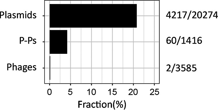
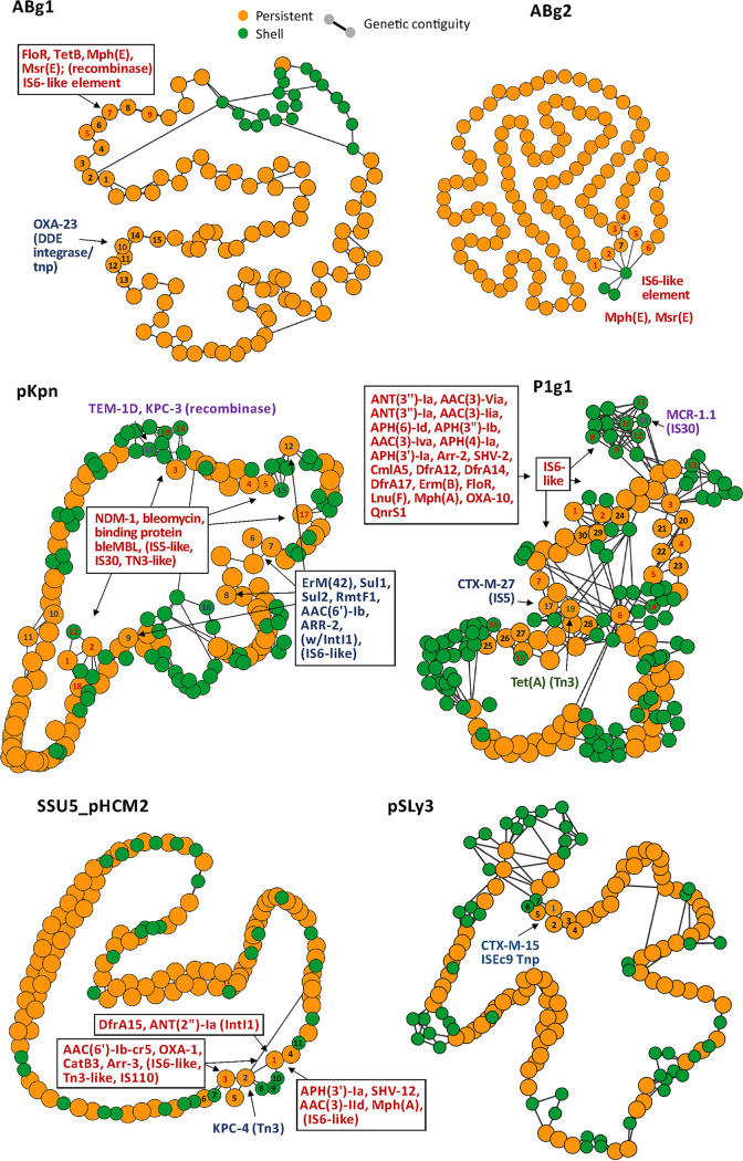
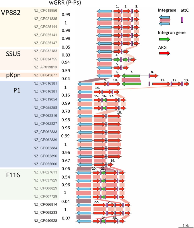
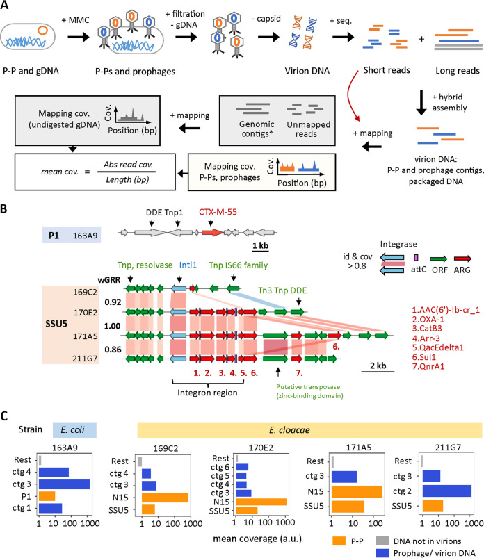
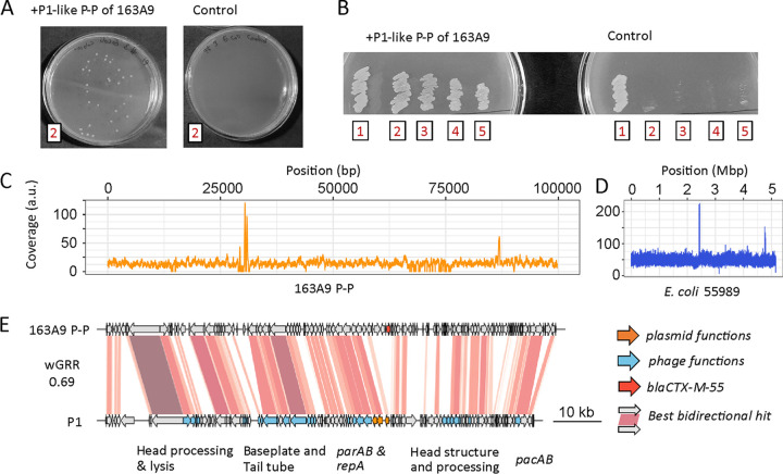
Antibiotic resistance is rapidly spreading via the horizontal transfer of resistance genes in mobile genetic elements. While plasmids are key drivers of this process, few integrative phages encode antibiotic resistance genes. Here, we find that phage-plasmids, elements that are both phages and plasmids, often carry antibiotic resistance genes. We found 60 phage-plasmids with 184 antibiotic resistance genes, providing resistance for broad-spectrum-cephalosporins, carbapenems, aminoglycosides, fluoroquinolones, and colistin. These genes are in a few hot spots, seem to have been cotranslocated with transposable elements, and are often in class I integrons, which had not been previously found in phages. We tried to induce six phage-plasmids with resistance genes (including four with resistance integrons) and succeeded in five cases. Other phage-plasmids and integrative prophages were coinduced in these experiments. As a proof of concept, we focused on a P1-like element encoding an extended spectrum β-lactamase, blaCTX-M-55. After induction, we confirmed that it is capable of infecting and converting four other E. coli strains. Its reinduction led to the further conversion of a sensitive strain, confirming that it is a fully functional phage. This study shows that phage-plasmids carry a large diversity of clinically relevant antibiotic resistance genes that they can transfer across bacteria. As plasmids, these elements seem plastic and capable of acquiring genes from other plasmids. As phages, they may provide novel paths of transfer for resistance genes because they can infect bacteria that are distant in time and space from the original host. As a matter of alarm, they may also mediate transfer to other types of phages. IMPORTANCE The dissemination of antimicrobial resistance is a major threat to global health. Here, we show that a group of temperate bacterial viruses (phages), termed phage-plasmids, commonly encode different and multiple types of resistance genes of high clinical importance, often in integrons. This is unexpected, as phages typically do not carry resistance genes and, hence, do not confer upon their hosts resistance via infection and genome integration. Our experiments with phage-plasmids isolated from clinical settings confirmed that they infect sensitive strains and render them antibiotic resistant. The spread of antibiotic resistance genes by phage-plasmids is worrisome because it dispenses cell-to-cell contact, which is necessary for canonical plasmid transfer (conjugation). Furthermore, their integrons become genetic platforms for the acquisition of novel resistance genes.
Keywords: antibiotic resistance; bacteriophages; integrons; phage genomics; plasmids; prophage induction.
Conflict of interest statement
The authors declare no conflict of interest.
Figures
Similar articles
-
In silico genomic insights into bacteriophages infecting ESBL-producing Escherichia coli from human, animal, and environmental sources.BMC Microbiol. 2025 Apr 8;25(1):200. doi: 10.1186/s12866-025-03913-9. BMC Microbiol. 2025. PMID: 40200154 Free PMC article.
-
Phage-plasmid hybrids as vectors for antibiotic resistance in environmental Escherichia coli.Sci Total Environ. 2025 Jan 10;959:178157. doi: 10.1016/j.scitotenv.2024.178157. Epub 2024 Dec 26. Sci Total Environ. 2025. PMID: 39729844
-
Antibiotic resistance genes, mobile elements, virulence genes, and phages in cultivated ESBL-producing Escherichia coli of poultry origin in Kwara State, North Central Nigeria.Int J Food Microbiol. 2023 Mar 16;389:110086. doi: 10.1016/j.ijfoodmicro.2023.110086. Epub 2023 Jan 21. Int J Food Microbiol. 2023. PMID: 36738714
-
Antimicrobial Resistance in Escherichia coli.Microbiol Spectr. 2018 Jul;6(4):10.1128/microbiolspec.arba-0026-2017. doi: 10.1128/microbiolspec.ARBA-0026-2017. Microbiol Spectr. 2018. PMID: 30003866 Free PMC article. Review.
-
Dynamics of antibiotic resistance genes in plasmids and bacteriophages.Crit Rev Microbiol. 2025 Mar;51(2):219-228. doi: 10.1080/1040841X.2024.2339262. Epub 2024 Apr 23. Crit Rev Microbiol. 2025. PMID: 38651513 Review.
Cited by
-
Prospects and Challenges of Bacteriophage Substitution for Antibiotics in Livestock and Poultry Production.Biology (Basel). 2024 Jan 4;13(1):28. doi: 10.3390/biology13010028. Biology (Basel). 2024. PMID: 38248459 Free PMC article. Review.
-
The Biotechnological Application of Bacteriophages: What to Do and Where to Go in the Middle of the Post-Antibiotic Era.Microorganisms. 2023 Sep 13;11(9):2311. doi: 10.3390/microorganisms11092311. Microorganisms. 2023. PMID: 37764155 Free PMC article. Review.
-
Isolation, susceptibility profiles and genomic analysis of a colistin-resistant Salmonella enterica serovar Kentucky strain COL-R.3 Biotech. 2023 May;13(5):140. doi: 10.1007/s13205-023-03559-2. Epub 2023 Apr 26. 3 Biotech. 2023. PMID: 37124985 Free PMC article.
-
One Health at Risk: Plasmid-Mediated Spread of mcr-1 Across Clinical, Agricultural, and Environmental Ecosystems.Antibiotics (Basel). 2025 May 15;14(5):506. doi: 10.3390/antibiotics14050506. Antibiotics (Basel). 2025. PMID: 40426572 Free PMC article. Review.
-
Genomic insights into bacteriophages: a new frontier in AMR detection and phage therapy.Brief Funct Genomics. 2025 Jan 15;24:elaf011. doi: 10.1093/bfgp/elaf011. Brief Funct Genomics. 2025. PMID: 40720171 Free PMC article. Review.
References
-
- Spera AM, Esposito S, Pagliano P. 2019. Emerging antibiotic resistance: carbapenemase-producing enterobacteria. Bad new bugs, still no new drugs. Infez Med 27:357–364. - PubMed
-
- Lin Q, Wang Y, Yu J, Li S, Zhang Y, Wang H, Lai X, Liu D, Mao L, Luo Y, Tang G, Chen Z, Sun Z. 2021. Bacterial characteristics of carbapenem-resistant Enterobacteriaceae (CRE) colonized strains and their correlation with subsequent infection. BMC Infect Dis 21:638. doi:10.1186/s12879-021-06315-0. - DOI - PMC - PubMed
Publication types
MeSH terms
Substances
LinkOut - more resources
Full Text Sources
Other Literature Sources
Medical
Molecular Biology Databases
Research Materials
