. 2022 Oct 26;13(5):e0163322.
doi: 10.1128/mbio.01633-22. Epub 2022 Sep 26.

Bacterial Membrane Vesicles as a Novel Strategy for Extrusion of Antimicrobial Bismuth Drug in Helicobacter pylori

Affiliations

Bacterial Membrane Vesicles as a Novel Strategy for Extrusion of Antimicrobial Bismuth Drug in Helicobacter pylori

Sumith Kumar et al. mBio. .

Abstract

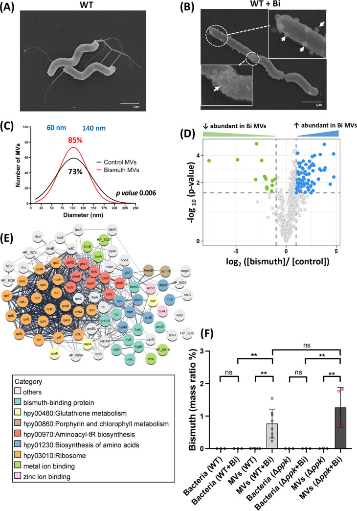
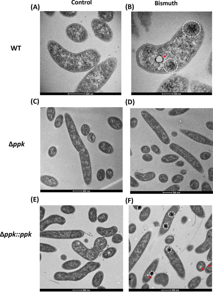
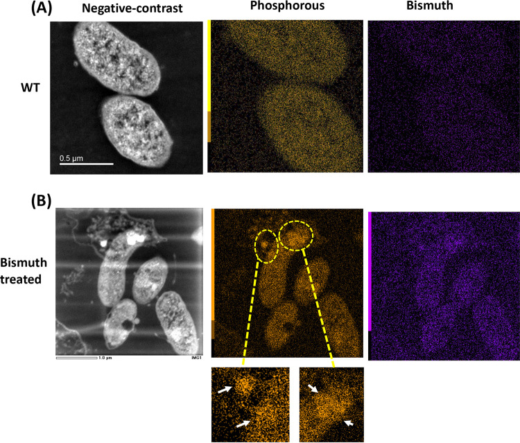
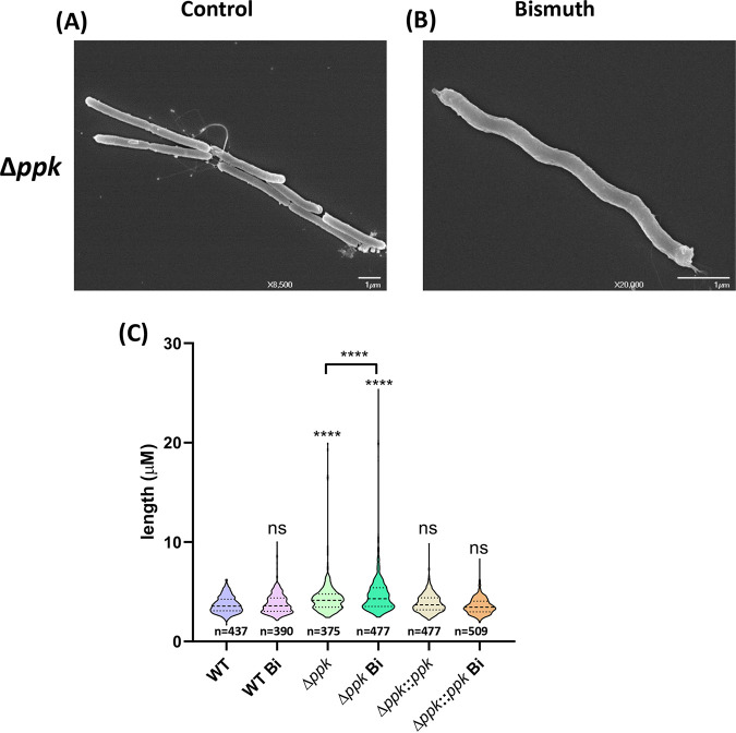
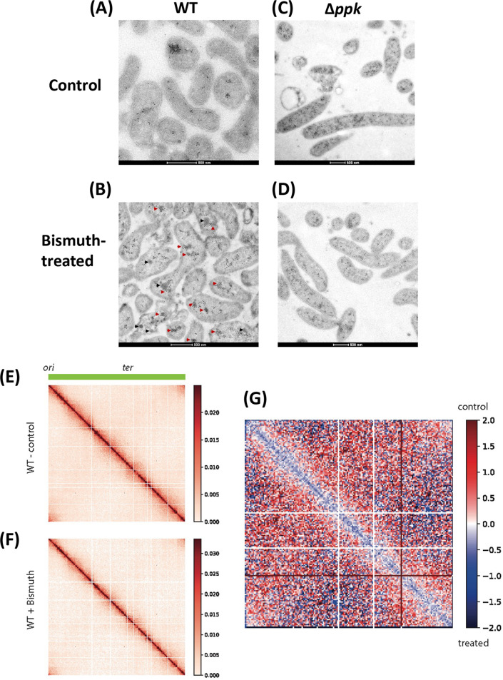
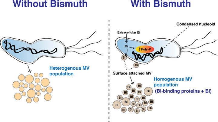
Bacterial antibiotic resistance is a major threat to human health. A combination of antibiotics with metals is among the proposed alternative treatments. Only one such combination is successfully used in clinics; it associates antibiotics with the metal bismuth to treat infections by Helicobacter pylori. This bacterial pathogen colonizes the human stomach and is associated with gastric cancer, killing 800,000 individuals yearly. The effect of bismuth in H. pylori treatment is not well understood in particular for sublethal doses such as those measured in the plasma of treated patients. We addressed this question and observed that bismuth induces the formation of homogeneously sized membrane vesicles (MVs) with unique protein cargo content enriched in bismuth-binding proteins, as shown by quantitative proteomics. Purified MVs of bismuth-exposed bacteria were strongly enriched in bismuth as measured by inductively coupled plasma optical emission spectrometry (ICP-OES), unlike bacterial cells from which they originate. Thus, our results revealed a novel function of MVs in bismuth detoxification, where secreted MVs act as tool to discard bismuth from the bacteria. Bismuth also induces the formation of intracellular polyphosphate granules that are associated with changes in nucleoid structure. Nucleoid compaction in response to bismuth was established by immunogold electron microscopy and refined by the first chromosome conformation capture (Hi-C) analysis of H. pylori. Our results reveal that even low doses of bismuth induce profound changes in H. pylori physiology and highlight a novel defense mechanism that involves MV-mediated bismuth extrusion from the bacteria and a probable local DNA protective response where polyphosphate granules are associated with nucleoid compaction. IMPORTANCE Bacterial resistance to antibiotics is a major threat to human health. Treatments combining antibiotics with metals were proposed to circumvent this hurdle. Only one such combination is successfully used in clinics associating antibiotics with the metal bismuth to treat infections by the human pathogen Helicobacter pylori. H. pylori causes 800,000 deaths by gastric cancer yearly. How bismuth impacts H. pylori and its response to this toxic metal were ill defined. We discovered that upon bismuth exposure, H. pylori secretes membrane vesicles that are enriched in bismuth. Bismuth also induces the formation of intracellular polyphosphate granules associated with compaction of the chromosome. Upon bismuth exposure, H. pylori displays both defense and protection mechanisms, with bismuth extrusion by vesicles and shielding of the chromosome.

Keywords: Helicobacter pylori; Hi-C; antibiotic resistance; bismuth; chromosome conformation capture; membrane vesicles; polyphosphate granules.


Conflict of interest statement

The authors declare no conflict of interest.

Figures

FIG 1
FIG 1
Characterization of MVs from bismuth-treated and untreated H. pylori cells. (A and B) Scanning electron microscopy (SEM) of control bacteria (A) and bismuth-treated bacteria (2.5 μM, 6 h) (B). Surface-attached MVs are visible in the bismuth-treated sample, indicated by white arrows in the insets. (C) Size distribution of MVs purified from control and bismuth-treated H. pylori cells. MV diameter was measured with the ImageJ software using TEM images of a total of 600 MVs for each condition, and the Gaussian equation was fitted to the histogram data to obtain a frequency distribution plot. The nonparametric Kolmogorov-Smirnov test shows statistically significant differences between the two distributions (P = 0.006). (D) Volcano plot representing the differential abundance of proteins purified from MVs from bismuth-treated H. pylori cells and from control bacteria. The x axis shows the bismuth/control fold change value in logarithmic scale. The y axis shows the −log10 of the adjusted P value showing statistical significance. The horizontal dashed line corresponds to a P of 0.01 used as the threshold cutoff. Vertical dotted lines correspond to log2 fold changes of ±2. Blue dots (87 proteins) and green dots (14 proteins) represent more abundant and less abundant proteins, respectively, in MVs of bismuth-treated sample. (E) STRING network analysis of differentially abundant proteins present in the vesicles of bismuth-treated bacteria. The thickness of the gray lines represents the strength of the interaction between proteins. The functional category of each protein is indicated by the color code. (F) ICP-OES metal content of purified MVs and their parent H. pylori cells exposed or not to bismuth. Wild-type (WT) bacteria and a mutant carrying a deletion of the ppk gene (Δppk) were analyzed. The amount of bismuth is expressed as a mass ratio percentage of dry mass (n ≥ 3). The limit of detection, 10 μg/L, corresponds to 10 ppb. Statistics using the Mann-Whitney test are shown. **, P < 0.01; ns, nonsignificant.
FIG 2
FIG 2
Transmission electron microscopy analysis of bismuth-treated H. pylori cells. (A) Untreated control bacteria exhibit a uniform membrane structure with an intact cytoplasm. (B) Bismuth-treated bacteria show an enrichment in electron-dense cytoplasmic structures (highlighted by white circles) as well as “vacuole-like” structures surrounded by electron-dense structures (red arrow). (C) Untreated Δppk mutant cells. (D) Bismuth-treated Δppk mutant cells, where no electron-dense structures are observed. (E) Untreated Δppk mutant complemented with a wild-type ppk copy. (F) Bismuth-treated Δppk mutant complemented with a wild-type ppk copy. In this strain, electron-dense structures are restored upon bismuth treatment (white circles and red arrowheads).
FIG 3
FIG 3
Elemental mapping of bismuth-treated H. pylori cells. (A) Scanning-transmission electron micrographs (STEM) of untreated bacteria. The negative-contrast image of control bacterial sections is shown together with the EDX map for phosphorus and bismuth. (B) STEM negative-contrast image of H. pylori cells exposed to bismuth. Electron-dense regions are seen as bright white regions. EDX maps of phosphorus and bismuth are shown. Accumulation of phosphorus in the cytoplasm of these bismuth-treated bacteria is highlighted by yellow open circles and white arrows in the inset panels; this accumulation overlaps with the electron-dense regions.
FIG 4
FIG 4
Ultrastructural analysis of the H. pylori Δppk mutant. (A and B) SEM of the H. pylori Δppk mutant under control conditions (A) or treated with bismuth (B). (C) Bacterial size distribution of wild-type, Δppk mutant, and complemented Δppk::ppk cells under control conditions or treated with bismuth. Images were taken by phase-contrast microscopy, and size measurements were performed with ImageJ software with the MicrobeJ plugin. Statistical analysis was carried out using a one-way ANOVA test (n > 400 for each condition). ****, P < 0.0001; ns, nonsignificant.
FIG 5
FIG 5
Bismuth exposure induces condensation of the H. pylori nucleoid. (A and B) TEM of H. pylori wild-type cells under control conditions (A) or treated with bismuth (B). (C and D) Sections of Δppk mutant cells under control conditions (C) or treated with bismuth (D). Wild-type cells, but not Δppk mutants, show condensation of the nucleoid structure upon bismuth treatment. Red arrowheads indicate the presence of DNA structures in the phosphorus-rich regions. Black arrowheads indicate the electron-dense structure not labeled with anti-DNA antibody. (E to G) Hi-C analysis of H. pylori chromosome structure of cells exposed or not to bismuth. (E and F) Normalized contact map of H. pylori exponentially growing cells not treated (E) or treated (F) with a sublethal concentration of bismuth. x and y axes indicate chromosomal coordinates binned in 5-kb bins (origin and terminus of replication are indicated at the top). (G) Ratio map (log2) of the two tested conditions (wild type not treated versus treated); color scales indicate contact enrichment in treated (blue) or not treated (red) cells (white indicates no difference between the two conditions).
FIG 6
FIG 6
Schematic representation of the consequences of H. pylori exposure to sublethal bismuth concentrations. Bismuth is taken up by H. pylori cells, induces the formation of poly-P granules in the cytoplasm, and causes nucleoid condensation. Bismuth induces the formation of homogenous membrane vesicles that are enriched in bismuth-binding proteins and in the metal bismuth. We conclude that H. pylori eliminates the toxic bismuth by packaging this metal together with bismuth-binding proteins in membrane vesicles and locally protects its chromosome from metal toxicity.

Similar articles

Cited by

References

    1. Ventola CL. 2015. The antibiotic resistance crisis: Part 1—causes and threats. P&T 40:277–283. - PMC - PubMed
    1. Ghosh C, Sarkar P, Issa R, Haldar J. 2019. Alternatives to conventional antibiotics in the era of antimicrobial resistance. Trends Microbiol 27:323–338. doi:10.1016/j.tim.2018.12.010. - DOI - PubMed
    1. Frei A, Zuegg J, Elliott AG, Baker M, Braese S, Brown C, Chen F, G Dowson C, Dujardin G, Jung N, King AP, Mansour AM, Massi M, Moat J, Mohamed HA, Renfrew AK, Rutledge PJ, Sadler PJ, Todd MH, Willans CE, Wilson JJ, Cooper MA, Blaskovich MAT. 2020. Metal complexes as a promising source for new antibiotics. Chem Sci 11:2627–2639. doi:10.1039/c9sc06460e. - DOI - PMC - PubMed
    1. Mjos KD, Orvig C. 2014. Metallodrugs in medicinal inorganic chemistry. Chem Rev 114:4540–4563. doi:10.1021/cr400460s. - DOI - PubMed
    1. Tursi A, Franceschi M, Allegretta L, Savarino E, De Bastiani R, Elisei W, Baldassarre G, Ferronato A, Scida S, Miraglia C, Penna A, Licci C, Rizzo GL, Pranzo G, Cassieri C, Brandimarte G, Picchio M, Di Mario F. 2018. Effectiveness and safety of Pylera in patients infected by Helicobacter pylori: a multicenter, retrospective, real life study. Dig Dis 36:264–268. doi:10.1159/000487391. - DOI - PubMed

Publication types