. 2022 Sep 26;21(1):41.
doi: 10.1186/s12941-022-00533-3.

Curcumin and capsaicin regulate apoptosis and alleviate intestinal inflammation induced by Clostridioides difficile in vitro

Affiliations

Curcumin and capsaicin regulate apoptosis and alleviate intestinal inflammation induced by Clostridioides difficile in vitro

Masoumeh Azimirad et al. Ann Clin Microbiol Antimicrob. .

Abstract

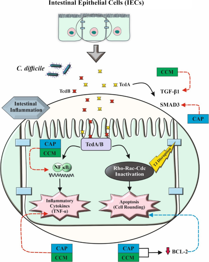
Background: The dramatic upsurge of Clostridioides difficile infection (CDI) by hypervirulent isolates along with the paucity of effective conventional treatment call for the development of new alternative medicines against CDI. The inhibitory effects of curcumin (CCM) and capsaicin (CAP) were investigated on the activity of toxigenic cell-free supernatants (Tox-S) of C. difficile RT 001, RT 126 and RT 084, and culture-filtrate of C. difficile ATCC 700057.

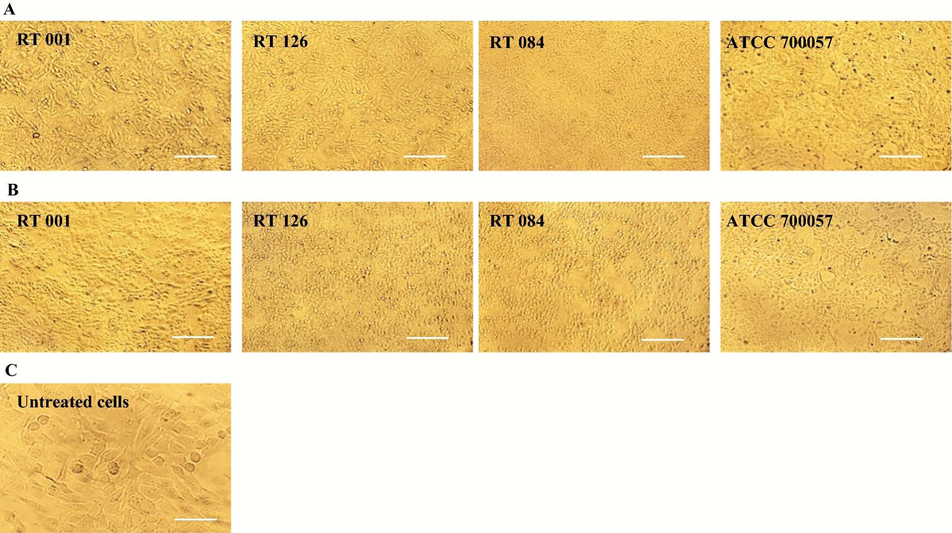
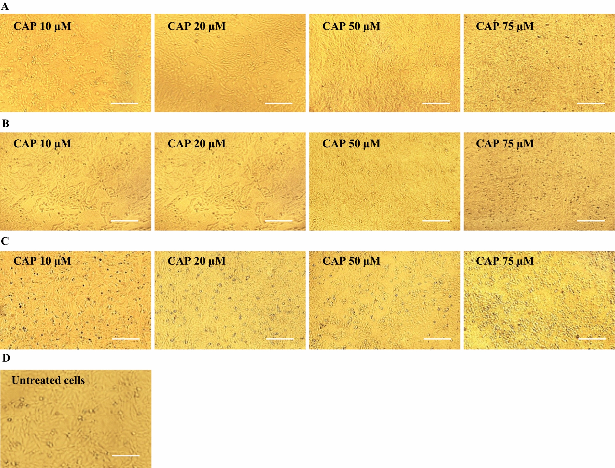
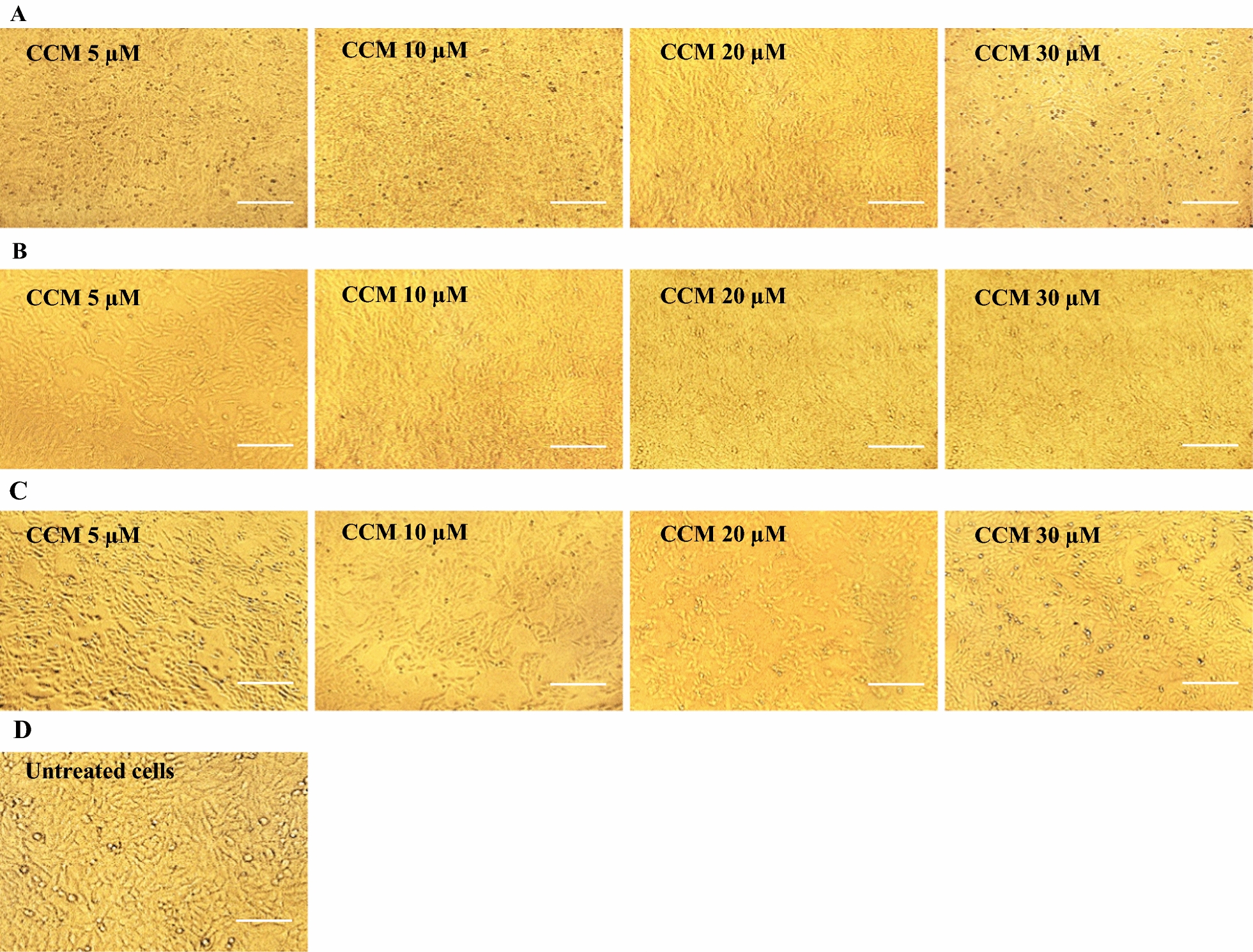
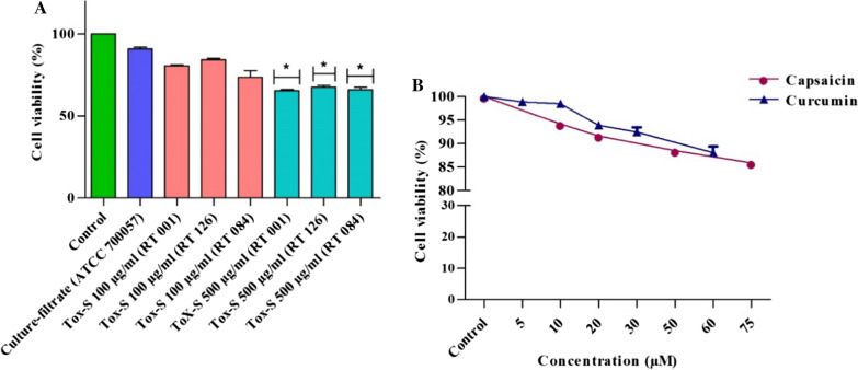
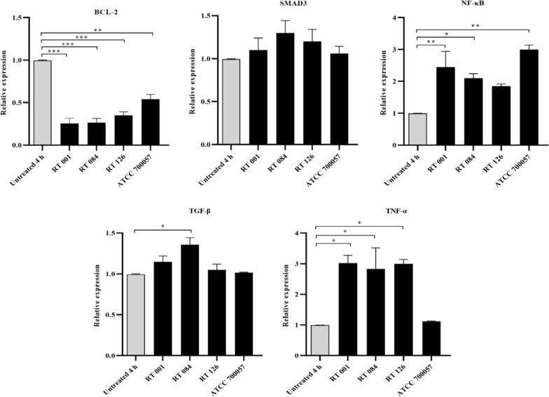
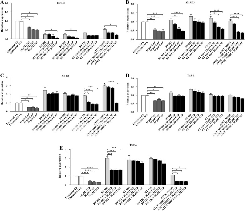
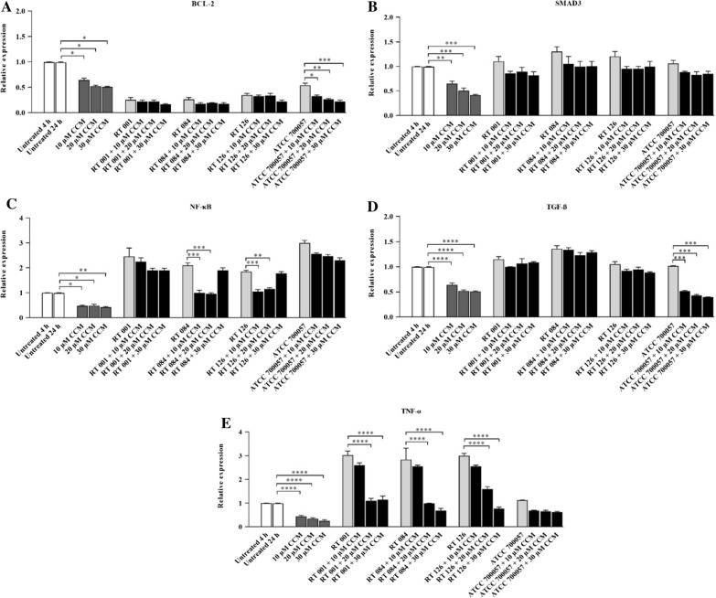
Methods: Cell viability of HT-29 cells exposed to varying concentrations of CCM, CAP, C. difficile Tox-S and culture-filtrate was assessed by MTT assay. Anti-inflammatory and anti-apoptotic effects of CCM and CAP were examined by treatment of HT-29 cells with C. difficile Tox-S and culture-filtrate. Expression of BCL-2, SMAD3, NF-κB, TGF-β and TNF-α genes in stimulated HT-29 cells was measured using RT-qPCR.

Results: C. difficile Tox-S significantly (P < 0.05) reduced the cell viability of HT-29 cells in comparison with untreated cells. Both CAP and CCM significantly (P < 0.05) downregulated the gene expression level of BCL-2, SMAD3, NF-κB and TNF-α in Tox-S treated HT-29 cells. Moreover, the gene expression of TGF-β decreased in Tox-S stimulated HT-29 cells by both CAP and CCM, although these reductions were not significantly different (P > 0.05).

Conclusion: The results of the present study highlighted that CCM and CAP can modulate the inflammatory response and apoptotic effects induced by Tox-S from different clinical C. difficile strains in vitro. Further studies are required to accurately explore the anti-toxin activity of natural components, and their probable adverse risks in clinical practice.

Keywords: Apoptosis; Capsaicin; Clostridioides difficile infection; Curcumin; Inflammation; Tox-S.


Conflict of interest statement

The authors declare that they have no conflicts of interest.

Figures

Fig. 1
Fig. 1
Cytopathic effect (CPE) of two different concentrations (100 and 500 μg/ml) of Tox-S from C. difficile (RT 001, RT 126, RT 084) and culture-filtrate of C. difficile ATCC 700057 on Vero cells using microscopy. A Microscopic cell morphology of Vero cells after treatment with 100 μg/ml Tox-S and culture-filtrate for 4 h at 37 °C; B Microscopic cell morphology of Vero cells after treatment with 500 μg/ml Tox-S and culture-filtrate for 4 h at 37 °C; C Untreated Vero cell monolayer. A, B Light microscopy × 20, Scale bar = 50 µm; C Light microscopy × 40, Scale bar = 50
Fig. 2
Fig. 2
Cytotoxic effect of capsaicin (CAP) on Vero cells. A Microscopic cell morphology of Vero cells after treatment with the indicated concentrations of CAP for 1 h at 37 °C; B Microscopic cell morphology of Vero cells after treatment with the indicated concentrations of CAP for 4 h at 37 °C; C Microscopic cell morphology of Vero cells after treatment with the indicated concentrations of CAP for 24 h at 37 °C; D Untreated Vero cell monolayer. Light microscopy × 20, Scale bar = 50 µm
Fig. 3
Fig. 3
Cytotoxic effect of curcumin (CCM) on Vero cells. A Microscopic cell morphology of Vero cells after treatment with the indicated concentrations of CCM for 1 h at 37 °C; B Microscopic cell morphology of Vero cells after treatment with the indicated concentrations of CCM for 4 h at 37 °C; C Microscopic cell morphology of Vero cells after treatment with the indicated concentrations of CCM for 24 h at 37 °C; D Untreated Vero cell monolayer. Light microscopy × 20, Scale bar = 50 µm
Fig. 4
Fig. 4
Cell viability determined by MTT assay. A Different concentrations (100 and 500 μg/ml) of Tox-S from C. difficile (RT 001, RT 126, RT 084) and culture-filtrate of C. difficile ATCC 700057 were added to HT-29 cells for 4 h at 37 °C; B Different concentrations of capsaicin (10, 20, 50 and 75 µM) and curcumin (5, 10, 20, 30 and 60 µM) were added to HT-29 cells for 24 h at 37 °C. Data were presented as mean ± SD from three independent experiments. A P value of < 0.05 was considered as significant (*P < 0.05) by unpaired student’s t test statistical analysis
Fig. 5
Fig. 5
Relative expression of BCL-2, SMAD3, NF-κB, TGF-β and TNF-α genes in HT-29 cells upon treatment with Tox-S (100 μg/ml) from C. difficile (RT 001, RT 126, RT 084) and culture-filtrate (100 μg/ml) of C. difficile ATCC 700,057 measured by using quantitative real-time PCR assay. Gene expression data were normalized to β-actin as the reference gene. Data were presented as mean ± SD from three independent experiments. A P value of < 0.05 was considered as significant (*P < 0.05; **P < 0.01; ***P < 0.001) by unpaired student’s t test and one-way ANOVA statistical analysis
Fig. 6
Fig. 6
Relative expression of BCL-2 A, SMAD3 B, NF-κB C, TGF-β D and TNF-α E genes in HT-29 cells upon treatment with Tox-S (100 μg/ml) from C. difficile (RT 001, RT 126, RT 084), culture-filtrate (100 μg/ml) of C. difficile ATCC 700057, and different concentrations (10, 20, 50 µM) of capsaicin (CAP) measured by using quantitative real-time PCR assay. Gene expression data were normalized to β-actin as the reference gene. Data were presented as mean ± SD from three independent experiments. A P value of < 0.05 was considered as significant (*P < 0.05; **P < 0.01) by unpaired student’s t test and one-way ANOVA statistical analysis
Fig. 7
Fig. 7
Relative expression of BCL-2 A, SMAD3 B, NF-κB C, TGF-β D and TNF-α E genes in HT-29 cells upon treatment with Tox-S (100 μg/ml) from C. difficile (RT 001, RT 126, RT 084), culture-filtrate (100 μg/ml) of C. difficile ATCC 700057, and different concentrations (10, 20, 30 µM) of curcumin (CCM) measured by using quantitative real-time PCR assay. Gene expression data were normalized to β-actin as the reference gene. Data were presented as mean ± SD from three independent experiments. A P value of < 0.05 was considered as significant (*P < 0.05; **P < 0.01) by unpaired student’s t test and one-way ANOVA statistical analysis
Fig. 8
Fig. 8
A schematic diagram which demonstrates the potential biological impact of capsaicin (CAP) and curcumin (CCM) on C. difficile toxin-mediated inflammation. Exposure of intestinal epithelial cells (IECs) to CCM and CAP can lead to inhibition of TGF-β1 and Smad3, respectively, which may result in maintaining the integrity of the intestinal epithelial tight junctions (TJs) barrier. In addition, CAP and CCM can alleviate host inflammatory responses induced by TcdA and TcdB toxins probably through downregulation of NF-κB and TNF-α. On the other hand, they may induce apoptosis via downregulation of BCL-2 as an anti-apoptotic factor. Notes: red arrows indicate inhibitory actions, whereas blue arrows indicate enhancing actions

Similar articles

Cited by

References

    1. Martin JS, Monaghan TM, Wilcox MH. Clostridium difficile infection: epidemiology, diagnosis and understanding transmission. Nat Rev Gastroenterol Hepatol. 2016;13(4):206–16. doi: 10.1038/nrgastro.2016.25. - DOI - PubMed
    1. Freeman J, et al. The changing epidemiology of Clostridium difficile infections. Clin Microbiol Rev. 2010;23(3):529–49. doi: 10.1128/CMR.00082-09. - DOI - PMC - PubMed
    1. Dingle KE, et al. Clinical Clostridium difficile: clonality and pathogenicity locus diversity. PLoS ONE. 2011;6(5):e19993. doi: 10.1371/journal.pone.0019993. - DOI - PMC - PubMed
    1. Rupnik M, Wilcox MH, Gerding DN. Clostridium difficile infection: new developments in epidemiology and pathogenesis. Nat Rev Microbiol. 2009;7(7):526–36. doi: 10.1038/nrmicro2164. - DOI - PubMed
    1. Crobach MJ, et al. Understanding Clostridium difficile colonization. Clin Microbiol Rev. 2018;31(2):e00021–e117. doi: 10.1128/CMR.00021-17. - DOI - PMC - PubMed

LinkOut - more resources