Review
. 2022 Sep-Oct;137(1-2):213-222.
doi: 10.1016/j.ymgme.2022.09.004. Epub 2022 Sep 18.

Clinical and biochemical footprints of inherited metabolic disorders: X. Metabolic myopathies

Affiliations
Review

Clinical and biochemical footprints of inherited metabolic disorders: X. Metabolic myopathies

Corrado Angelini et al. Mol Genet Metab. 2022 Sep-Oct.

Abstract

Metabolic myopathies are characterized by the deficiency or dysfunction of essential metabolites or fuels to generate energy for muscle contraction; they most commonly manifest with neuromuscular symptoms due to impaired muscle development or functioning. We have summarized associations of signs and symptoms in 358 inherited metabolic diseases presenting with myopathies. This represents the tenth of a series of articles attempting to create and maintain a comprehensive list of clinical and metabolic differential diagnoses according to system involvement.

Keywords: Exercise intolerance; Hypotonia; Rhabdomyolysis; Skeletal muscle; Weakness.


Conflict of interest statement

Declaration of Competing Interest The authors have no conflicts of interest to declare.

Figures

Figure 1.
Figure 1.
Hierarchical structure of skeletal muscle. A) Sarcomere morphology and sliding mechanism (scalebar 0.5 nm): Actin (red), Myosin (blue) and Titin (yellow) filaments are shown in the relaxed state (I) and during the contraction (II). The jagged sides represent the Z-lines. The central space without actin filaments is the H zone. B) Transmission Electron Microscopy (TEM) image of myofibrils (scalebar = 1 nm). C) Phase Contrast Microscope (PCM) image of skeletal muscle fibers. Dark violet elliptical elements are the myocytes nuclei (scalebar = 50 μm). D) Histological image of a fascicle cross-section. Larger white bands are the perimysium membranes. Circular structures are the muscle fibers, while the darker violet dots are the myocytes nuclei (scalebar = 100 μm). E) Histological image of a portion of muscle cross-section. In the upper part, the epimysium membrane is visible (scalebar = 0.5 mm). Adapted from „Hierarchical fibrous structures for muscle-inspired soft-actuators: A review‟ Applied Materials Today 20 (2020) 100772, (reproduced under permission).
Figure 2.
Figure 2.
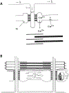
A) Excitation-contraction coupling requires energy from ATP and Ca2+ions released from Sarcoplasmic reticulum (SR) cisterna stored in calsequestrin. B) The SR is divided in two domains, the longitudinal and the junctional SR. The longitudinal SR is composed of numerous tubules inter-connected with each other forming a network around each myofibril. At their ends, the longitudinal tubules form a single dilated sac called terminal cisterna. Two terminal cisternae and one T-tubule form a triad. Both the longitudinal and the junctional SR show a specific spatial organization, being regularly aligned with specific regions associated with glycolytic pathway enzymes.
Figure 3.
Figure 3.
Occurrence (%) of symptoms associated with metabolic myopathies in 10 categories of IMDs. The percentages for myopathy involvement were calculated using as the denominator the total number of IMDs in each category presenting with any metabolic myopathy. Heat scale ranges from red (0%) for diseases with no particular symptoms reported to violet (100%) for diseases with particular symptoms occurring with highly frequency. For definition of 10 categories of disorders with metabolic myopathies see Supplemental Table S2. For interpretation of the references to color in this figure legend, the reader is referred to the web version of this article.

Similar articles

Cited by

References

    1. Ferreira CR, Hoffmann GF, Blau N, Clinical and biochemical footprints of inherited metabolic diseases. I. Movement disorders. Mol Genet Metab 127 (2019) 28–30. - PMC - PubMed
    1. Ferreira CR, Cassiman D, Blau N, Clinical and biochemical footprints of inherited metabolic diseases. II. Metabolic liver diseases. Mol Genet Metab 127 (2019) 117–121. - PMC - PubMed
    1. Horvath GA, Stowe RM, Ferreira CR, Blau N, Clinical and biochemical footprints of inherited metabolic diseases. III. Psychiatric presentations. Mol Genet Metab 130 (2020) 1–6. - PMC - PubMed
    1. Ferreira CR, Blau N, Clinical and biochemical footprints of inherited metabolic diseases. IV. Metabolic cardiovascular disease. Mol Genet Metab 132 (2021) 112–118. - PMC - PubMed
    1. Horvath GA, Blau N, Ferreira CR, Clinical and biochemical footprints of inherited metabolic disease. V. Cerebral palsy phenotypes. Mol Genet Metab (2021) DOI: 10.1016/j.ymgme.2021.03.008. - DOI - PMC - PubMed

Publication types

MeSH terms