. 2022 Sep 11:(187):10.3791/64240.
doi: 10.3791/64240.

Detection of Homologous Recombination Intermediates via Proximity Ligation and Quantitative PCR in Saccharomyces cerevisiae

Affiliations

Detection of Homologous Recombination Intermediates via Proximity Ligation and Quantitative PCR in Saccharomyces cerevisiae

Diedre Reitz et al. J Vis Exp. .

Abstract

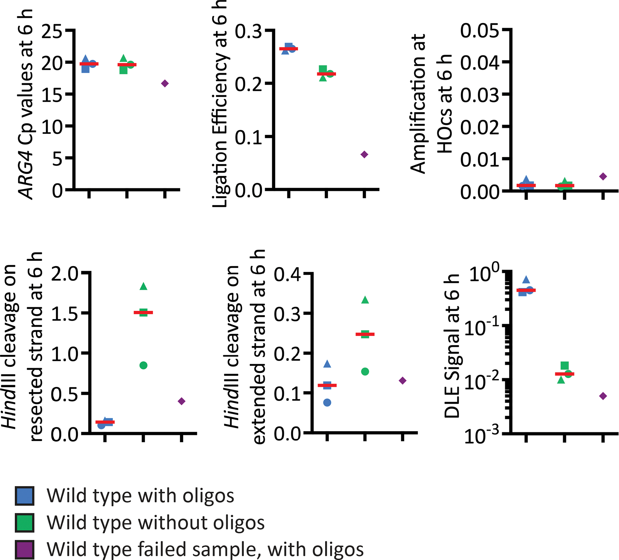
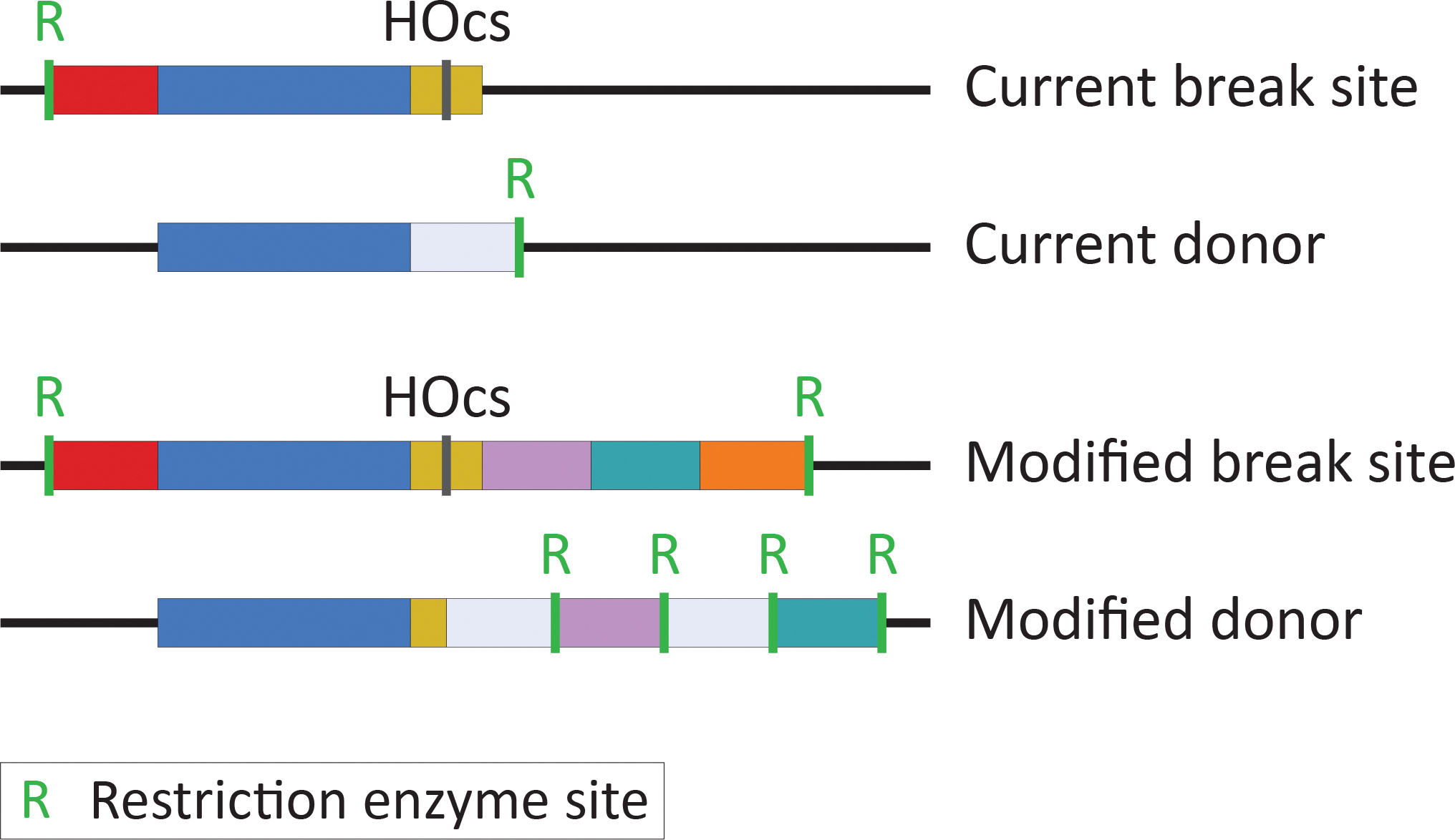
DNA damage, including DNA double-stranded breaks and inter-strand cross-links, incurred during the S and G2 phases of the cell cycle can be repaired by homologous recombination (HR). In addition, HR represents an important mechanism of replication fork rescue following stalling or collapse. The regulation of the many reversible and irreversible steps of this complex pathway promotes its fidelity. The physical analysis of the recombination intermediates formed during HR enables the characterization of these controls by various nucleoprotein factors and their interactors. Though there are well-established methods to assay specific events and intermediates in the recombination pathway, the detection of D-loop formation and extension, two critical steps in this pathway, has proved challenging until recently. Here, efficient methods for detecting key events in the HR pathway, namely DNA double-stranded break formation, D-loop formation, D-loop extension, and the formation of products via break-induced replication (BIR) in Saccharomyces cerevisiae are described. These assays detect their relevant recombination intermediates and products with high sensitivity and are independent of cellular viability. The detection of D-loops, D-loop extension, and the BIR product is based on proximity ligation. Together, these assays allow for the study of the kinetics of HR at the population level to finely address the functions of HR proteins and regulators at significant steps in the pathway.


Conflict of interest statement

DISCLOSURES:

The authors have nothing to disclose.

Figures

Figure 1:
Figure 1:. Homologous recombination and resolution sub-pathways.
Following DNA damage that results in a one- or two-ended DSB (shown) or an ssDNA gap, 5’ to 3’ resection of the DNA ends reveals 3’ ssDNA overhangs on which the Rad51 filament forms, aided by its accessory factors. Rad51 then searches the genome for an intact duplex DNA (i.e., the donor) on which to template the repair event. This process culminates in DNA strand invasion, in which the broken strand Watson-Crick base pairs with the complementary strand of the double-stranded DNA donor, displacing the opposite strand and forming the nascent D-loop. This D-loop can either be reversed to allow a Rad51 homology search to select a different donor or extended by a DNA polymerase to replace the bases lost during the DNA damage event. Three HR sub-pathways are available to resolve this extended D-loop intermediate into a product. First, the extended D-loop can be disrupted by a helicase, permitting the newly extended end of the break to anneal to the second end in a process termed synthesis-dependent strand annealing (SDSA). Fill-in DNA synthesis and ligation then lead to product formation. Alternatively, the second end of the break can anneal to the displaced donor strand, forming a double-Holliday junction (dHJ). Nucleolytic resolution of the dHJ results in either a crossover (CO) or non-crossover (NCO), whereas dHJ dissolution (not shown) results in only NCO products. Lastly, failure to engage the second end of the DSB results in break-induced replication (BIR), a mutagenic process in which thousands of base pairs are copied from the donor onto the broken strand. This process can extend as far as the converging replication fork or the end of the chromosome.
Figure 2:
Figure 2:. The premise of the D-loop capture (DLC), D-loop extension (DLE), and break-induced replication (BIR) product formation assays.
DSB formation is driven by a site-specific endonuclease under the control of the GAL1 promoter. DSB induction leads to the formation of a nascent D-loop. In the DLC assay, inter-strand crosslinking of the DNA preserves this structure, which is then extracted. Restriction enzyme site restoration is achieved via hybridization with a long oligonucleotide, and then the DNA is digested and ligated to form a product that can be quantified by quantitative PCR (qPCR). The DLE assay differs in that the DNA is not cross-linked, and instead, the intramolecular ligation product forms between the two ends of the ssDNA on one side of the break, the 3’ end having been extended by a DNA polymerase. qPCR is again used to quantify the formation of the chimeric ligation product. The detection of D-loop extension via the DLE assay likewise requires restriction enzyme site restoration. In contrast, the double-stranded BIR product is detected using the DLE assay primers without the hybridizing oligonucleotides. R indicates that a restriction enzyme site is competent for enzyme cleavage; (R) indicates a restriction enzyme site that cannot be cut.
Figure 3:
Figure 3:. Representative results from DLC assay analysis of D-loops at 2 h post-DSB induction.
Samples were collected, prepared, and analyzed by qPCR as described in this protocol. Blue symbols represent results for the standard wild-type strain with hybridizing oligos for n = 3. Green symbols represent results for the wild-type strain without hybridizing oligos for n = 3. The thick red line shows the median. The purple symbols represent samples without psoralen crosslinking but with hybridizing oligos for n = 2. Symbols indicate that the samples are derived from the same culture. Inter-experimental differences in crosslinking efficiency can introduce variability into certain qPCR controls but are not problematic as long as there is no inter-sample variability in these qPCR controls within an experiment.
Figure 4:
Figure 4:. Representative results from DLE assay analysis 6 h post-DSB induction.
Samples were collected, prepared, and analyzed by qPCR as described in this protocol. Blue symbols represent results for the standard wild-type strain with hybridizing oligos for n = 3. Green symbols represent results for the wild-type strain without hybridizing oligos for n = 3. The thick red line shows the median. Note that the with- and without-hybridizing oligos samples are derived from the same cultures. The purple diamond represents a failed sample without hybridizing oligos for n = 1. Symbols indicate the samples are derived from the same culture.
Figure 5:
Figure 5:. Representative results from psoralen crosslink reversal.
(A) Psoralen-DNA mono-adducts (*) and inter-strand crosslinks (X) specifically occur on dsDNA and prevent its amplification by DNA polymerases, unlike ssDNA templates. This difference introduces a bias in the quantification of dsDNA- and ssDNA-containing templates by qPCR. This bias can be overcome upon reversal of the psoralen crosslink. (B) Representative Cp values of dsDNA (loading, circular), ssDNA, and mixed ds-ssDNA (DLC) amplicons obtained 4 h post-DSB induction. Data represent individual values and the median of four biological replicates. (C) Amplification recovery upon crosslink reversal, calculated from the Cp values in (B). (D) The ssDNA amplification relative to the dsDNA loading control with and without psoralen crosslink reversal. Upon reversal, the ssDNA amplicon amplifies at the expected 0.5 of the dsDNA loading control. (E) The dsDNA circularization control relative to the dsDNA loading control with and without psoralen crosslink reversal. (F) The DLC signal relative to the dsDNA circularization control.
Figure 6:
Figure 6:. Current DLC/DLE assay system and the proposed modifications.
Above: Current DLC/DLE assay break site and donor are shown. Below: Planned modifications to the DLC/DLE assay break site and donor. (I) The 117 bp HO endonuclease cut site is indicated in yellow. To prevent confounding effects while monitoring D-loop disruption, the left side of the HOcs (74 bp) will be introduced into the donor, such that recombination between the two creates a perfectly matched D-loop lacking a 3’ flap. (II) To make the system repairable and, thus, more physiological, DNA homologous to the donor (indicated in teal and lilac) will be inserted into the right side of the HOcs. (III) Invasion and extension by the strand to the right of the HOcs will be monitored using sequences unique to that side of the break (indicated in orange). (IV) Additional evenly spaced restriction enzyme sites and sequences unique to the donor will allow D-loop extension (via invasion from the left side of the HOcs) to be monitored at more distant sites. In this modified system, synthesis-dependent strand annealing (SDSA) or double-Holliday junction (dHJ) formation can occur at the sites shown in teal or lilac.

References

    1. Symington LS, Gautier J Double-strand break end resection and repair pathway choice. Annual Review of Genetics. 45 (1), 247–271 (2011). - PubMed
    1. Heyer W-D. Regulation of recombination and genomic maintenance. Cold Spring Harbor Perspectives in Biology. 7 (8), a016501 (2015). - PMC - PubMed
    1. Mimitou EP, Symington LS Sae2, Exo1 and Sgs1 collaborate in DNA double-strand break processing. Nature. 455 (7214), 770–774 (2008). - PMC - PubMed
    1. Bzymek M, Thayer NH, Oh SD, Kleckner N, Hunter N Double Holliday junctions are intermediates of DNA break repair. Nature. 464 (7290), 937–941 (2010). - PMC - PubMed
    1. Chen H, Lisby M, Symington LS RPA coordinates DNA end resection and prevents formation of DNA hairpins. Molecular Cell. 50 (4), 589–600 (2013). - PMC - PubMed

Publication types

MeSH terms