

Application of optical coherence tomography angiography to assess systemic severity in patients with hereditary transthyretin amyloidosis
- PMID: 36156600
- PMCID: PMC9512205
- DOI: 10.1371/journal.pone.0275180
Application of optical coherence tomography angiography to assess systemic severity in patients with hereditary transthyretin amyloidosis
Abstract
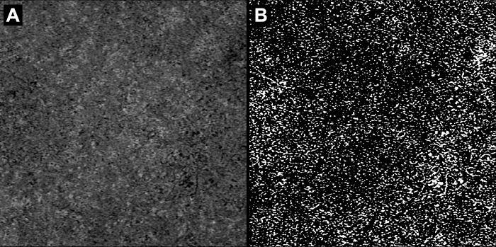
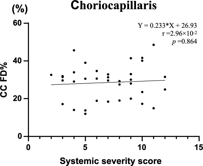
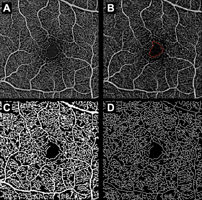
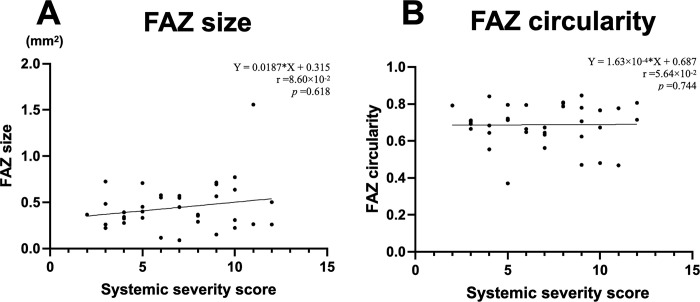
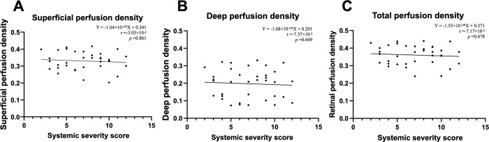
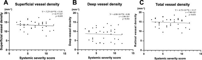
Hereditary transthyretin amyloidosis is an autosomal dominant form of amyloidosis caused by an abnormality in transthyretin, with various ocular manifestations. Among these, ocular amyloid angiopathy has attracted attention because of its direct link to visual impairment and its correlation with systemic severity. We hypothesized that optical coherence tomography angiographic parameters would be useful biomarkers of amyloidosis systemic severity and investigated their correlation with the systemic severity score. The primary outcome was the correlation between the systemic severity score and choriocapillaris flow deficit percentage. Secondary outcomes were the correlations between the systemic severity score and retinal optical coherence tomography angiographic parameters, including foveal avascular zone size and circularity and superficial/deep/total retinal perfusion and vessel densities. The choroidal and retinal vasculature was quantified in 36 eyes from 36 patients (age, 51.8±12.1 years; disease duration, 13.4±6.2 years). Ten eyes had a history of vitrectomy for vitreous opacity. Choriocapillaris flow deficit percentage was not significantly correlated with the systemic severity score (Spearman's rank correlation: r = 2.96×10⁻², p = 0.863). Similarly, foveal avascular zone size and circularity, and superficial/deep/total retinal perfusion and vessel densities were not significantly correlated with the systemic severity score. These results may indicate that optical coherence tomography angiographic parameters are not sufficient to predict amyloidosis severity.
Conflict of interest statement
The authors have declared that no competing interests exist.
Figures
Similar articles
-
Subclinical retinal angiopathy associated with hereditary transthyretin amyloidosis - assessed with optical coherence tomography angiography.Amyloid. 2021 Mar;28(1):66-71. doi: 10.1080/13506129.2020.1827381. Epub 2020 Sep 30. Amyloid. 2021. PMID: 32996337
-
OCULAR ANGIOGRAPHIC FEATURES IN JAPANESE PATIENTS WITH VAL30MET HEREDITARY TRANSTHYRETIN AMYLOIDOSIS.Retina. 2022 Jan 1;42(1):210-215. doi: 10.1097/IAE.0000000000003291. Retina. 2022. PMID: 34483312
-
Deposits on Retinal Surface Seen on OCT in Ocular Amyloidosis.Ophthalmol Retina. 2021 Oct;5(10):1005-1008. doi: 10.1016/j.oret.2020.12.028. Epub 2021 Jan 7. Ophthalmol Retina. 2021. PMID: 33422693
-
Quantitative Retinal Optical Coherence Tomography Angiography in Patients With Diabetes Without Diabetic Retinopathy.Invest Ophthalmol Vis Sci. 2017 Jan 1;58(1):190-196. doi: 10.1167/iovs.16-20531. Invest Ophthalmol Vis Sci. 2017. PMID: 28114579
-
VISUAL FUNCTION AND OPTICAL COHERENCE TOMOGRAPHY ANGIOGRAPHY FEATURES IN CHILDREN BORN PRETERM.Retina. 2019 Nov;39(11):2233-2239. doi: 10.1097/IAE.0000000000002301. Retina. 2019. PMID: 30180146
Cited by
-
Progression of Capillary Hypoperfusion in Advanced Stages of Nonproliferative Diabetic Retinopathy: 6-month Analysis of RICHARD Study.Ophthalmol Sci. 2024 Oct 16;5(2):100632. doi: 10.1016/j.xops.2024.100632. eCollection 2025 Mar-Apr. Ophthalmol Sci. 2024. PMID: 39639890 Free PMC article.
References
MeSH terms
Substances
Supplementary concepts
LinkOut - more resources
Full Text Sources
Research Materials
