

Normal Corneal Thickness and Endothelial Cell Density in Rhesus Macaques (Macaca mulatta)
- PMID: 36156731
- PMCID: PMC9526363
- DOI: 10.1167/tvst.11.9.23
Normal Corneal Thickness and Endothelial Cell Density in Rhesus Macaques (Macaca mulatta)
Abstract
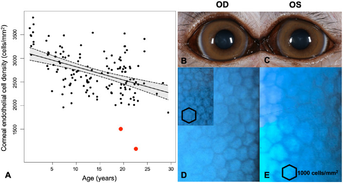
Purpose: To define the normal range of central corneal thickness (CCT) and corneal endothelial cell density (ECD) in rhesus macaques (Macaca mulatta) and the effects of age, body weight, sex, and intraocular pressure (IOP) on these parameters.
Methods: Ophthalmic examinations were performed on 144 rhesus macaques without anterior segment pathology. The CCT was measured via ultrasound pachymetry (USP) and specular microscopy, and the ECD was semiautomatically and manually counted using specular microscopy. Rebound tonometry was used to measure IOP. Linear regression and mixed-effects linear regression models were used to evaluate the effects of age, body weight, sex, and IOP on CCT and ECD.
Results: We included 98 females and 46 males with an age range of 0.2 to 29.4 years. The mean CCT by USP and specular microscopy were 483 ± 39 and 463 ± 33 µm, respectively, and were statistically different (P < 0.001). The ECDs were 2717 ± 423 and 2747 ± 438 cells/mm2 by semiautomated and manual analysis, respectively. Corneal endothelial degeneration was identified in one aged rhesus macaque.
Conclusions: The mean USP and specular microscopy CCT values differed significantly, whereas the semiautomatic and manual ECD did not. The CCT was associated with the IOP and sex, whereas the ECD was associated with body weight and age (P < 0.05). As in humans, corneal disease in rhesus macaques is uncommon.
Translational relevance: Establishing reference values is fundamental to use rhesus macaques as a model for corneal disease or to identify toxicity in studies of ocular drugs or devices.
Conflict of interest statement
Disclosure:
Figures


Similar articles
-
Corneal thickness and endothelial cell density measured by non-contact specular microscopy and pachymetry in Rhesus macaques (Macaca mulatta) with laser-induced ocular hypertension.Exp Eye Res. 2003 Jun;76(6):671-7. doi: 10.1016/s0014-4835(03)00055-1. Exp Eye Res. 2003. PMID: 12742349
-
Comparison of Pentacam Scheimpflug camera with ultrasound pachymetry and noncontact specular microscopy in measuring central corneal thickness.Curr Eye Res. 2007 Feb;32(2):89-94. doi: 10.1080/02713680601115010. Curr Eye Res. 2007. PMID: 17364741
-
Comparison between laser scanning in vivo confocal microscopy and noncontact specular microscopy in assessing corneal endothelial cell density and central corneal thickness.Cornea. 2011 Jul;30(7):754-9. doi: 10.1097/ICO.0b013e3182000c5d. Cornea. 2011. PMID: 21150426
-
Human corneal thickness and its impact on intraocular pressure measures: a review and meta-analysis approach.Surv Ophthalmol. 2000 Mar-Apr;44(5):367-408. doi: 10.1016/s0039-6257(00)00110-7. Surv Ophthalmol. 2000. PMID: 10734239 Review.
-
Age-Associated Pathology in Rhesus Macaques (Macaca mulatta).Vet Pathol. 2016 Mar;53(2):399-416. doi: 10.1177/0300985815620628. Epub 2016 Feb 10. Vet Pathol. 2016. PMID: 26864889 Free PMC article. Review.
Cited by
-
Safety and biocompatibility of a novel biodegradable aflibercept-drug delivery system in rhesus macaques.Drug Deliv. 2025 Dec;32(1):2460671. doi: 10.1080/10717544.2025.2460671. Epub 2025 Mar 4. Drug Deliv. 2025. PMID: 40038090 Free PMC article.
-
Tolerability and tropism of recombinant adeno-associated virus vectors in the African green monkey (Chlorocebus sabaeus) anterior chamber.Gene Ther. 2023 Sep;30(9):714-722. doi: 10.1038/s41434-023-00407-z. Epub 2023 May 24. Gene Ther. 2023. PMID: 37221271 Free PMC article.
-
Squishy matters - Corneal mechanobiology in health and disease.Prog Retin Eye Res. 2024 Mar;99:101234. doi: 10.1016/j.preteyeres.2023.101234. Epub 2024 Jan 2. Prog Retin Eye Res. 2024. PMID: 38176611 Free PMC article. Review.
-
Choroidal Features of Cynomolgus Macaques in Relation to Age and Axial Length.Invest Ophthalmol Vis Sci. 2025 Jul 1;66(9):17. doi: 10.1167/iovs.66.9.17. Invest Ophthalmol Vis Sci. 2025. PMID: 40626809 Free PMC article.
References
Publication types
MeSH terms
Grants and funding
LinkOut - more resources
Full Text Sources
Research Materials
