

The combination of a 3D-Printed porous Ti-6Al-4V alloy scaffold and stem cell sheet technology for the construction of biomimetic engineered bone at an ectopic site
- PMID: 36157052
- PMCID: PMC9493059
- DOI: 10.1016/j.mtbio.2022.100433
The combination of a 3D-Printed porous Ti-6Al-4V alloy scaffold and stem cell sheet technology for the construction of biomimetic engineered bone at an ectopic site
Abstract
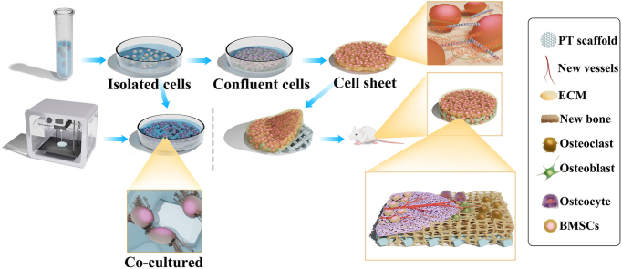
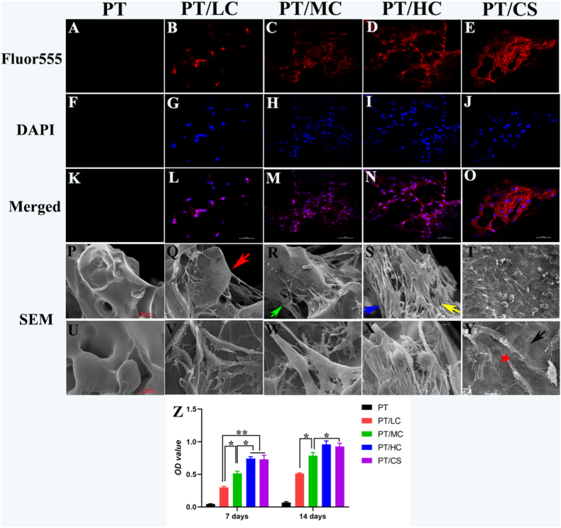
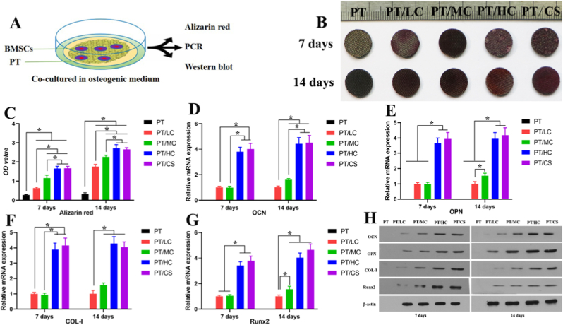
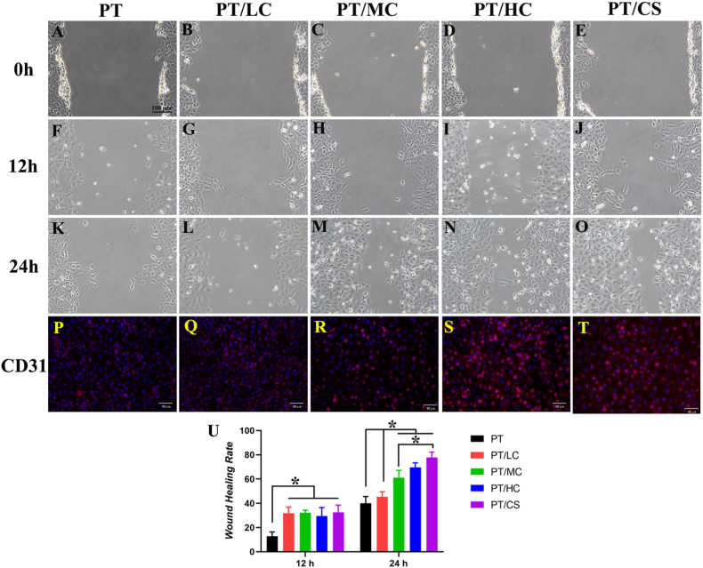
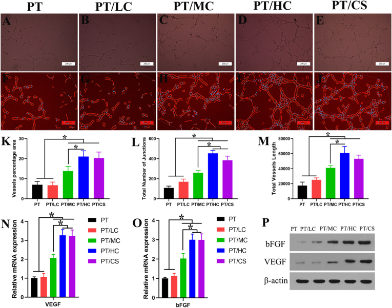
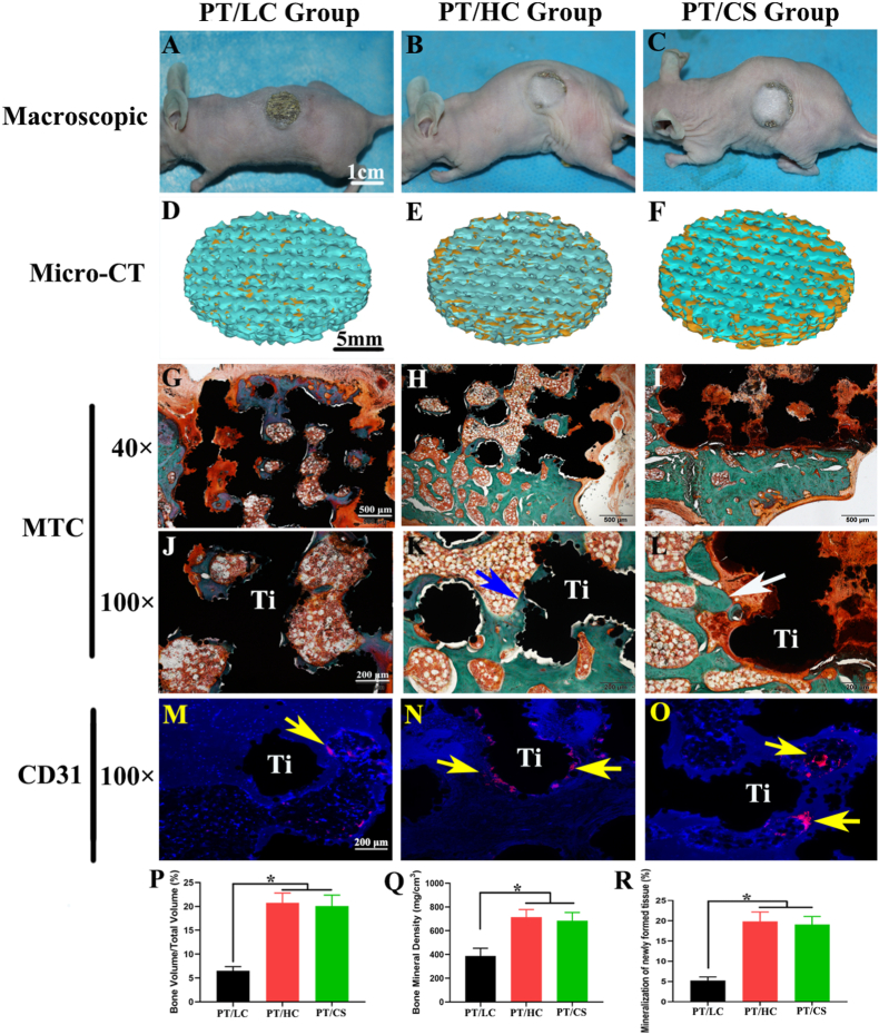
Cell sheet technology has been widely used in bone tissue engineering and regenerative medicine. However, controlling the shape and volume of large pieces of engineered bone tissue remains impossible without additional suitable scaffolds. Three-dimensional (3D) printed titanium (Ti) alloy scaffolds are mostly used as implant materials for repairing bone defects, but the unsatisfactory bioactivities of traditional Ti-based scaffolds severely limit their clinical applications. Herein, we hypothesize that the combination of bone marrow mesenchymal stem cell (BMSC) sheet technology and 3D porous Ti-6Al-4V (PT) alloy scaffolds could be used to fabricate biomimetic engineered bone. First, various concentrations of BMSCs were directly cocultured with PT scaffolds to obtain complexes of osteoblastic cell sheets and scaffolds. Then, as an experimental control, an osteoblastic BMSC sheet was prepared by continuous culturing under osteogenic conditions for 2 weeks without passaging and used to wrap the scaffolds. The BMSC sheet was composed of several layers of extracellular matrix (ECM) and a mass of BMSCs. The BMSCs exhibited excellent adherent, proliferative and osteogenic potential when cocultured with PT scaffolds, which may be attributed to the ability of the 3D microstructure of scaffolds to facilitate the biological behaviors of cells, as confirmed by the in vitro results. Moreover, the presence of BMSCs and ECM increased the angiogenic potential of PT scaffolds by the secretion of VEGF. Micro-CT and histological analysis confirmed the in vivo formation of biomimetic engineered bone when the complex of cocultured BMSCs and PT scaffolds and the scaffolds wrapped by prepared BMSC sheets were implanted subcutaneously into nude mice. Therefore, the combination of BMSC sheet technology and 3D-printed PT scaffolds could be used to construct customized biomimetic engineered bone, offering a novel and promising strategy for the precise repair of bone defects.
Keywords: 3D-printed porous titanium alloy scaffold; Angiogenesis; Biomimetic engineered bone; Cell sheet technology; Osteogenesis.
© 2022 The Authors. Published by Elsevier Ltd.
Conflict of interest statement
The authors declare that they have no known competing financial interests or personal relationships that could have appeared to influence the work reported in this paper.
Figures
Similar articles
-
Biomimetic Ti-6Al-4V alloy/gelatin methacrylate hybrid scaffold with enhanced osteogenic and angiogenic capabilities for large bone defect restoration.Bioact Mater. 2021 Mar 21;6(10):3437-3448. doi: 10.1016/j.bioactmat.2021.03.010. eCollection 2021 Oct. Bioact Mater. 2021. PMID: 33817419 Free PMC article.
-
Stem Cell-Seeded 3D-Printed Scaffolds Combined with Self-Assembling Peptides for Bone Defect Repair.Tissue Eng Part A. 2022 Feb;28(3-4):111-124. doi: 10.1089/ten.TEA.2021.0055. Epub 2021 Dec 30. Tissue Eng Part A. 2022. PMID: 34157886
-
Integrating 3D Printing and Biomimetic Mineralization for Personalized Enhanced Osteogenesis, Angiogenesis, and Osteointegration.ACS Appl Mater Interfaces. 2018 Dec 12;10(49):42146-42154. doi: 10.1021/acsami.8b17495. Epub 2018 Dec 3. ACS Appl Mater Interfaces. 2018. PMID: 30507136 Free PMC article.
-
3D-printed porous Ti6Al4V scaffolds for long bone repair in animal models: a systematic review.J Orthop Surg Res. 2022 Feb 2;17(1):68. doi: 10.1186/s13018-022-02960-6. J Orthop Surg Res. 2022. PMID: 35109907 Free PMC article.
-
Three-dimensional (3D) printed scaffold and material selection for bone repair.Acta Biomater. 2019 Jan 15;84:16-33. doi: 10.1016/j.actbio.2018.11.039. Epub 2018 Nov 24. Acta Biomater. 2019. PMID: 30481607 Review.
Cited by
-
Advanced surface modification techniques for titanium implants: a review of osteogenic and antibacterial strategies.Front Bioeng Biotechnol. 2025 Mar 19;13:1549439. doi: 10.3389/fbioe.2025.1549439. eCollection 2025. Front Bioeng Biotechnol. 2025. PMID: 40177619 Free PMC article. Review.
-
Long Bone Defect Filling with Bioactive Degradable 3D-Implant: Experimental Study.Biomimetics (Basel). 2023 Mar 28;8(2):138. doi: 10.3390/biomimetics8020138. Biomimetics (Basel). 2023. PMID: 37092390 Free PMC article.
-
Fabrication and properties of PLA/β-TCP scaffolds using liquid crystal display (LCD) photocuring 3D printing for bone tissue engineering.Front Bioeng Biotechnol. 2024 Feb 19;12:1273541. doi: 10.3389/fbioe.2024.1273541. eCollection 2024. Front Bioeng Biotechnol. 2024. PMID: 38440328 Free PMC article.
-
Raising the Bar: Progress in 3D-Printed Hybrid Bone Scaffolds for Clinical Applications: A Review.Cell Transplant. 2024 Jan-Dec;33:9636897241273562. doi: 10.1177/09636897241273562. Cell Transplant. 2024. PMID: 39517106 Free PMC article. Review.
-
New insight into biodegradable macropore filler on tuning mechanical properties and bone tissue ingrowth in sparingly dissolvable bioceramic scaffolds.Mater Today Bio. 2023 Dec 28;24:100936. doi: 10.1016/j.mtbio.2023.100936. eCollection 2024 Feb. Mater Today Bio. 2023. PMID: 38234459 Free PMC article.
References
-
- Ruales-Carrera E., Engler M., Vaz P., Ozcan M., Volpato C.A.M. Esthetic and functional rehabilitation of bilateral congenital absence of maxillary lateral incisors: minimally invasive surgical and prosthetic approach. J. Esthetic Restor. Dent. 2019;31(1):5–12. - PubMed
-
- Wang Z., Weng Y., Lu S., Zong C., Qiu J., Liu Y., et al. Osteoblastic mesenchymal stem cell sheet combined with Choukroun platelet-rich fibrin induces bone formation at an ectopic site. J. Biomed. Mater. Res. B Appl. Biomater. 2015;103(6):1204–1216. - PubMed
-
- Biggemann J., Pezoldt M., Stumpf M., Greil P., Fey T. Modular ceramic scaffolds for individual implants. Acta Biomater. 2018;80:390–400. - PubMed
-
- Sun Q., Li Z., Liu B., Yuan X., Guo S., Helms J.A. Improving intraoperative storage conditions for autologous bone grafts: an experimental investigation in mice. J Tissue Eng Regen Med. 2019;13(12):2169–2180. - PubMed
-
- Ikeguchi R., Aoyama T., Kakinoki R., Ueda M., Kasai Y., Maekawa T., et al. A clinical trial for Kienbock disease by cultured autologous multipotent mesenchymal stromal cells augmented with vascularized bone grafts: a report of five cases. J. Orthop. Sci. 2019;24(4):750–756. - PubMed
LinkOut - more resources
Full Text Sources
