

Emerging roles of keratinocytes in nociceptive transduction and regulation
- PMID: 36157074
- PMCID: PMC9500148
- DOI: 10.3389/fnmol.2022.982202
Emerging roles of keratinocytes in nociceptive transduction and regulation
Abstract
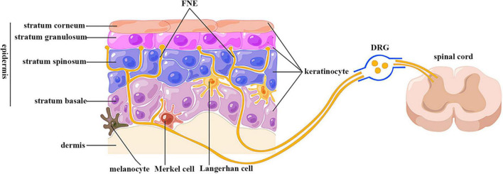
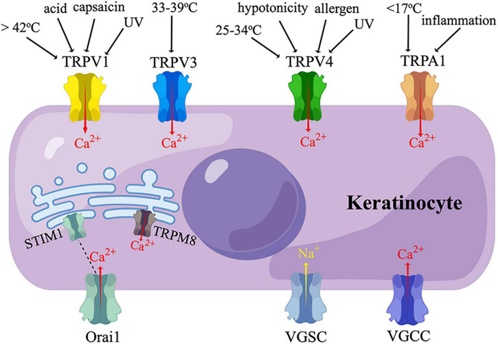
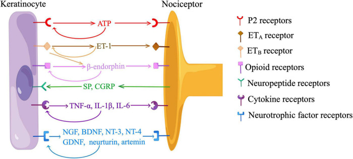
Keratinocytes are the predominant block-building cells in the epidermis. Emerging evidence has elucidated the roles of keratinocytes in a wide range of pathophysiological processes including cutaneous nociception, pruritus, and inflammation. Intraepidermal free nerve endings are entirely enwrapped within the gutters of keratinocyte cytoplasm and form en passant synaptic-like contacts with keratinocytes. Keratinocytes can detect thermal, mechanical, and chemical stimuli through transient receptor potential ion channels and other sensory receptors. The activated keratinocytes elicit calcium influx and release ATP, which binds to P2 receptors on free nerve endings and excites sensory neurons. This process is modulated by the endogenous opioid system and endothelin. Keratinocytes also express neurotransmitter receptors of adrenaline, acetylcholine, glutamate, and γ-aminobutyric acid, which are involved in regulating the activation and migration, of keratinocytes. Furthermore, keratinocytes serve as both sources and targets of neurotrophic factors, pro-inflammatory cytokines, and neuropeptides. The autocrine and/or paracrine mechanisms of these mediators create a bidirectional feedback loop that amplifies neuroinflammation and contributes to peripheral sensitization.
Keywords: free nerve ending; keratinocyte; neuroinflammation; neuropeptide; neurotransmitter; peripheral sensitization.
Copyright © 2022 Xu, Yu, Xu and Xu.
Conflict of interest statement
The authors declare that the research was conducted in the absence of any commercial or financial relationships that could be construed as a potential conflict of interest.
Figures
Similar articles
-
Intra-epidermal nerve endings progress within keratinocyte cytoplasmic tunnels in normal human skin.Exp Dermatol. 2020 Apr;29(4):387-392. doi: 10.1111/exd.14081. Epub 2020 Feb 14. Exp Dermatol. 2020. PMID: 32003039
-
Keratinocytes Communicate with Sensory Neurons via Synaptic-like Contacts.Ann Neurol. 2020 Dec;88(6):1205-1219. doi: 10.1002/ana.25912. Epub 2020 Oct 10. Ann Neurol. 2020. PMID: 32951274
-
Keratinocytes express cytokines and nerve growth factor in response to neuropeptide activation of the ERK1/2 and JNK MAPK transcription pathways.Regul Pept. 2013 Sep 10;186:92-103. doi: 10.1016/j.regpep.2013.08.001. Epub 2013 Aug 17. Regul Pept. 2013. PMID: 23958840 Free PMC article.
-
Anatomical contacts between sensory neurons and epidermal cells: an unrecognized anatomical network for neuro-immuno-cutaneous crosstalk.Br J Dermatol. 2023 Feb 10;188(2):176-185. doi: 10.1093/bjd/ljac066. Br J Dermatol. 2023. PMID: 36763869 Review.
-
What about physical contacts between epidermal keratinocytes and sensory neurons?Exp Dermatol. 2018 Jan;27(1):9-13. doi: 10.1111/exd.13411. Epub 2017 Oct 19. Exp Dermatol. 2018. PMID: 28767170 Review.
Cited by
-
Communicating pain: emerging axonal signaling in peripheral neuropathic pain.Front Neuroanat. 2024 Jul 9;18:1398400. doi: 10.3389/fnana.2024.1398400. eCollection 2024. Front Neuroanat. 2024. PMID: 39045347 Free PMC article. Review.
-
IL-33/ST2 induces macrophage-dependent ROS production and TRPA1 activation that mediate pain-like responses by skin incision in mice.Theranostics. 2024 Aug 19;14(13):5281-5302. doi: 10.7150/thno.97856. eCollection 2024. Theranostics. 2024. PMID: 39267790 Free PMC article.
-
NMDA Receptors Regulate Oxidative Damage in Keratinocytes during Complex Regional Pain Syndrome in HaCaT Cells and Male Rats.Antioxidants (Basel). 2024 Feb 18;13(2):244. doi: 10.3390/antiox13020244. Antioxidants (Basel). 2024. PMID: 38397842 Free PMC article.
-
Review of sensory systems deployed by epidermal keratinocytes.Front Cell Dev Biol. 2025 Jun 2;13:1598326. doi: 10.3389/fcell.2025.1598326. eCollection 2025. Front Cell Dev Biol. 2025. PMID: 40530329 Free PMC article. Review.
-
Evaluation of Kynu, Defb2, Camp, and Penk Expression Levels as Psoriasis Marker in the Imiquimod-Induced Psoriasis Model.Mediators Inflamm. 2024 Jul 16;2024:5821996. doi: 10.1155/2024/5821996. eCollection 2024. Mediators Inflamm. 2024. PMID: 39045230 Free PMC article.
References
-
- Anand P., Terenghi G., Warner G., Kopelman P., Williams-Chestnut R. E., Sinicropi D. V. (1996). The role of endogenous nerve growth factor in human diabetic neuropathy. Nat. Med. 2 703–707. - PubMed
Publication types
Grants and funding
LinkOut - more resources
Full Text Sources
Research Materials