. 2022 Sep 9:3:877553.
doi: 10.3389/falgy.2022.877553. eCollection 2022.

Gibberellin-regulated proteins: Emergent allergens

Affiliations

Gibberellin-regulated proteins: Emergent allergens

T Iizuka et al. Front Allergy. .

Abstract

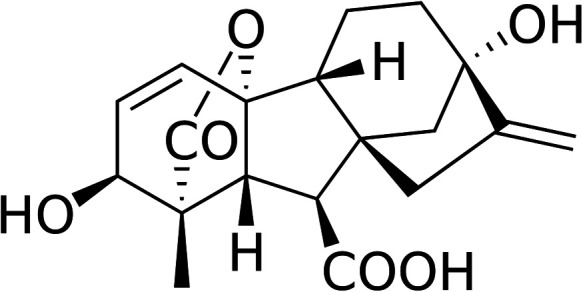
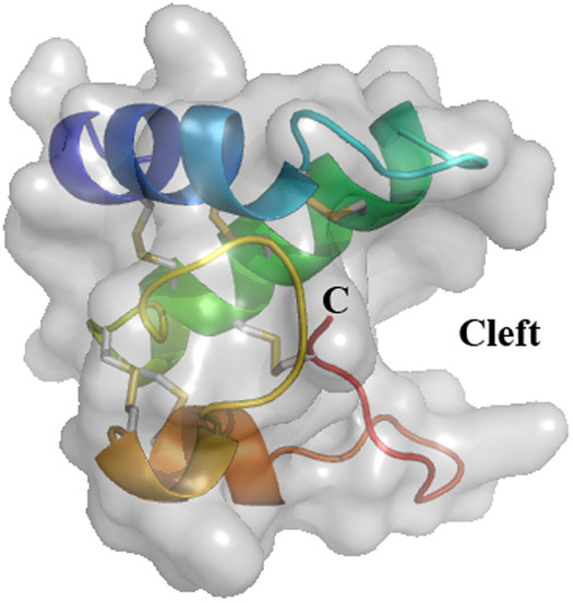
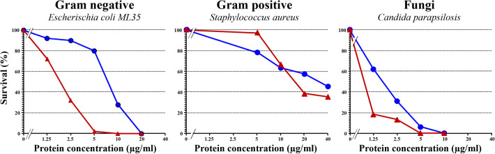
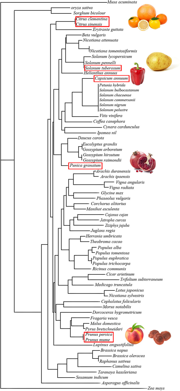
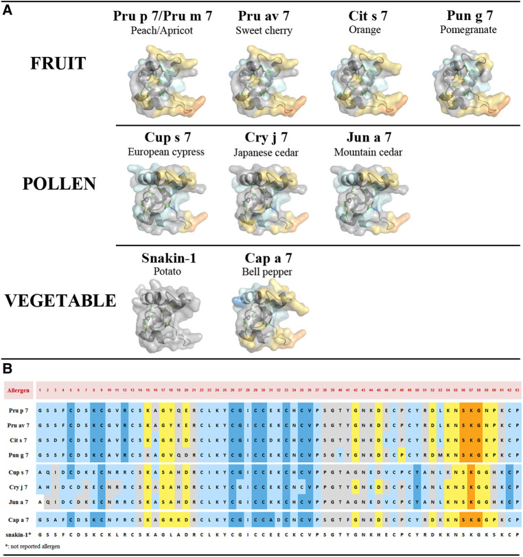
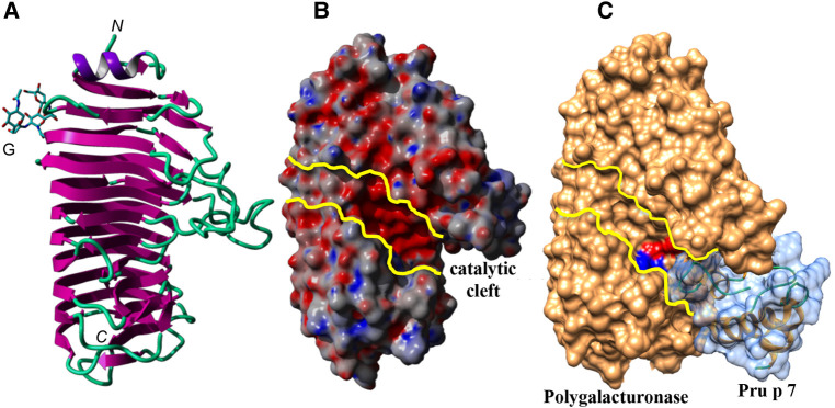
About 10 years ago, a protein family was shown for the first time to contain allergenic members, gibberellin-regulated protein (GRP). The first reported member was from peach, Pru p 7. One can hypothesize that it was not detected before because its physicochemical characteristics overlap with those of lipid transfer protein (LTP), a well-known allergen, or because the exposure to GRP increased due to an increase in the gibberellin phythormone level in plant food, either exogenous or endogenous. Like LTPs, GRPs are small cationic proteins with disulfide bridges, are resistant to heat and proteolytic cleavage, and are involved in the defense of the plant. Besides peach, GRP allergens have been described in Japanese apricot (Pru m 7), sweet cherry (Pru av 7), orange (Cit s 7), pomegranate (Pun g 7), bell pepper (Cap a 7), strawberry (Fra a GRP), and also in pollen with a restriction to Cupressaceae tree family (Cup s 7, Cry j 7, and Jun a 7). IgE cross-reactivities were described between GRPs, and the reported peach/cypress and citrus/cypress syndromes may therefore be explained because of these GRP cross-reactivities. GRPs are clinically relevant, and severe adverse reactions may sometimes occur in association with cofactors. More than 60% and up to 95% sequence identities are calculated between various allergenic GRPs, and three-dimensional models show a cleft in the molecule and predict at least three epitopic regions. The structure of the protein and its properties and the matrix effect in the original allergenic source should be unraveled to understand why, despite the ubiquity of the protein family in plants, only a few members are able to sensitize patients.

Keywords: 3D structure; food allergy; gibberellin-regulated protein; pollen allergy; pollen food allergy syndrome.


Conflict of interest statement

The authors declare that the research was conducted in the absence of any commercial or financial relationships that could be construed as a potential conflict of interest.

Figures

Figure 1
Figure 1
Gibberellin acid (GA3), one of the active phythormone in plants. The commercially available GA3 is one of the most used active gibberellin for plant treatment.
Figure 2
Figure 2
Three-dimensional structure of snakin-1 (PDB 5E5Q). Ribbon representation with surface obtained from crystallography data show three α-helices colored in blue (S2–C13), green (L18–K34), and orange (N43–E46 and C47–K53). The cleft is indicated where a putative ligand could bind. The six disulfide bridges are shown in gray/yellow small bars. C: C-terminal end of the protein. N-terminal is masked by the first α-helix (dark blue).
Figure 3
Figure 3
Antimicrobial activity of recombinant Pru p 7 (blue circles) in comparison with snakin-2 (red triangles), a cysteine-rich protein from potato reported to exhibit antimicrobial properties and thus considered as a positive control in the experiment. Various concentrations of recombinant Pru p 7 and snakin-2 were mixed with cell suspension in potassium phosphate buffer (10 mM, pH 6.0) and incubated at 37°C for bacteria (E. coli and S. aureus) and 30°C for fungi (C. parapsilosis) for 1 h. After incubation, the reaction mixture was diluted and plated on agar plates. Tryptic soy broth plates were used for bacteria and Sabouraud agar plates for fungi. After incubation, colonies were counted for calculating the survival rates. A representative experiment is shown and each point was performed in simplicate.
Figure 4
Figure 4
Unrooted tree built up from the multiple alignment of plant food GRPs using the neighboring joining method, showing the phylogenetic relationships among the GRPs of different plant families. GRPs with similar amino-acid sequences are grouped in closely related clusters. Plant food with red frame contains studied GRPs.
Figure 5
Figure 5
(A) Three-dimensional structure modeling of allergenic GRPs inferred by homology modeling using snakin-1 (5E5Q from PDB) as a template with SWISS-MODEL. At least three conformational epitopic regions are predicted using the software DiscoTope 2.0. They are colored yellow and orange. (B) GRP allergen sequences with predicted AA involved in epitopic regions. The color codes correspond to DiscoTope propensity scores (62). The highest the score is the highest the propensity to be an epitope is. In ascending order: blue (−20 or less), light blue (−20 to −15), gray (−15 to −12.5), yellow (−12.5 to −5), and orange (greater than −5).
Figure 6
Figure 6
(A) Ribbon diagram of the endopolygalacturonase of F. moniliforme. The N-glycan chain linked to the β-prism backbone is indicated. N and C indicate the N-terminal and C-terminal ends of the polypeptide chain, respectively. (B) Molecular surface of the endopolygalacturonase showing the catalytic cleft, delineated by yellow lines. (C) Docking of Pru p 7 (colored light blue) to the endopolygalacturonase of F. moniliforme (colored orange). The amino-acid residues involved in the catalytic cleavage of polygalacturonase chains, are colored red (D191, D212, D213) and blue (R267, K269), respectively. Docking experiments of the modeled Pru p 7 to the endopolygalacturonase of F. moniliforme (PDB code 1HG8) (73) used as a target, were performed with GRAMM_X (74, 75) and displayed with Chimera.

References

    1. Inomata N, Miyakawa M, Aihara M. Eyelid edema as a predictive factor for sensitization to Pru p 7 in peach allergy. J Dermatol. (2016) 43(8):900–5. 10.1111/1346-8138.13316 - DOI - PubMed
    1. Tuppo L, Alessandri C, Pomponi D, Picone D, Tamburrini M, Ferrara R, et al. Peamaclein—a new peach allergenic protein: similarities, differences and misleading features compared to Pru p 3. Clin Exp Allergy. (2013) 43(1):128–40. 10.1111/cea.12028 - DOI - PubMed
    1. Tanaka N, Konishi H, Khan MM, Komatsu S. Proteome analysis of rice tissues by two-dimensional electrophoresis: an approach to the investigation of gibberellin regulated proteins. Mol Genet Genomics. (2004) 270(6):485–96. 10.1007/s00438-003-0929-9 - DOI - PubMed
    1. Nahirnak V, Almasia NI, Hopp HE, Vazquez-Rovere C. Snakin/GASA proteins: involvement in hormone crosstalk and redox homeostasis. Plant Signal Behav. (2012) 7(8):1004–8. 10.4161/psb.20813 - DOI - PMC - PubMed
    1. Segura A, Moreno M, Madueno F, Molina A, Garcia-Olmedo F. Snakin-1, a peptide from potato that is active against plant pathogens. Mol Plant Microbe Interact. (1999) 12(1):16–23. 10.1094/MPMI.1999.12.1.16 - DOI - PubMed