Review
. 2022 Aug 16:10:988014.
doi: 10.3389/fcell.2022.988014. eCollection 2022.

ER-mitochondria contact sites; a multifaceted factory for Ca2+ signaling and lipid transport

Affiliations
Review

ER-mitochondria contact sites; a multifaceted factory for Ca2+ signaling and lipid transport

Maria Livia Sassano et al. Front Cell Dev Biol. .

Abstract

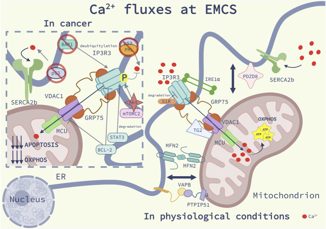
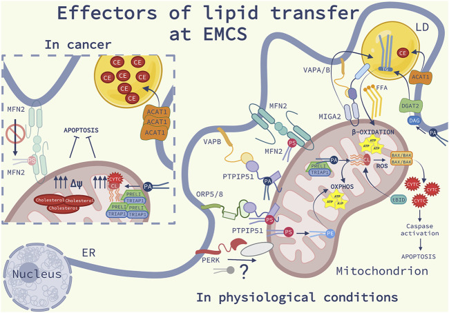
Membrane contact sites (MCS) between organelles of eukaryotic cells provide structural integrity and promote organelle homeostasis by facilitating intracellular signaling, exchange of ions, metabolites and lipids and membrane dynamics. Cataloguing MCS revolutionized our understanding of the structural organization of a eukaryotic cell, but the functional role of MSCs and their role in complex diseases, such as cancer, are only gradually emerging. In particular, the endoplasmic reticulum (ER)-mitochondria contacts (EMCS) are key effectors of non-vesicular lipid trafficking, thereby regulating the lipid composition of cellular membranes and organelles, their physiological functions and lipid-mediated signaling pathways both in physiological and diseased conditions. In this short review, we discuss key aspects of the functional complexity of EMCS in mammalian cells, with particular emphasis on their role as central hubs for lipid transport between these organelles and how perturbations of these pathways may favor key traits of cancer cells.

Keywords: Ca2+ signaling; ER-mitochondria contact sites; cancer; lipid transfer protein; lipids.


Conflict of interest statement

The authors declare that the research was conducted in the absence of any commercial or financial relationships that could be construed as a potential conflict of interest.

Figures

FIGURE 1
FIGURE 1
Ca2+ fluxes at EMCS: Schematic representation of the main modulators of Ca2+ homeostasis at ER-mitochondria contact sites (EMCS) in physiological conditions (on the right) and in cancer (on the left), as discussed in the main text. On the right side of the image, are represented the main Ca2+ regulatory systems at EMCS: the inositol 1,4,5-trisphosphate receptors 3 (IP3R3)- glucose-related regulated protein 75 (Grp75)-voltage-dependent anion channel 1 (VDAC1) signaling complex, delivering Ca2+ to the mitochondria, the Ca2+ uniporter (MCU), allowing Ca2+entry into the mitochondrial matrix, and the sarco/endoplasmic reticulum Ca2+ ATPase 2b (SERCA2b), which pumps Ca2+ from the cytosol to the ER. Maintenance of mitochondrial Ca2+ homeostasis through the integrity of the EMCS, promotes mitochondrial bioenergetics. Sigma-1Receptor (S1R) binds to IP3R3 and decreases its degradation; the ER stress sensor Inositol-requiring enzyme 1 (IRE1α) interacts and brings IP3R3 at EMCS. Transglutaminase type 2 (TG2) binds to Grp75 and promotes mitochondrial Ca2+ uptake. The complex formed by the protein tyrosine phosphatase interacting protein 51 (PTPIP51) and the vesicle-associated membrane protein-associated protein B (VAPB), PDZ domain-containing protein 8 (PDZD8) and Mitofusin 2 (MFN2) homodimers favor Ca2+ fluxes by regulating the EMCS proximity. On the left side of the image we describe Ca2+ dysregulation in cancer. The tumor suppressor p53 at EMCS binds to SERCA pump preventing its ROS-mediated inactivation. P53 interacts with promyelocytic leukemia (PML) protein, promoting the dephosphorylation and inactivation of the proto-oncogene serine/threonine kinase Akt, which together with the Rapamycin complex 2 (mTORC2) phosphorylate and inhibit IP3R3. The proto-oncogene B-cell lymphoma 2 (Bcl-2) interacts and inhibits VDAC1 and IP3R3 suppressing Ca2+ fluxes. IP3R3 stability is regulated by the oncogenic signal transducer and activator of transcription 3 (STAT3) which promotes its degradation and the tumor suppressor deubiquitylase BRCA1-associated protein 1 (BAP1), which prevents its ubiquitylation. The mechanisms described here contribute to a reduction of mitochondrial Ca2+ import, and consequently to a lower oxidative phosphorylation (OXPHOS) and reduced apoptosis sensitivity.
FIGURE 2
FIGURE 2
Effectors of lipid transfer at EMCS: Schematic representation of the lipid homeostasis at ER-mitochondria contact sites (EMCS) and lipid droplets (LD), in physiological conditions (on the right) and in cancer (on the left). On the right side of the image, are represented the main lipid transfer proteins (LTP) discussed in the review (see the main text for further explanations); Mitofusin 2 (MFN2) homodimers, bind and transfer phosphatidylserine (PS), the oxysterol-binding protein-related protein (ORP) 5 and 8-protein- tyrosine phosphatase interacting protein 51 (PTPIP51) complex, the vesicle-associated membrane protein-associated protein B (VAPB)- PTPIP51 complex, responsible of the transfer of phosphatidic acid (PA). The Ser/Thr kinase RNA-dependent protein kinase (PKR)-like ER kinase (PERK) is shown to interact with an unknown mitochondrial protein, possibly facilitating phospholipid (PL) transport. In the inner mitochondrial membrane (IMM) PS is converted into PE and PA and transferred into the mitochondrial intermembrane space (IMS) by TP53-regulated inhibitor of apoptosis gene 1 TRIAP1-PRELI complex. PA is converted into cardiolipin (CL), an anionic PL which promotes mitochondrial oxidative phosphorylation (OXPHOS). In response to cell death stimuli, CL interacts with truncated BH3 interacting-domain death agonist (tBID) promoting BAX/BAK oligomerization, release of cytochrome c (CYTC), triggering caspase activation and apoptosis. Mitoguardin 2 (MIGA2)-VAPA/B complex tethers the EMCS to LD and facilitates the production of triglycerides (TG). TG is synthesized into the ER from PA via the diacylglycerol acyltransferase 2 (DGAT2) enriched at EMCS. The enzyme acyl-CoA cholesterol acyltransferase-1 (ACAT1) localized at EMCS synthetizes cholesteryl esters (CE). TG and CE are stored in LD. TG hydrolysis provides free fatty acids (FFA) as fuel for β-oxidation occurring in the mitochondria. On the left panel, some molecular mechanisms involving lipid signaling at EMCS, which are altered in cancer cells, are illustrated: ACAT1 expression is increased, consequently leading to the accumulation of elevated levels of CE into LD; the complex TRIPA1-PRELI is upregulated, PA is converted into CL, impairing the release of CYTC and the sensitivity to apoptosis; cholesterol is highly accumulated into the IMM, affecting the mitochondrial potential (Δψ) and favoring resistance to apoptosis. Deficiency of MFN2 compromises the transferring of PS at EMCS causing ER stress, inflammation, fibrosis and cancer.

References

    1. Acín-Pérez R., Fernández-Silva P., Peleato M. L., Pérez-Martos A., Enriquez J. A. (2008). Respiratory active mitochondrial supercomplexes. Mol. Cell 32, 529–539. 10.1016/j.molcel.2008.10.021 - DOI - PubMed
    1. Acoba M. G., Senoo N., Claypool S. M. (2020). Phospholipid ebb and flow makes mitochondria go. J. Cell Biol. 219, e202003131. 10.1083/jcb.202003131 - DOI - PMC - PubMed
    1. Ardail D., Lerme F., Louisot P. (1991). Involvement of contact sites in phosphatidylserine import into liver mitochondria. J. Biol. Chem. 266, 7978–7981. 10.1016/s0021-9258(18)92926-1 - DOI - PubMed
    1. Avalle L., Camporeale A., Morciano G., Caroccia N., Ghetti E., Orecchia V., et al. (2019). STAT3 localizes to the ER, acting as a gatekeeper for ER-mitochondrion Ca2+ fluxes and apoptotic responses. Cell Death Differ. 26, 932–942. 10.1038/s41418-018-0171-y - DOI - PMC - PubMed
    1. Baker C. D., Basu Ball W., Pryce E. N., Gohil V. M. (2016). Specific requirements of nonbilayer phospholipids in mitochondrial respiratory chain function and formation. Mol. Biol. Cell 27, 2161–2171. 10.1091/mbc.E15-12-0865 - DOI - PMC - PubMed