

Epidermal growth factor receptor (EGFR) expression in the serum of patients with triple-negative breast carcinoma: prognostic value of this biomarker
- PMID: 36158981
- PMCID: PMC9458269
- DOI: 10.3332/ecancer.2022.1431
Epidermal growth factor receptor (EGFR) expression in the serum of patients with triple-negative breast carcinoma: prognostic value of this biomarker
Abstract
Background: Epidermal growth factor receptor (EGFR) overexpression has been considered a poor prognostic factor in breast cancer.
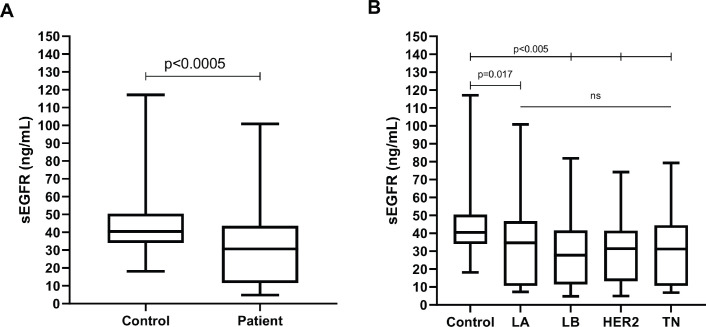
Methodology: A prospective study of 206 women with breast cancer analysed by stages (I, II, III and IV) and by immunohistochemical subtype (Luminal A, Luminal B, HER2+ and triple-negative (TN)); 89 healthy controls with normal recent mammography were included. The EGFR measured in the serum (sEGFR) was detected by the Enzyme-Linked Immunosorbent Assay (ELISA) method (R&D Systems kit DY231) collected by blood before any treatment in patients. Kaplan-Meier method and Cox regression were carried out to obtain the prognostic value, considering significance if p < 0.05.
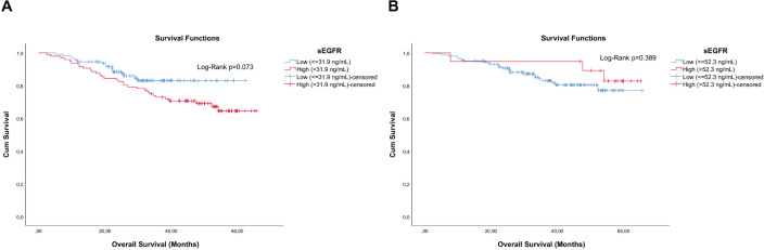
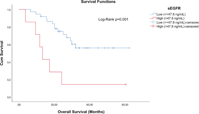
Results: With a median follow-up of 36.6 months, 47 deaths occurred. Multivariable Cox regression showed difference of overall survival (OS) associated with sEGFR levels (sEGFR ≤ or > 47.8 ng/mL) in patients with TN cancers, but not of Luminal A, Luminal B or HER2+ subtypes; adjusted by stage, the death risk increased by approximately 415% [hazard ratio (HR): 5.149 (1.900-13.955), p = 0.001] for patients with sEGFR > 47.8 ng/mL compared to patients with a lower sEGFR value. There was no significant correlation of sEGFR with staging, histological tumour grade (G1/G2/G3), Ki67 (< or ≥14%) or body mass index.
Conclusions: Increased sEGFR expression in patients with TN tumours is a significant predictor of lower OS and its quantification is inexpensive and straightforward.
Keywords: breast cancer; prognosis; sEGFR; tEGFR.
© the authors; licensee ecancermedicalscience.
Conflict of interest statement
The authors declare that there are no conflicts of interest.
Figures
Similar articles
-
Analysis of the prognostic value of soluble epidermal growth factor receptor plasma concentration in advanced non-small-cell lung cancer patients.Clin Lung Cancer. 2011 Sep;12(5):320-7. doi: 10.1016/j.cllc.2011.03.031. Epub 2011 May 11. Clin Lung Cancer. 2011. PMID: 21729651
-
Soluble epidermal growth factor receptor (sEGFR) and carcinoembryonic antigen (CEA) concentration in patients with non-small cell lung cancer: correlation with survival after erlotinib and gefitinib treatment.Ecancermedicalscience. 2010;4:178. doi: 10.3332/ecancer.2010.178. Epub 2010 Nov 3. Ecancermedicalscience. 2010. PMID: 22276032 Free PMC article.
-
Soluble epidermal growth factor receptor (sEGFR/sErbB1) as a potential risk, screening, and diagnostic serum biomarker of epithelial ovarian cancer.Cancer Epidemiol Biomarkers Prev. 2003 Feb;12(2):103-13. Cancer Epidemiol Biomarkers Prev. 2003. PMID: 12582019
-
Prognostic and predictive impact of soluble epidermal growth factor receptor (sEGFR) protein in the serum of patients treated with chemotherapy for metastatic breast cancer.Anticancer Res. 2006 Mar-Apr;26(2B):1479-87. Anticancer Res. 2006. PMID: 16619561 Clinical Trial.
-
Potential prognostic tumor biomarkers in triple-negative breast carcinoma.Beijing Da Xue Xue Bao Yi Xue Ban. 2012 Oct 18;44(5):666-72. Beijing Da Xue Xue Bao Yi Xue Ban. 2012. PMID: 23073572 Review.
Cited by
-
Targeting Receptor Tyrosine Kinases as a Novel Strategy for the Treatment of Triple-Negative Breast Cancer.Technol Cancer Res Treat. 2024 Jan-Dec;23:15330338241234780. doi: 10.1177/15330338241234780. Technol Cancer Res Treat. 2024. PMID: 38389413 Free PMC article. Review.
-
Identification of Hub of the Hub-Genes From Different Individual Studies for Early Diagnosis, Prognosis, and Therapies of Breast Cancer.Bioinform Biol Insights. 2024 Sep 4;18:11779322241272386. doi: 10.1177/11779322241272386. eCollection 2024. Bioinform Biol Insights. 2024. PMID: 39239087 Free PMC article.
References
LinkOut - more resources
Full Text Sources
Research Materials
Miscellaneous