. 2022 Jul 1;12(3):e2022134.
doi: 10.5826/dpc.1203a134. eCollection 2022 Jul.

An Updated Algorithm Integrated With Patient Data for the Differentiation of Atypical Nevi From Early Melanomas: the idScore 2021

Affiliations

An Updated Algorithm Integrated With Patient Data for the Differentiation of Atypical Nevi From Early Melanomas: the idScore 2021

Linda Tognetti et al. Dermatol Pract Concept. .

Abstract

Introduction: It is well known that multiple patient-related risk factors contribute to the development of cutaneous melanoma, including demographic, phenotypic and anamnestic factors.

Objectives: We aimed to investigate which MM risk factors were relevant to be incorporated in a risk scoring-classifier based clinico-dermoscopic algorithm.

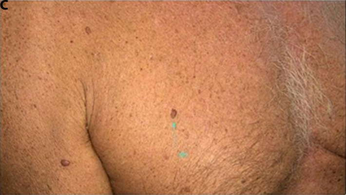
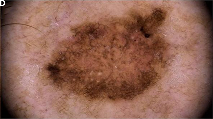
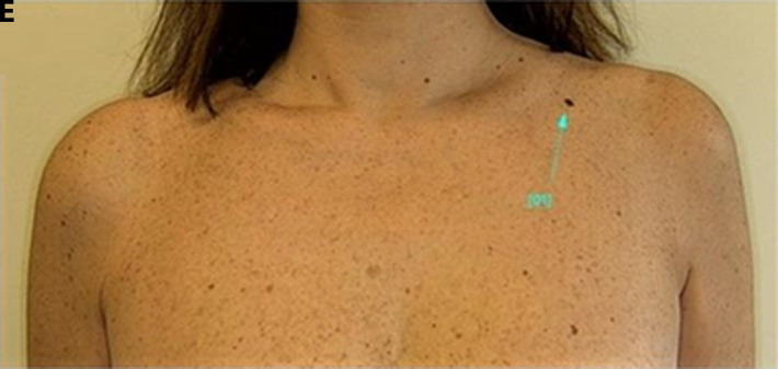
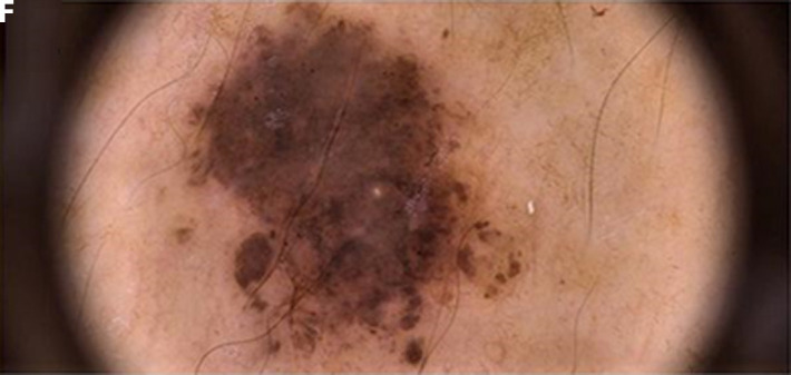
Methods: This retrospective study was performed on a monocentric dataset of 374 atypical melanocytic skin lesions sharing equivocal dermoscopic features, excised in the suspicion of malignancy. Dermoscopic standardized images of 258 atypical nevi (aN) and 116 early melanomas (eMM) were collected along with objective lesional data (i.e., maximum diameter, specific body site and body area) and 7 dermoscopic data. All cases were combined with a series of 10 MM risk factors, including demographic (2), phenotypic (5) and anamnestic (3) ones.

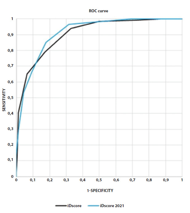
Results: The proposed iDScore 2021 algorithm is composed by 9 variables (age, skin phototype I/II, personal/familiar history of MM, maximum diameter, location on the lower extremities (thighs/legs/ankles/back of the feet) and 4 dermoscopic features (irregular dots and globules, irregular streaks, blue gray peppering, blue white veil). The algorithm assigned to each lesion a score from 0 to 18, reached an area under the ROC curve of 92% and, with a score threshold ≥ 6, a sensitivity (SE) of 98.2% and a specificity (SP) of 50.4%, surpassing the experts in SE (+13%) and SP (+9%).

Conclusions: An integrated checklist combining multiple anamnestic data with selected relevant dermoscopic features can be useful in the differential diagnosis and management of eMM and aN exhibiting with equivocal features.

Keywords: atypical nevi; dermoscopy; melanoma; risk factors.


Conflict of interest statement

Competing interests: None.

Figures

Figure 1
Figure 1
Examples of atypical melanocytic skin lesions (aMSLs) on the upper back from the case study. A 71 years-old male, phototype II, personal history of melanoma, with a 11 mm aMSL, iDScore 2021 = 15 (iDScore 2018 = 9): histological examination revealed an early melanoma (MM T1aN0M0, thickness 0.7mm) (aA and B). A 61 years-old male, phototype II, with 7mm aMSL: the iDScore 2021 was 9 (iDScore 2018 = 7) and the histological examination revealed a nevus with moderate atypia (C and D). A 50 years-old male, 11 mm, phototype II, > 11 nevi/right arm, 1st relative history of MM, sunburns before the age of 14, with a 10mm aMSL: the iDScore 2021 was 10 (iDScore 2018 = 8) and the histological examination revealed a nevus with severe atypia (E and F).
Figure 1
Figure 1
Examples of atypical melanocytic skin lesions (aMSLs) on the upper back from the case study. A 71 years-old male, phototype II, personal history of melanoma, with a 11 mm aMSL, iDScore 2021 = 15 (iDScore 2018 = 9): histological examination revealed an early melanoma (MM T1aN0M0, thickness 0.7mm) (aA and B). A 61 years-old male, phototype II, with 7mm aMSL: the iDScore 2021 was 9 (iDScore 2018 = 7) and the histological examination revealed a nevus with moderate atypia (C and D). A 50 years-old male, 11 mm, phototype II, > 11 nevi/right arm, 1st relative history of MM, sunburns before the age of 14, with a 10mm aMSL: the iDScore 2021 was 10 (iDScore 2018 = 8) and the histological examination revealed a nevus with severe atypia (E and F).
Figure 2
Figure 2
Examples of atypical melanocytic skin lesions (aMSLs) of the chest from the case study. A 80 years-old male, phototype III, familiar history of melanoma, with a 7 mm aMSL: iDScore 2021 was 12 (iDScore 2018 = 8) and the histological analysis revealed an in situ melanoma (A and B). A 66 years-old male, phototype II, familiar history of MM, with a 10mm aMSL: the iDScore 2021 was 10 (iDScore 2018 = 8); the histological analysis revealed a nevus with moderate atypia (C and D). A 47 years-old female, phototype III, with a 7.7 mm aMSL, the iDScore 2021 was 6 (iDScore 2018 = 5) and the histological examination revealed a compound nevus (E and F).
Figure 2
Figure 2
Examples of atypical melanocytic skin lesions (aMSLs) of the chest from the case study. A 80 years-old male, phototype III, familiar history of melanoma, with a 7 mm aMSL: iDScore 2021 was 12 (iDScore 2018 = 8) and the histological analysis revealed an in situ melanoma (A and B). A 66 years-old male, phototype II, familiar history of MM, with a 10mm aMSL: the iDScore 2021 was 10 (iDScore 2018 = 8); the histological analysis revealed a nevus with moderate atypia (C and D). A 47 years-old female, phototype III, with a 7.7 mm aMSL, the iDScore 2021 was 6 (iDScore 2018 = 5) and the histological examination revealed a compound nevus (E and F).
Figure 3
Figure 3
Receiver operating characteristic (ROC) of the 3 integrated algorithms: iDScore 2018 (black) and iDScore 2021 (blue) obtained on the iDScore database 2018–2020 of 324 atypical melanocytic skin lesions. The segments of the curves represent cases obtaining the same score.

Similar articles

Cited by

References

    1. Tognetti L, Cevenini G, Moscarella E, et al. An integrated clinical-dermoscopic risk scoring system for the differentiation between early melanoma and atypical nevi: the iDScore. J Eur Acad Dermatol Venereol. 2018;32(12):2162–2170. doi: 10.1111/jdv.15106. - DOI - PubMed
    1. Rubegni P, Tognetti L, Argenziano G, et al. A risk scoring system for the differentiation between melanoma with regression and regressing nevi. J Dermatol Sci. 2016;83(2):138–144. doi: 10.1016/j.jdermsci.2016.04.012. - DOI - PubMed
    1. Kittler H, Guitera P, Riedl E, et al. Identification of Clinically Featureless Incipient Melanoma Using Sequential Dermoscopy Imaging. Arch Dermatol. 2006;142(9):1113–1119. doi: 10.1001/archderm.142.9.1113. - DOI - PubMed
    1. Puig S, Argenziano G, Zalaudek I, et al. Melanomas that failed dermoscopic detection: a combined clinicodermoscopic approach for not missing melanoma. Dermatol Surg. 2007;33(10):1262–1273. doi: 10.1111/j.1524-4725.2007.33264.x. - DOI - PubMed
    1. Argenziano G, Zalaudek I, Ferrara G, et al. Dermoscopy features of melanoma incognito: indications for biopsy. J Am Acad Dermatol. 2007;56(3):508–13. doi: 10.1016/j.jaad.2006.10.029. - DOI - PubMed

LinkOut - more resources