. 2022 Jul 1;12(3):e2022113.
doi: 10.5826/dpc.1203a113. eCollection 2022 Jul.

Facial Dermatoses in Patients With Blepharitis: a Cross-sectional Prospective Analysis

Affiliations

Facial Dermatoses in Patients With Blepharitis: a Cross-sectional Prospective Analysis

Semih Güder et al. Dermatol Pract Concept. .

Abstract

Introduction: The relationship between facial dermatoses and blepharitis has been known for a long time.

Objectives: We aimed to investigate the frequency of accompanying facial dermatoses in patients with blepharitis and their relationship with the severity of blepharitis.

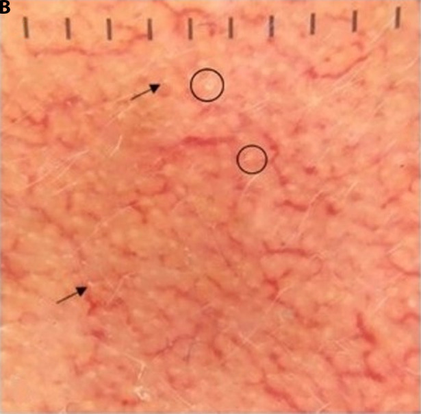
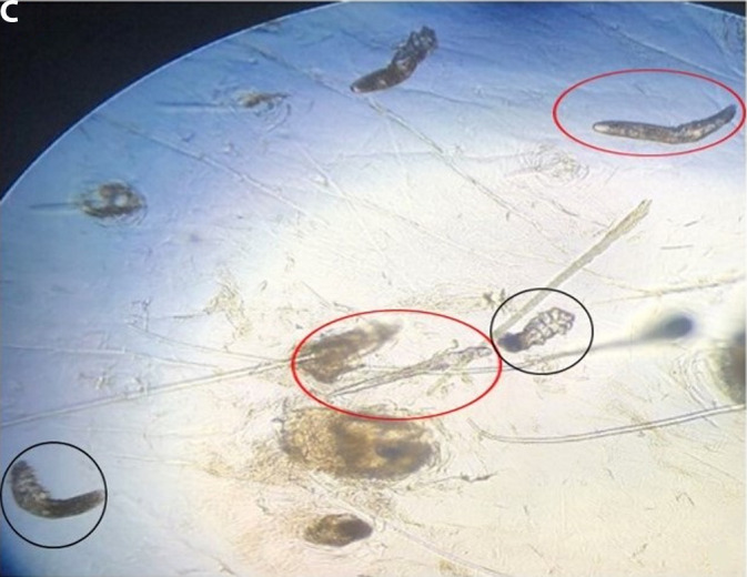
Methods: In this cross-sectional study, 95 patients with blepharitis were examined for attending facial dermatoses. The type of blepharitis, the severity of blepharitis, and the degree of dry eye were determined in the patients. Dermoscopic and microscopic examinations were used in the diagnosis of facial dermatoses. The history of allergic rhinitis was questioned because Demodex species frequently accompany blepharitis, facial dermatoses, and allergic rhinitis patients. Mann-Whitney U test was used compare 2 independent groups. In comparing categorical variables, Pearson chi-Squared, Fishere Exact, and Fisher-Freeman-Holton tests were used.

Results: At least 1 facial dermatosis was detected in 84.2% patients, and we did not see any facial dermatosis in 15.8% ones. No patients had acne, which is one of the most common facial dermatoses. The most common facial dermatosis detected in our patients was facial demodicosis (57.9%). It was followed by seborrheic dermatitis (22.1%) and rosacea (12.6%), respectively. In addition, 2.1% of the patients had atopic eyelid dermatitis, 23.2% had a history of allergic rhinitis, and 63.2% had ocular demodicosis.

Conclusions: It is essential to perform dermatological examinations of all patients with blepharitis in terms of accompanying facial dermatoses and their early diagnosis.

Keywords: blepharitis; demodicosis; facial dermatoses; rosacea; seborrheic dermatitis.


Conflict of interest statement

Competing interests: None.

Figures

Figure 1
Figure 1
Dermoscopic findings of facial demodicosis (FD). (A)Pityriasis folliculorum on the right cheek area of a young man. The white yellowish structures are Demodex tails (arrows). (B) Cheek area of middle-aged woman. The white yellowish structures are Demodex tails (arrows), and Demodex follicular openings (black circles) are seen. Reticular dilated vessels are remarkable in the patient with a history of intermittent steroidal cream use. (C) Diffuse eyelash demodicosis in an elderly man; white yellowish structures are Demodex tails (arrows); increased vascularity less pronounced possibly due to age-related atrophy. (D and E) Dermoscopy of the cheek region of 2 different patients, lateral dermoscopic examination technique. In the patient on the left, Demodex follicular openings (black circles) and reticular dilated vessels are observed in addition to Demodex tails (arrows). (F) Lateral dermoscopic technique for FD.
Figure 1
Figure 1
Dermoscopic findings of facial demodicosis (FD). (A)Pityriasis folliculorum on the right cheek area of a young man. The white yellowish structures are Demodex tails (arrows). (B) Cheek area of middle-aged woman. The white yellowish structures are Demodex tails (arrows), and Demodex follicular openings (black circles) are seen. Reticular dilated vessels are remarkable in the patient with a history of intermittent steroidal cream use. (C) Diffuse eyelash demodicosis in an elderly man; white yellowish structures are Demodex tails (arrows); increased vascularity less pronounced possibly due to age-related atrophy. (D and E) Dermoscopy of the cheek region of 2 different patients, lateral dermoscopic examination technique. In the patient on the left, Demodex follicular openings (black circles) and reticular dilated vessels are observed in addition to Demodex tails (arrows). (F) Lateral dermoscopic technique for FD.
Figure 2
Figure 2
Dermoscopic findings of seborrheic dermatitis (SD). (A) Interfollicular pale erythema (red circle) and oily scale (black circle) in the scalp region. (B) Oily scale on eyebrows (circle) (C) Eyelash of the patient with SD. Thin scales (white circles) attached to the eyelashes are seen (no accompanying Demodex). (D) Eyelash demodicosis in a patient with SD. The white yellowish structures are Demodex tails (arrows), and dilated, and arborized vessels are visible. (E) A dermoscopic view of the nasolabial fold. Fine white nonspecific scales are seen (circles). (F) Dermoscopic view of the cheek of a patient with SD accompanied by facial demodicosis. Demodex follicular openings (circles) and Demodex tails (arrows) are visible.
Figure 2
Figure 2
Dermoscopic findings of seborrheic dermatitis (SD). (A) Interfollicular pale erythema (red circle) and oily scale (black circle) in the scalp region. (B) Oily scale on eyebrows (circle) (C) Eyelash of the patient with SD. Thin scales (white circles) attached to the eyelashes are seen (no accompanying Demodex). (D) Eyelash demodicosis in a patient with SD. The white yellowish structures are Demodex tails (arrows), and dilated, and arborized vessels are visible. (E) A dermoscopic view of the nasolabial fold. Fine white nonspecific scales are seen (circles). (F) Dermoscopic view of the cheek of a patient with SD accompanied by facial demodicosis. Demodex follicular openings (circles) and Demodex tails (arrows) are visible.
Figure 3
Figure 3
The appearance of eyelid and eyelashes of a patient with atopic eyelid dermatitis and allergic rhinoconjunctivitis; microscopic views of facial and ocular demodicosis. (A) A middle-aged man with atopic eyelid dermatitis, presumably secondary to scratching trauma. Prominent skin lines (red circle) and polypoid structures (black circles) are seen. (B) Eyelashes of the patient with allergic rhinoconjunctivitis. The white yellowish structures are Demodex tails (arrows). Thin white scales wrapped around the lashes are also observed (circles). (C) Microscopic view of Demodex folliculorum (black circles) and its larvae (red circles) appear on standardized skin surface biopsy (SSSB) (×40). (D) Microscopic view of the eyelash: Demodex folliculorum eggs (black circle) and larvae (red circle) (×40).
Figure 3
Figure 3
The appearance of eyelid and eyelashes of a patient with atopic eyelid dermatitis and allergic rhinoconjunctivitis; microscopic views of facial and ocular demodicosis. (A) A middle-aged man with atopic eyelid dermatitis, presumably secondary to scratching trauma. Prominent skin lines (red circle) and polypoid structures (black circles) are seen. (B) Eyelashes of the patient with allergic rhinoconjunctivitis. The white yellowish structures are Demodex tails (arrows). Thin white scales wrapped around the lashes are also observed (circles). (C) Microscopic view of Demodex folliculorum (black circles) and its larvae (red circles) appear on standardized skin surface biopsy (SSSB) (×40). (D) Microscopic view of the eyelash: Demodex folliculorum eggs (black circle) and larvae (red circle) (×40).

Similar articles

Cited by

References

    1. Bron AJ, Tiffany JM. The Evolution of Lid Margin Changes in Blepharitis. In: Lass JH, editor. Advances in Corneal Research: Selected Transactions of the World Congress on the Cornea IV. Boston, MA: Springer US; 1997. pp. 3–18.
    1. Hsu JI, Pflugfelder SC, Kim SJ. Ocular complications of atopic dermatitis. Cutis. 2019;104(3):189–193. - PubMed
    1. Arici C, Mergen B, Bahar Tokman H, et al. Investigation of the Demodex Lid Infestation with in Vivo Confocal Microscopy versus Light Microscopy in Patients with Seborrheic Blepharitis. Ocul Immunol Inflamm. 2021:1–5. doi: 10.1080/09273948.2020.1857792. - DOI - PubMed
    1. Auw-Haedrich C, Reinhard T. Chronische Blepharitis. Pathogenese, klinischer Verlauf und therapeutische Ansätze. [Chronic blepharitis. Pathogenesis, clinical features, and therapy]. Ophthalmologe. 2007;104(9):817–826. doi: 10.1007/s00347-007-1608-8. quiz 827–828. - DOI - PubMed
    1. Yan Y, Yao Q, Lu Y, et al. Association Between Demodex Infestation and Ocular Surface Microbiota in Patients With Demodex Blepharitis. Front Med (Lausanne) 2020;7:592759. doi: 10.3389/fmed.2020.592759. - DOI - PMC - PubMed

LinkOut - more resources