

Building bridges: simultaneous multimodal neuroimaging approaches for exploring the organization of brain networks
- PMID: 36159712
- PMCID: PMC9506627
- DOI: 10.1117/1.NPh.9.3.032202
Building bridges: simultaneous multimodal neuroimaging approaches for exploring the organization of brain networks
Abstract
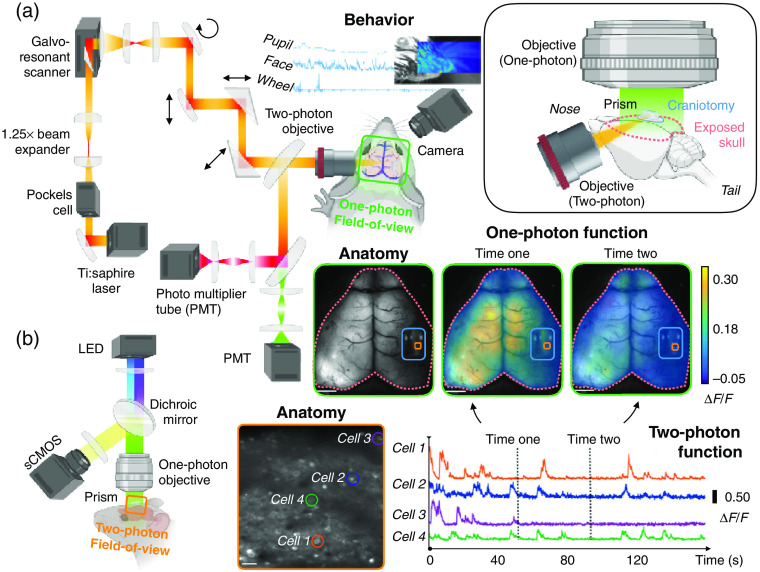
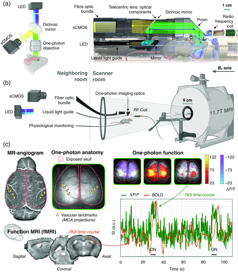
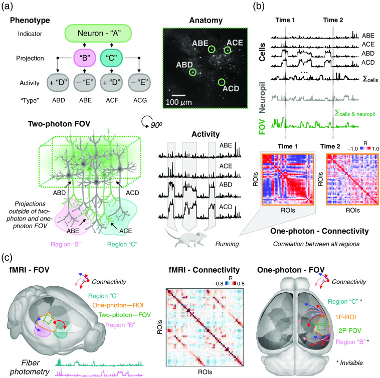
Brain organization is evident across spatiotemporal scales as well as from structural and functional data. Yet, translating from micro- to macroscale (vice versa) as well as between different measures is difficult. Reconciling disparate observations from different modes is challenging because each specializes within a restricted spatiotemporal milieu, usually has bounded organ coverage, and has access to different contrasts. True intersubject biological heterogeneity, variation in experiment implementation (e.g., use of anesthesia), and true moment-to-moment variations in brain activity (maybe attributable to different brain states) also contribute to variability between studies. Ultimately, for a deeper and more actionable understanding of brain organization, an ability to translate across scales, measures, and species is needed. Simultaneous multimodal methods can contribute to bettering this understanding. We consider four modes, three optically based: multiphoton imaging, single-photon (wide-field) imaging, and fiber photometry, as well as magnetic resonance imaging. We discuss each mode as well as their pairwise combinations with regard to the definition and study of brain networks.
Keywords: brain networks; fiber photometry; magnetic resonance imaging; multiphoton imaging; simultaneous imaging; single-photon imaging.
© 2022 The Authors.
Figures
Similar articles
-
Elucidating the complementarity of resting-state networks derived from dynamic [18F]FDG and hemodynamic fluctuations using simultaneous small-animal PET/MRI.Neuroimage. 2021 Aug 1;236:118045. doi: 10.1016/j.neuroimage.2021.118045. Epub 2021 Apr 10. Neuroimage. 2021. PMID: 33848625 Free PMC article.
-
Multimodal Connectomics in Psychiatry: Bridging Scales From Micro to Macro.Biol Psychiatry Cogn Neurosci Neuroimaging. 2018 Sep;3(9):767-776. doi: 10.1016/j.bpsc.2018.03.017. Epub 2018 Apr 19. Biol Psychiatry Cogn Neurosci Neuroimaging. 2018. PMID: 29779726 Review.
-
Hybrid fiber optic-fMRI for multimodal cell-specific recording and manipulation of neural activity in rodents.Neurophotonics. 2022 Jul;9(3):032206. doi: 10.1117/1.NPh.9.3.032206. Epub 2022 Mar 21. Neurophotonics. 2022. PMID: 35355657 Free PMC article. Review.
-
Simultaneous GCaMP6-based fiber photometry and fMRI in rats.J Neurosci Methods. 2017 Sep 1;289:31-38. doi: 10.1016/j.jneumeth.2017.07.002. Epub 2017 Jul 4. J Neurosci Methods. 2017. PMID: 28687521 Free PMC article.
-
Multiscale imaging informs translational mouse modeling of neurological disease.Neuron. 2022 Nov 16;110(22):3688-3710. doi: 10.1016/j.neuron.2022.09.006. Epub 2022 Oct 4. Neuron. 2022. PMID: 36198319 Review.
Cited by
-
Emerging trends in the development of flexible optrode arrays for electrophysiology.APL Bioeng. 2023 Sep 7;7(3):031503. doi: 10.1063/5.0153753. eCollection 2023 Sep. APL Bioeng. 2023. PMID: 37692375 Free PMC article. Review.
-
Functional network properties derived from wide-field calcium imaging differ with wakefulness and across cell type.Neuroimage. 2022 Dec 1;264:119735. doi: 10.1016/j.neuroimage.2022.119735. Epub 2022 Nov 5. Neuroimage. 2022. PMID: 36347441 Free PMC article.
-
Calcium imaging: A versatile tool to examine Huntington's disease mechanisms and progression.Front Neurosci. 2022 Nov 3;16:1040113. doi: 10.3389/fnins.2022.1040113. eCollection 2022. Front Neurosci. 2022. PMID: 36408400 Free PMC article. Review.
-
Special Section Guest Editorial: Hybrid Photonic/X Neurointerfaces.Neurophotonics. 2022 Jul;9(3):032201. doi: 10.1117/1.NPh.9.3.032201. Epub 2022 Sep 30. Neurophotonics. 2022. PMID: 36196247 Free PMC article.
