. 2022 Sep 8:13:1008764.
doi: 10.3389/fimmu.2022.1008764. eCollection 2022.

LY6G6D is a selectively expressed colorectal cancer antigen that can be used for targeting a therapeutic T-cell response by a T-cell engager

Affiliations

LY6G6D is a selectively expressed colorectal cancer antigen that can be used for targeting a therapeutic T-cell response by a T-cell engager

Leticia Corrales et al. Front Immunol. .

Abstract

Colorectal cancer (CRC) is one of the most common cancers worldwide and demands more effective treatments. We sought to identify tumor selective CRC antigens and their therapeutic potential for cytotoxic T-cell targeting by transcriptomic and immunohistochemical analysis. LY6G6D was identified as a tumor selectively expressed CRC antigen, mainly in the microsatellite stable (MSS) subtype. A specific anti LY6G6D/CD3 T cell engager (TcE) was generated and demonstrated potent tumor cell killing and T cell activation in vitro. Ex vivo treatment of primary patient-derived CRC tumor slice cultures with the LY6G6D/CD3 TcE led to IFNγ secretion in LY6G6D positive tumor samples. In vivo, LY6G6D/CD3 TcE monotherapy demonstrated tumor regressions in pre-clinical mouse models of engrafted human CRC tumor cells and PBMCs. Lastly, 2D and 3D cocultures of LY6G6D positive and negative cells were used to explore the bystander killing of LY6G6D negative cells after specific activation of T cells by LY6G6D positive cells. LY6G6D/CD3 TcE treatment was shown to lyse target negative cells in the vicinity of target positive cells through a combined effect of IFNγ, TNFα and Fas/FasL. In summary, LY6G6D was identified as a selectively expressed CRC antigen that can be utilized to potently re-direct and activate cytotoxic T-cells to lyse LY6G6D expressing CRC using a TcE. This effect can be spread to target negative neighboring tumor cells, potentially leading to improved therapeutic efficacy.

Keywords: CD3; CRC (colorectal cancer); LY6G6D; TcE (T cell engager); immunotherapy.


Conflict of interest statement

LC, SH, KM, NS, IT, KF, BT, CW, KB, PC, AV, and PA are employees of Boehringer Ingelheim affiliates. MB received research funding and speakers fee by Boehringer Ingelheim and Bristol Myers Squibb. The remaining authors declare that the research was conducted in the absence of any commercial or financial relationships that could be construed as a potential conflict of interest.

Figures

Figure 1
Figure 1
LY6G6D is a tumor antigen specifically expressed in CRC. (A-C) LY6G6D IHC staining. Representative images of CRC samples (A) and normal colon tissue (B). Graph depicting the percentage of positive cells in CRC samples (n=41). (D) Schematic graphic of LY6G6D/CD3 TcE. The Fab-scFv TcE format contains a N-terminal 2C11A8 derived Fab linked with via (G4S)4 linker to a C-terminal CD3ϵ specific scFv derived from the murine clone SP34. (E) Surface plasmon resonance analysis of LY6G6D specific TcE. TcE was immobilized on CM5 chip, recombinant LY6G6D protein was used as analyte, and data were analyzed by a 1:1 Langmuir model. KD=2.5 nM is for LY6G6D, ka=6.9x10⁴ 1/M*s and kd=1.7x10⁻⁴ 1/s.
Figure 2
Figure 2
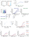
In vitro efficacy and specificity of LY6G6D/CD3 TcE. (A) LY6G6D expression in parental and LY6G6D transfected HEK293 cells (B) Jurkat cells and HEK293 LY6G6D- or HEK293 LY6G6D+ cells were co-cultivated in the presence of increasing concentrations of LY6G6D/CD3 TcE for 24 hours. (C) LY6G6D density in HEK293 LY6G6D+ and HEK293 LY6G6D- cells pre-incubated with PI-PLC for 30 min. (D) PI-PLC pre-incubated HEK293 cells and Jurkat cells were co-cultivated in presence of increasing concentrations of LY6G6D/CD3 TcE for 24 hours. (E) Correlation between LY6G6D density and activation of Jurkat cells. (F) Activation of Jurkat cells after 48 hours of incubation with LY6G6D-positive (HT55, LS1034, CL-14 and NCI-H508) or negative (SK-CO1 and NCM460) tumor cells and increasing concentrations of LY6G6D/CD3 TcE. (G, H) LY6G6D-positive (HT55, LS1034, CL-14 and NCI-H508) or negative (SK-CO1 and NCM460) tumor cells were co-incubated with purified T cells and increasing amounts of concentrations of LY6G6D/CD3 TcE. Killing of tumor cells (G) and activation of T cells (H) were assessed after 72 hours of incubation.
Figure 3
Figure 3
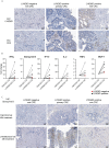
Ex vivo activation of T cells in LY6G6D positive CRC Precision Cut Tumor Slice Cultures. (A) Baseline expression of LY6G6D and CD3 expression in representative samples. (B) Fresh tumor tissue slides from eight CRC patients were incubated for 48 hours with 1 nM LY6G6D/CD3 TcE or control TNP/CD3 TcE. After 48 hours IFN-γ, Granzyme B, IP-10, Il-2, TNFα and MCP-1 were quantified in the cell culture supernatant. Each dot shows one CRC patient sample whereas red dots indicate LY6G6D negative tumor samples. (C) Representative images of Granzyme B staining in tumor slides after 48 hours of incubation with 1 nM LY6G6D/CD3 TcE or control TNP/CD3.
Figure 4
Figure 4
In vivo efficacy of LY6G6D/CD3 TcE. (A-C) Anti-tumor activity of LY6G6D/CD3 TcE in established LS1034 xenograft tumors in NSG mice. Mice were humanized with human PBMCs at randomization, and treated with 0.5 mg/kg LY6G6D/CD3 TcE or control TNP/CD3 five days per week. Each graph shows data from one out of three independent PBMC donors. Days of treatment are annotated by gray arrows. **p<0.01.
Figure 5
Figure 5
LY6G6D/CD3 TcE induces bystander killing of LY6G6D- cells in vitro. (A, B) HEK293 LY6G6D+ and LY6G6D- were labeled with cell tracer, plate isolated (100%) or cocultured at 1:1 ratio (A) or at different ratios (B). Isolated or cocultured HEK293 cells were then co-incubated with purified T cells and increasing amounts of concentrations of LY6G6D/CD3 TcE. After 72 hours, the total number of HEK293 LY6G6D+ and LY6G6D- were counted and the percentage of viable cells were calculated by normalizing with non-treated cells in each condition. (C) Activation of T cells after 48 hours of co-incubation with HEK293 LY6G6D+ and LY6G6D- cells isolated or cocultured under different ratios. (D, E) LS1034 and NCM460D tumor cells were labeled with cell tracer, plate isolated (100%) or cocultured at 1:1 ratio. Tumor cells were then co-incubated with purified T cells and increasing amounts of concentrations of LY6G6D/CD3 TcE. The total number of tumor cells (D) and T cell activation (E) was analyzed after 48 hours of incubation. (F, G) HEK293 LY6G6D+ (blue) and LY6G6D- (red) were labeled, plate isolated (100%) or cocultured and co-incubated with purified T cells, increasing amounts of LY6G6D/CD3 TcE and caspase 3/7 green dye. Confocal microscopy images of cultures were acquired over time (F), and apoptotic or double stained HEK293 LY6G6D+ (blue/green) and LY6G6D- (red/green) cells were quantified (G).
Figure 6
Figure 6
LY6G6D/CD3 TcE induces bystander killing of LY6G6D- cells in 3D spheroids. (A) Image analysis of spheroids containing NCM460D cells or a 1:1 coculture of NCM460D and LS1034 cells. Spheroids were incubated with PBMCs (1:5 ratio, T:E) and increasing amounts of LY6G6D/CD3 TcE for 7 days. Total green signal at day 7 from NCM460D cells is shown. (B-E) Spheroids containing a 1:1 coculture of NCM460D and LS1034 cells, PBMCs (1:5 ratio, T:E), increasing amounts LY6G6D/CD3 TcE and 10 µg/ml CD178 monoclonal antibody (Invitrogen) (B), 10µg/ml Entanercept (C), 1µg/ml human IFNγ antibody with 1µg/ml human IFNGR antibody (D) or a combination with all blocking compounds (E). Total green signal at day 7 from NCM460D cells is shown. (F) Image analysis of spheroids containing LS1034 cells. Spheroids were incubated with PBMCs (1:5 ratio, T:E), increasing amounts of LY6G6D/CD3 TcE for 7 days or a combination with all blocking compounds like in Figure (E) Total green signal at day 7 from LS1034 cells is shown.

References

    1. Sung H, Ferlay J, Siegel RL, Laversanne M, Soerjomataram I, Jemal A, et al. . Global cancer statistics 2020: GLOBOCAN estimates of incidence and mortality worldwide for 36 cancers in 185 countries. CA Cancer J Clin (2021) 71:209–49. doi: 10.3322/caac.21660 - DOI - PubMed
    1. Picard E, Verschoor CP, Ma GW, Pawelec G. Relationships between immune landscapes, genetic subtypes and responses to immunotherapy in colorectal cancer. Front Immunol (2020) 11:369. doi: 10.3389/fimmu.2020.00369 - DOI - PMC - PubMed
    1. Brenner H, Kloor M, Pox CP. Colorectal cancer. Lancet (2014) 383:1490–502. doi: 10.1016/s0140-6736(13)61649-9 - DOI - PubMed
    1. Sadanandam A, Lyssiotis CA, Homicsko K, Collisson EA, Gibb WJ, Wullschleger S, et al. . A colorectal cancer classification system that associates cellular phenotype and responses to therapy. Nat Med (2013) 19:619–25. doi: 10.1038/nm.3175 - DOI - PMC - PubMed
    1. Le DT, Uram JN, Wang H, Bartlett BR, Kemberling H, Eyring AD, et al. . PD-1 blockade in tumors with mismatch-repair deficiency. N Engl J Med (2015) 372:2509–20. doi: 10.1056/nejmoa1500596 - DOI - PMC - PubMed