

Effect of Angelica polysaccharide on mouse myeloid-derived suppressor cells
- PMID: 36159871
- PMCID: PMC9500156
- DOI: 10.3389/fimmu.2022.989230
Effect of Angelica polysaccharide on mouse myeloid-derived suppressor cells
Abstract
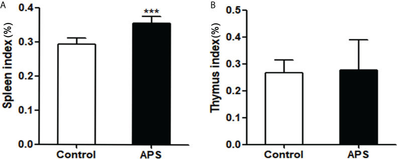
Angelica polysaccharide (APS) is a polysaccharide extracted from Angelica sinensis and it is one of the main active components of Angelica sinensis. Many studies have demonstrated that APS can promote the activation and function of a variety of immune cells and is recognized as an immune enhancer, but the regulatory effect of APS on myeloid-derived suppressor cells (MDSC) is still unclear. In this study, we investigated the effects of APS on MDSC proliferation, differentiation and function through in vivo and in vitro experiments. In vitro, our results showed that APS promoted the proliferation, differentiation and immunosuppressive function of MDSC through STAT1 and STAT3 signaling pathways, and positively correlated with the expression level of Mannose receptor (MR, also known as CD206) and in a concentration-dependent manner on APS. In vivo, APS up-regulated T cells, γδT cells, CD8+T cells, natural killer cells, monocytes/macrophages, and granulocytes in the peripheral blood and spleen of mice to varying degrees and was accompanied by the same degree of increase in the proportion of MDSC. That reminds to the clinician that when applying APS as treatment they should pay attention to its possible side effects of increasing the quantity and function of MDSC, in order to increase its efficacy.
Keywords: Angelica polysaccharide; differentiation; immunosuppression; mannose receptor; myeloid-derived suppressor cells; proliferation.
Copyright © 2022 Shen, Zhang, Zhang, Qin, Liu, Liang, Chen and Peng.
Conflict of interest statement
The authors declare that the research was conducted in the absence of any commercial or financial relationships that could be construed as a potential conflict of interest.
Figures


Similar articles
-
The angelica Polysaccharide: a review of phytochemistry, pharmacology and beneficial effects on systemic diseases.Int Immunopharmacol. 2024 May 30;133:112025. doi: 10.1016/j.intimp.2024.112025. Epub 2024 Apr 26. Int Immunopharmacol. 2024. PMID: 38677093 Review.
-
Structure characterization and anti-leukemia activity of a novel polysaccharide from Angelica sinensis (Oliv.) Diels.Int J Biol Macromol. 2019 Jan;121:161-172. doi: 10.1016/j.ijbiomac.2018.09.213. Epub 2018 Oct 2. Int J Biol Macromol. 2019. PMID: 30290264
-
Angelica polysaccharides inhibit the growth and promote the apoptosis of U251 glioma cells in vitro and in vivo.Phytomedicine. 2017 Sep 15;33:21-27. doi: 10.1016/j.phymed.2017.06.007. Epub 2017 Jul 8. Phytomedicine. 2017. PMID: 28887916
-
Leukemia cells apoptosis by a newly discovered heterogeneous polysaccharide from Angelica sinensis (Oliv.) Diels.Carbohydr Polym. 2020 Aug 1;241:116279. doi: 10.1016/j.carbpol.2020.116279. Epub 2020 Apr 30. Carbohydr Polym. 2020. PMID: 32507223
-
Extraction, structure, pharmacological activities and drug carrier applications of Angelica sinensis polysaccharide.Int J Biol Macromol. 2021 Jul 31;183:2337-2353. doi: 10.1016/j.ijbiomac.2021.05.213. Epub 2021 Jun 6. Int J Biol Macromol. 2021. PMID: 34090852 Review.
Cited by
-
Exploring the immunometabolic potential of Danggui Buxue Decoction for the treatment of IBD-related colorectal cancer.Chin Med. 2024 Aug 29;19(1):117. doi: 10.1186/s13020-024-00978-y. Chin Med. 2024. PMID: 39210410 Free PMC article. Review.
-
Progress and prospect of polysaccharides as adjuvants in vaccine development.Virulence. 2024 Dec;15(1):2435373. doi: 10.1080/21505594.2024.2435373. Epub 2024 Dec 5. Virulence. 2024. PMID: 39601191 Free PMC article. Review.
-
What can traditional Chinese medicine do for adult neurogenesis?Front Neurosci. 2023 Apr 12;17:1158228. doi: 10.3389/fnins.2023.1158228. eCollection 2023. Front Neurosci. 2023. PMID: 37123359 Free PMC article. Review.
-
Harnessing the power of traditional Chinese medicine monomers and compound prescriptions to boost cancer immunotherapy.Front Immunol. 2023 Nov 15;14:1277243. doi: 10.3389/fimmu.2023.1277243. eCollection 2023. Front Immunol. 2023. PMID: 38035069 Free PMC article. Review.
-
Unraveling the web of defense: the crucial role of polysaccharides in immunity.Front Immunol. 2024 Oct 25;15:1406213. doi: 10.3389/fimmu.2024.1406213. eCollection 2024. Front Immunol. 2024. PMID: 39524445 Free PMC article. Review.
References
Publication types
MeSH terms
Substances
LinkOut - more resources
Full Text Sources
Research Materials
Miscellaneous