. 2022 Aug 29;2(9):100280.
doi: 10.1016/j.crmeth.2022.100280. eCollection 2022 Sep 19.

Functional microvascularization of human myocardium in vitro

Affiliations

Functional microvascularization of human myocardium in vitro

Oisín King et al. Cell Rep Methods. .

Abstract

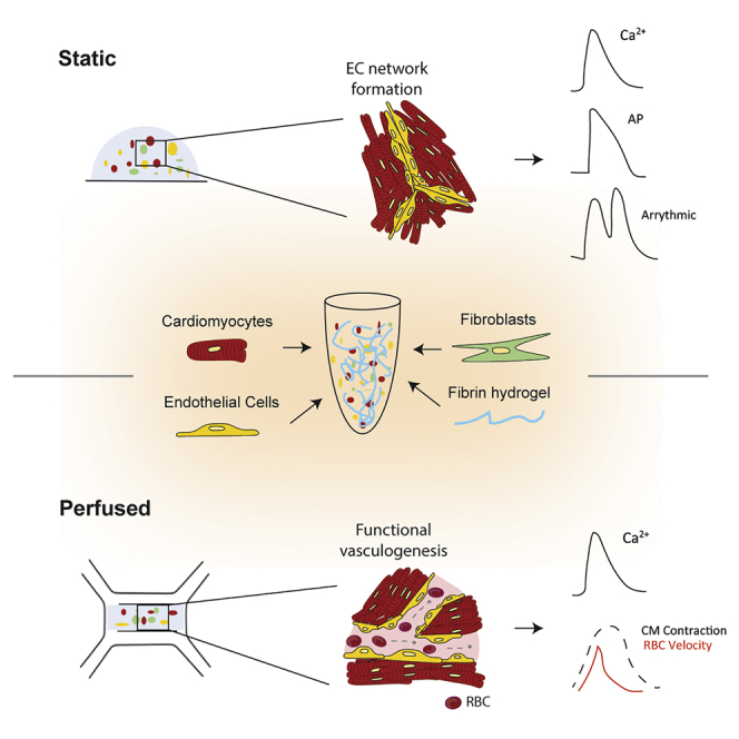
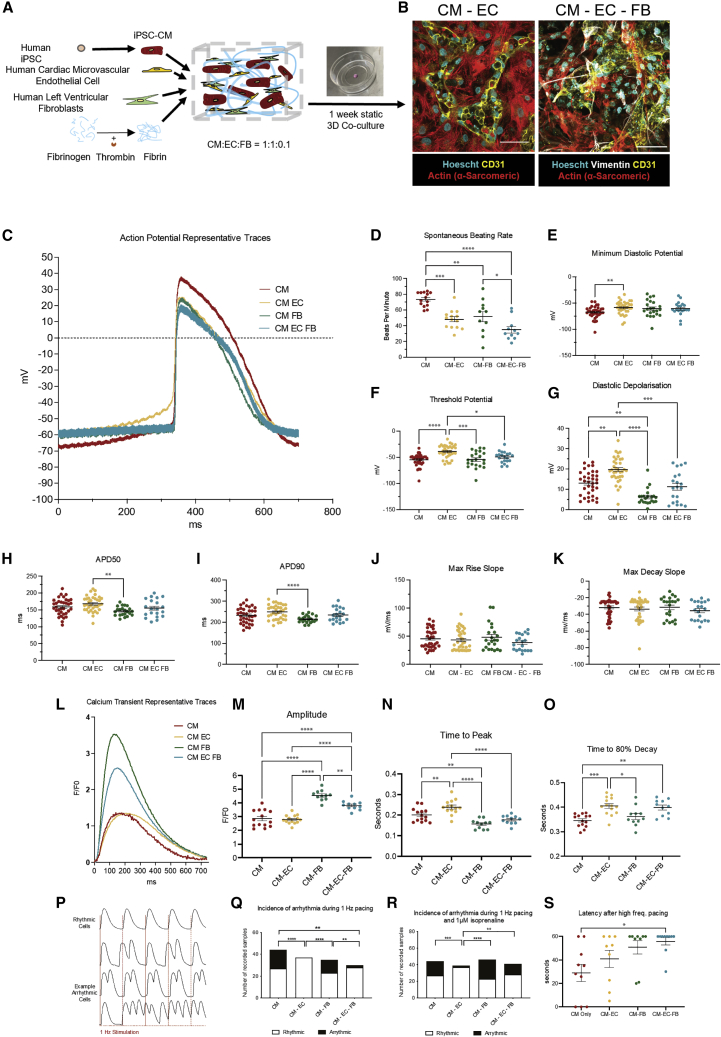
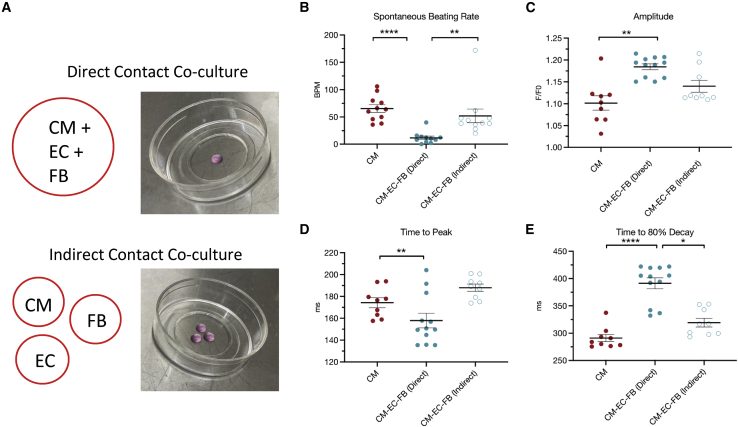
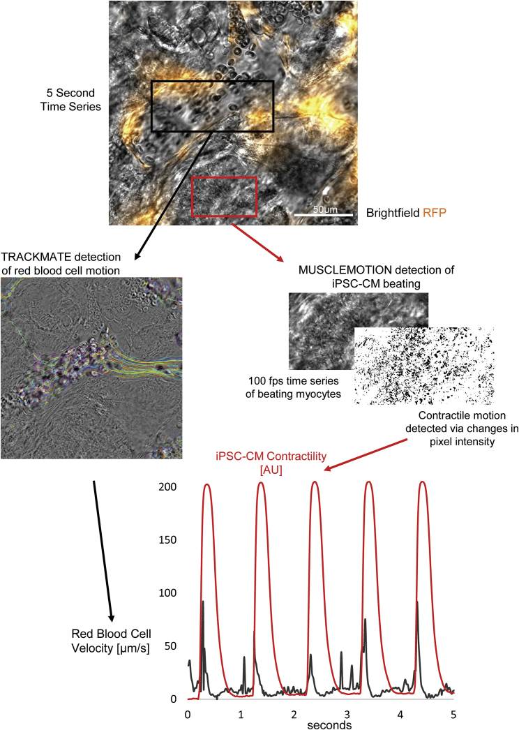
In this study, we report static and perfused models of human myocardial-microvascular interaction. In static culture, we observe distinct regulation of electrophysiology of human induced pluripotent stem cell derived-cardiomyocytes (hiPSC-CMs) in co-culture with human cardiac microvascular endothelial cells (hCMVECs) and human left ventricular fibroblasts (hLVFBs), including modification of beating rate, action potential, calcium handling, and pro-arrhythmic substrate. Within a heart-on-a-chip model, we subject this three-dimensional (3D) co-culture to microfluidic perfusion and vasculogenic growth factors to induce spontaneous assembly of perfusable myocardial microvasculature. Live imaging of red blood cells within myocardial microvasculature reveals pulsatile flow generated by beating hiPSC-CMs. This study therefore demonstrates a functionally vascularized in vitro model of human myocardium with widespread potential applications in basic and translational research.

Keywords: E-C coupling; cardiac physiology; cardiomyocyte; electrophysiology; endothelial cell; microphysiological systems; microvasculature; organ-on-chip; stem cell-derived models; tissue engineering.


Conflict of interest statement

The authors declare no competing interests.

Figures

None
Graphical abstract
Figure 1
Figure 1
Endothelial and fibroblast 3D co-culture modulates iPSC-CM excitation-contraction coupling and reduces pro-arrhythmic substrate (A) Schematic of cellular encapsulation of hiPSC-CMs, hCMVECs, and hLVFBs in fibrin hydrogel. (B) Physical cell-cell association between CMs (actin (α-sarcomeric)), ECs (CD31), and FBs (vimentin) after 1 week of co-culture. Scale bar: 50 μm. (C) Representative traces of action potential morphology in spontaneous co-cultures measured by sharp microelectrode. (D and E) Spontaneous beating rate. (E) Minimum diastolic potential. (F) Membrane potential at threshold of action potential upstroke. (G) Diastolic depolarization (membrane potential depolarization between minimum and threshold potentials). (H) 50% of action potential duration. (I) 90% of action potential duration. (J) Maximum rate of action potential upstroke. (K) Maximum rate of action potential repolarization. (L) Representative traces of iPSC-CM Ca2+ handling. (M) Calcium transient amplitude (F1/F0). (N) Time to calcium transient peak. (O) Time to 80% decay of calcium transient. (P) Example traces of rhythmic versus arrhythmic iPSC-CMs during 1 Hz electrical stimulation. (Q) iPSC-CM arrhythmogenesis while electrically simulating at 1 Hz. (R) iPSC-CM arrhythmogenesis while electrically simulating at 1 Hz and 1 μm isoprenaline treatment. (S) iPSC-CM latency after high-frequency pacing protocol. Data are shown as mean ± SEM. ∗p < 0.05, ∗∗p < 0.01, ∗∗∗p < 0.001, ∗∗∗∗p < 0.0001. Each datapoint represents 1 cell, 3 beats/cell, N = 5.
Figure 2
Figure 2
Direct contact co-culture is required for non-myocyte regulation of iPSC-CM electrophysiology (A) Experimental outline showing combined encapsulation of all cells (direct contact) or separation of each cell type (indirect contact). (B) Quantification of iPSC-CM spontaneous beating rate. (C) iPSC-CM calcium transient amplitude. (D) iPSC-CM calcium transient time to peak. (E) iPSC-CM calcium transient time to 80% decay. Data are shown as mean ± SEM. Each datapoint represents 1 dish, containing average of 3 cells, 3 beats/cell, N = 3.
Figure 3
Figure 3
Generation of perfusable myocardial microvasculature via microfluidic and vasculogenic culture (A) Schematic of heart-on-a-chip (i) layout, (ii) design, (iii) cell seeding, and (iv) connection to perfusion. (B) Representative image of microfluidic chip connected to syringe for perfusion (tubing shortened for display). (C) Vasculogenic culture protocol. Ri, Rho/ROCK inhibitor; VEGF, vascular endothelial growth factor; Ang-1, angiopoietin 1. (D) Representative image of cellular distribution in microfluidic chips (EC = RFP-HUVEC). Scale bar: 1 mm. (E) Confocal z stack demonstrating myocardial microvasculature with open lumen. White asterisk represents open luminal space (EC = hCMVEC). Scale bar: 50 μm (Video S3). (F) Live-cell image of perfused erythrocyte in myocardial microvasculature lumen (EC = RFP-HUVEC). Scale bar: 50 μm (Video S4). (G) Representative Ca2+ transient traces in heart on a chip under vascularized and/or perfused conditions. (H–J) Quantification of Ca2+ transient (H) amplitude and (I and J) kinetics under vascularized and perfused conditions. Data are shown as mean ± SEM. Each datapoint represents 1 chip, containing average of 3 regions of interest (ROIs; 5–15 cells), 3 beats/ROI.
Figure 4
Figure 4
Beating iPSC-CMs induce pulsatile intra-microvascular flow profile Simultaneous recording and quantification of iPSC-CM contractility (via MUSCLEMOTION [Sala et al., 2018]) and erythrocyte velocity (via TRACKMATE [Tinevez et al., 2017]) reveals surge in erythrocyte velocity as iPSC-CMs contract (Video S5).

References

    1. Ashikawa K., Kanatsuka H., Suzuki T., Takishima T. Phasic blood flow velocity pattern in epimyocardial microvessels in the beating canine left ventricle. Circ. Res. 1986;59:704–711. doi: 10.1161/01.RES.59.6.704. - DOI - PubMed
    1. Baeyens N., Nicoli S., Coon B.G., Ross T.D., Van den Dries K., Han J., Lauridsen H.M., Mejean C.O., Eichmann A., Thomas J.L., et al. Vascular remodeling is governed by a VEGFR3-dependent fluid shear stress set poin. Elife. 2015;4:1–35. doi: 10.7554/ELIFE.04645. - DOI - PMC - PubMed
    1. Beauchamp P., Jackson C.B., Ozhathil L.C., Agarkova I., Galindo C.L., Sawyer D.B., Suter T.M., Zuppinger C. 3D co-culture of hiPSC-derived cardiomyocytes with cardiac fibroblasts improves tissue-like features of cardiac spheroids. Front. Mol. Biosci. 2020;7:14. doi: 10.3389/fmolb.2020.00014. - DOI - PMC - PubMed
    1. Berry C., Duncker D.J. Coronary microvascular disease: the next frontier for cardiovascular research. Cardiovasc. Res. 2020;116:737–740. https://pubmed.ncbi.nlm.nih.gov/32149331/ - PMC - PubMed
    1. Blache U., Ehrbar M. Inspired by nature: hydrogels as versatile tools for vascular engineering. Adv. Wound Care. 2018;7:232–246. doi: 10.1089/wound.2017.0760. - DOI - PMC - PubMed

Publication types

LinkOut - more resources