. 2022 Sep 14:2022:5774735.
doi: 10.1155/2022/5774735. eCollection 2022.

CTA Study of Ruptured Aneurysms of the Posterior Communicating Artery

Affiliations

CTA Study of Ruptured Aneurysms of the Posterior Communicating Artery

Zibo Zhou et al. Stroke Res Treat. .

Abstract

Background: Only a few reported studies have used computed tomography angiography (CTA) to image ruptured aneurysms at the junction of the internal carotid artery (ICA) and posterior communicating artery (PcomA) in the context of the adjacent arteries. Therefore, we performed such a study using a GE Workstation.

Methods: The parameters of each aneurysm and its adjacent arteries were measured. Then, statistical assessments were performed to compare the parameters of the aneurysm side and the lesion-free (control) side.

Results: Sixty-three patients were included in this study. The average age was 62.1 ± 11.0 years, and the ratio of males to females was 0.8 : 1. The measurement results showed that the mean aneurysmal height was 5.2 ± 2.3 mm, the mean width was 4.7 ± 2.2 mm, and the mean neck width was 4.5 ± 1.9 mm. On the aneurysm side, the intradural ICA diameter was 4.34 ± 0.90 mm, and the diameter of the ICA at its termination was 3.55 ± 0.72 mm. A fetal-type PcomA was found in 52.4% of aneurysms. The other measured parameters were also provided. Statistical results showed that the height of the aneurysm was larger than the width (P < 0.05). The intradural ICA diameter, the ICA diameter at termination, the intradural ICA length, and the angle between the ICA and PcomA were larger in the aneurysm group than in the control group (P < 0.05).

Conclusions: This CTA study showed that the ruptured PcomA aneurysm was often wide-necked, nonspherical, and approximately 5 mm in size. In the presence of a ruptured PcomA aneurysm, the affected intradural ICA became thicker and longer than the contralateral control ICA, and the aneurysm significantly reduced the angle between the ICA and the PcomA.


Conflict of interest statement

The authors declare that they have no competing interests.

Figures
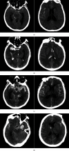
Figure 1
Figure 1
The mFS, a CT-based rating scale is as follows; (a) Grade 1 consists of thin, minimal, or diffuse SAH without IVH; (b) Grade 2 consists of minimal or thin SAH with IVH; (c) Grade 3 is defined by a thick cisternal clot without IVH; (d) Grade 4 is defined by a cisternal clot with IVH. Abbreviations: CT: computed tomography; IVH: intraventricular hemorrhage; mFS: modified Fisher scale; SAH: subarachnoid hemorrhage.
Figure 2
Figure 2
CTA-based measurement parameters are as follows: (a) No. 1 is the height of the aneurysm, No. 2 is the width of the aneurysm, and No. 3 is the width of the aneurysmal neck; (b) No. 4 is the diameter of the intradural ICA at its origin, No. 5 is the diameter of the ICA at its termination, and No. 6 is the diameter of the MCA at its origin; (c) No. 7 is the diameter of the PcomA at its origin, No. 8 is the diameter of the PcomA at its termination, No. 9 is the diameter of P2 at its origin, and No. 10 is the diameter of P1 at its termination; (d) No. 11 is the length of the intradural ICA, No. 12 is the length of the PcomA, and No. 13 is the angle between the ICA and the PcomA. Abbreviations: CTA: computed tomography angiography; ICA: internal carotid artery; MCA: middle cerebral artery; PCA: posterior cerebral artery; PcomA: posterior communicating artery; P1: first segment of PCA.
Figure 3
Figure 3
Aneurysm projection in CTA is as folows; (a–b) CTA showing that the aneurysm projected laterally (arrows). (c–d); CTA showing that the aneurysm projected posteriorly (arrows). Abbreviations: CTA: computed tomography angiography; ICA: internal carotid artery.
Figure 4
Figure 4
PcomA development as shown by CTA is as follows: (a) Grade I is defined by the absence of the PcomA; the arrow indicates the aneurysm. (b) Grade II is defined by the presence of a thin PcomA (asterisk); the arrow indicates the aneurysm. (c–d) Grade III is defined by the presence of a thick fetal-type PcomA (asterisk); the arrow indicates the aneurysm. Abbreviations: CTA: computed tomography angiography; ICA: internal carotid artery; PCA: posterior cerebral artery; PcomA: posterior communicating artery.

References

    1. Taweesomboonyat C., Tunthanathip T., Kaewborisutsakul A., et al. Outcome of ruptured posterior communicating artery aneurysm treatment comparing between clipping and coiling techniques. World Neurosurgery . 2019;125:e183–e188. doi: 10.1016/j.wneu.2019.01.037. - DOI - PubMed
    1. Niibo T., Takizawa K., Sakurai J., et al. Prediction of the difficulty of proximal vascular control using 3D-CTA for the surgical clipping of internal carotid artery-posterior communicating artery aneurysms. Journal of Neurosurgery . 2020;134(3):1165–1172. doi: 10.3171/2020.1.JNS192728. - DOI - PubMed
    1. Gabrovsky N. Microanatomical bases for intraoperative division of the posterior communicating artery. Acta Neurochirurgica . 2002;144(11):1205–1211. doi: 10.1007/s00701-002-1002-x. - DOI - PubMed
    1. Ijichi S., Ijichi N., Niina K., Nakamura F., Osame M., Tokito S. Angiographic morphology of the posterior communicating artery and basilar in patients with ICA-PComA aneurysm. Neurologia Medico-Chirurgica . 1991;31(4):189–193. doi: 10.2176/nmc.31.189. - DOI - PubMed
    1. Can A., Ho A. L., Emmer B. J., Dammers R., Dirven C. M., Du R. Association between vascular anatomy and posterior communicating artery aneurysms. World Neurosurgery . 2015;84(5):1251–1255. doi: 10.1016/j.wneu.2015.05.078. - DOI - PubMed

LinkOut - more resources