. 2022 Sep 15:2022:4155565.
doi: 10.1155/2022/4155565. eCollection 2022.

Acetylation of Atp5f1c Mediates Cardiomyocyte Senescence via Metabolic Dysfunction in Radiation-Induced Heart Damage

Affiliations

Acetylation of Atp5f1c Mediates Cardiomyocyte Senescence via Metabolic Dysfunction in Radiation-Induced Heart Damage

Zhimin Zeng et al. Oxid Med Cell Longev. .

Abstract

Objective: Ionizing radiation (IR) causes cardiac senescence, which eventually manifests as radiation-induced heart damage (RIHD). This study is aimed at exploring the mechanisms underlying IR-induced senescence using acetylation proteomics.

Methods: Irradiated mouse hearts and H9C2 cells were harvested for senescence detection. Acetylation proteomics was used to investigate alterations in lysine acetylation. Atp5f1c acetylation after IR was verified using coimmunoprecipitation (Co-IP). Atp5f1c lysine 55 site acetylation (Atp5f1c K55-Ac) point mutation plasmids were used to evaluate the influence of Atp5f1c K55-Ac on energy metabolism and cellular senescence. Deacetylation inhibitors, plasmids, and siRNA transfection were used to determine the mechanism of Atp5f1c K55-Ac regulation.

Results: The mice showed cardiomyocyte and cardiac aging phenotypes after IR. We identified 90 lysine acetylation sites from 70 protein alterations in the heart in response to IR. Hyperacetylated proteins are primarily involved in energy metabolism. Among them, Atp5f1c was hyperacetylated, as confirmed by Co-IP. Atp5f1c K55-Ac decreased ATP enzyme activity and synthesis. Atp5f1c K55 acetylation induced cardiomyocyte senescence, and Sirt4 and Sirt5 regulated Atp5f1c K55 deacetylation.

Conclusion: Our findings reveal a mechanism of RIHD through which Atp5f1c K55-Ac leads to cardiac aging and Sirt4 or Sirt5 modulates Atp5f1c acetylation. Therefore, the regulation of Atp5f1c K55-Ac might be a potential target for the treatment of RIHD.


Conflict of interest statement

The authors declare that they have no competing interests.

Figures

Figure 1
Figure 1
Ionizing radiation causes cardiac senescence. (a, b) Echocardiograph images and LVEF of mouse hearts in the control mice (control group) and in mice 1 and 5 months (1-month and 5-month groups, respectively) after local heart irradiation at a dose of 16 Gy. (c) q-PCR analysis mRNA of fibrosis-associated factors in the 1-month, 5-month, and control groups (p < 0.05, ∗∗p < 0.01, ∗∗∗p < 0.001; NS: not significant). (d) q-PCR analysis of the mRNA levels of inflammation-associated factors in the 1-month, 5-month, and control groups (p < 0.05, ∗∗p < 0.01, ∗∗∗p < 0.001, NS: not significant). (e) q-PCR analysis of telomere length in cardiac tissue from 1 month to 5 months, and control mice (p < 0.05).
Figure 2
Figure 2
Ionizing radiation causes cardiomyocyte senescence. (a) Senescence-associated β-galactosidase (SA-βgal) staining of sham-irradiated and irradiated H9C2 cells. (b)–(d) Western blotting and quantification analyses of p21 and p16 protein expression in H9C2 cells in the control group and after 6h, 12h, and 24h of irradiation (p < 0.05, ∗∗p < 0.01). (e) q-PCR analysis of the mRNA levels of inflammation- and fibrosis-associated factors in H9C2 cells of the control, 6h, 12h, and 24h irradiation groups (p < 0.05, ∗∗p < 0.01, ∗∗∗p < 0.001, ∗∗∗∗p < 0.0001). Bar: 100μm; h: hour.
Figure 3
Figure 3
Ionizing radiation induces lysine residue hyperacetylation, thus modifying cardiac metabolic enzymes. (a) Radiation induces protein acetylation in mouse hearts after 5 months. (b) Radiation induces protein acetylation in H9C2 cells at different time points. (c) Experimental flow chart of proteomic analysis. (d) Clusters of Orthologous Groups/KOG functional classification chart of proteins corresponding to differentially expressed modification sites. (e) GO enrichment bubble plot of proteins corresponding to differentially expressed modification sites in the biological process category. (f) KEGG pathway enrichment bubble plot of proteins corresponding to differentially expressed modification sites. (g) PPI network of the DEPs. Red nodes represent upregulated proteins, and blue nodes represent downregulated proteins. (h) The CytoHubba of Degree method were used to extract the top 10 hub proteins from the PPI network. (i) The CytoHubba of DMNC method was used to extract the top 10 hub proteins from the PPI network.
Figure 4
Figure 4
Ionizing radiation induces Atp5f1c acetylation in irradiated heart and cardiomyocytes. (a) Sequence alignment of mouse, rat, human, chimpanzee, monkey, and bovine Atp5f1c proteins. (b, c) Ionizing radiation induced Atp5f1c acetylation in heart tissue, as verified by Co-IP. (d) Ionizing radiation induced Atp5f1c acetylation in cardiomyocytes, as verified by Co-IP. K-Ac: lysine (K) acetylation; Co-IP: coimmunoprecipitation; WB: western blotting.
Figure 5
Figure 5
Atp5f1c is hyperacetylated at the 55th lysine site, which leads to metabolic disorders and senescence. (a) The expression of anti-His antibody after His-tagged Atp5f1c WT, K55R, and K55Q point mutation plasmids transfection in H9C2 cells. (b) Atp5f1c acetylation level verified by Co-IP after transfection of Atp5f1c WT, K55R, and K55Q point mutation plasmids in H9C2 cells. (c, d) Senescence-associated β-galactosidase (SABG) staining and statistical analysis after transfection of Atp5f1c WT, K55R, and K55Q point mutation plasmids in H9C2 cells (p < 0.05, ∗∗p < 0.01, ∗∗∗p < 0.001; NS: not significant). (e) q-PCR analysis of the mRNA levels of inflammation- and fibrosis-associated factors after transfection of Atp5f1c WT, K55R, and K55Q point mutation plasmids in H9C2 cells (p < 0.05, ∗∗p < 0.01, ∗∗∗p < 0.001, ∗∗∗∗p < 0.0001). (f)–(i) Western blotting and quantification analyses of p21 and p16 protein expression after transfection of Atp5f1c WT, K55R, and K55Q point mutation plasmids in H9C2 cells; relative protein levels were normalized to GAPDH or actin expression (p < 0.05). (j, k) ATP synthase activity and ATP production after transfection of Atp5f1c WT, K55R, and K55Q plasmids into H9C2 cells (p < 0.05, ∗∗p < 0.01, ∗∗∗p < 0.001; NS: not significant). Co-IP: Coimmunoprecipitation; WB: western blotting; WT: wild type.
Figure 6
Figure 6
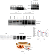
Sirt4 and Sirt5 mediated Atp5f1c K55 deacetylation. (a) Effect of trichostatin A (TSA) and nicotinamide (NAM) on Atp5f1c acetylation levels in H9C2 cells. (b) The expression of HA-tag after HA-tagged Sirt3, Sirt4, and Sirt5 plasmids was transfected into H9C2 cells. (c) Atp5f1c acetylation verified by Co-IP after HA-tagged Sirt3, Sirt4, and Sirt5 plasmids was transfected into H9C2 cells. (d) Atp5f1c acetylation level verified by Co-IP after siRNA of Sirt3, Sirt 4, and Sirt 5 was transfected into H9C2 cells. (e) q-PCR analysis of the mRNA levels of Sirt3, Sirt4, and Sirt 5 in H9C2 cells of the control, 6, 12, and 24 h irradiation groups (p < 0.05, ∗∗p < 0.01, ∗∗∗p < 0.001). (f) Schematic diagram of the study design. WB: western blotting; h: hour; WB: western blotting; RIHD: radiation-induce heart damage.

Similar articles

Cited by

References

    1. Stewart F. A., Seemann I., Hoving S., Russell N. S. Understanding radiation-induced cardiovascular damage and strategies for intervention. Clinical Oncology . 2013;25(10):617–624. doi: 10.1016/j.clon.2013.06.012. - DOI - PubMed
    1. Moding E. J., Kastan M. B., Kirsch D. G. Strategies for optimizing the response of cancer and normal tissues to radiation. Drug Discovery . 2013;12(7):526–542. doi: 10.1038/nrd4003. - DOI - PMC - PubMed
    1. de Groot C., Beukema J. C., Langendijk J. A., et al. Radiation-induced myocardial fibrosis in long-term esophageal cancer survivors. International Journal of Radiation Oncology • Biology • Physics . 2021;110(4):1013–1021. doi: 10.1016/j.ijrobp.2021.02.007. - DOI - PubMed
    1. Le Pechoux C., Pourel N., Barlesi F., et al. Postoperative radiotherapy versus no postoperative radiotherapy in patients with completely resected non-small-cell lung cancer and proven mediastinal N2 involvement (Lung ART, IFCT 0503): an open-label, randomised, phase 3 trial. Oncologia . 2022;23(1):104–114. doi: 10.1016/S1470-2045(21)00606-9. - DOI - PubMed
    1. Laugaard Lorenzen E., Christian Rehammar J., Jensen M. B., Ewertz M., Brink C. Radiation-induced risk of ischemic heart disease following breast cancer radiotherapy in Denmark, 1977-2005. Oncologia . 2020;152:103–110. doi: 10.1016/j.radonc.2020.08.007. - DOI - PubMed

LinkOut - more resources