. 2022 Sep 26;17(1):95.
doi: 10.1186/s11671-022-03723-2.

Divergent Effects of Laser Irradiation on Ensembles of Nitrogen-Vacancy Centers in Bulk and Nanodiamonds: Implications for Biosensing

Affiliations

Divergent Effects of Laser Irradiation on Ensembles of Nitrogen-Vacancy Centers in Bulk and Nanodiamonds: Implications for Biosensing

Domingo Olivares-Postigo et al. Nanoscale Res Lett. .

Abstract

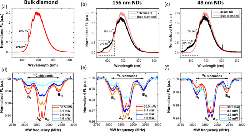
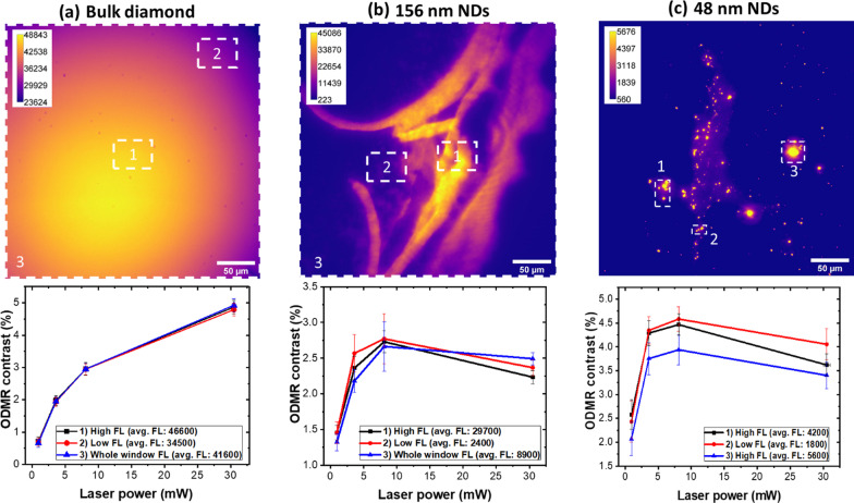
Ensembles of negatively charged nitrogen-vacancy centers (NV-) in diamond have been proposed for sensing of magnetic fields and paramagnetic agents, and as a source of spin-order for the hyperpolarization of nuclei in magnetic resonance applications. To this end, strongly fluorescent nanodiamonds (NDs) represent promising materials, with large surface areas and dense ensembles of NV-. However, surface effects tend to favor the less useful neutral form, the NV0 centers, and strategies to increase the density of shallow NV- centers have been proposed, including irradiation with strong laser power (Gorrini in ACS Appl Mater Interfaces. 13:43221-43232, 2021). Here, we study the fluorescence properties and optically detected magnetic resonance (ODMR) of NV- centers as a function of laser power in strongly fluorescent bulk diamond and in nanodiamonds obtained by nanomilling of the native material. In bulk diamond, we find that increasing laser power increases ODMR contrast, consistent with a power-dependent increase in spin-polarization. Conversely, in nanodiamonds we observe a non-monotonic behavior, with a decrease in ODMR contrast at higher laser power. We hypothesize that this phenomenon may be ascribed to more efficient NV-→NV0 photoconversion in nanodiamonds compared to bulk diamond, resulting in depletion of the NV- pool. A similar behavior is shown for NDs internalized in macrophage cells under the typical experimental conditions of imaging bioassays. Our results suggest strong laser irradiation is not an effective strategy in NDs, where the interplay between surface effects and local microenvironment determine the optimal experimental conditions.

Keywords: 13C; Bulk diamond; Cells; Charge dynamics; Charge stability; Nanodiamonds; Nanomilling; Nitrogen-vacancy centers; Photoconversion; Polarization; Spin dynamics.


Conflict of interest statement

The authors declare that they have no competing interests.

Figures

Fig. 1
Fig. 1
Preparation of NDs and schematic of the wide-field ODMR set-up. a NDs, 156 nm and 48 nm in size were obtained by milling bulk, 13C-enriched HPHT fluorescent diamond. b Schematics of the inverted wide-field ODMR microscope. Green laser light is focused onto the sample. The fluorescence is collected by the objective through a dichroic mirror to the high-sensitivity CMOS camera. The image shows an actual fluorescence image of NDs deposited on a glass slide. MW delivery is obtained with a loop adjacent to the sample. The panel shows the temporal diagram of the CW-ODMR experiment. The laser is kept ON during the entire measurement, while the camera detects the fluorescence image synchronously with the MW irradiation. c Photo of the wide-field ODMR microscope set-up
Fig. 2
Fig. 2
Fluorescence and ODMR spectra. Fluorescence and ODMR spectra for the a,d bulk diamond, b,e 156 nm and c,f 59 nm NDs, respectively. a,b,c NV0 and NV fluorescence spectra are characterized by ZPL lines at 575 nm (NV0) and 638 nm (NV). Due to surface effects, NDs have a larger NV0 component than bulk diamond, where the NV0 band is practically undetectable. Panels d,e,f show ODMR spectra at different laser power with fixed MW power. In the ODMR spectra, the NV central lines (AL and AR) provide a measure of the spin polarization of g,ms=0 ground state. The MW range was set to 2.75–3 GHz to show the NV central lines and the 13C-coupled sidebands (BL and BR). In bulk diamond, ODMR contrast increases with increasing laser power. Conversely, in the NDs, the ODMR contrast increases up to 8.1 mW and then, decreases at higher laser powers. Both FL spectra and ODMR spectra were acquired under continuous laser irradiation. The small peak at ~ 800 nm in the FL spectra is an artifact of the Raman spectrometer
Fig. 3
Fig. 3
Comparison of ODMR contrast at different laser powers for bulk diamond and NDs. ODMR curves are extracted from three different regions of interest (ROIs) containing the bright spots that indicate presence of NV centers (see wide-field fluorescence images). As laser power increases, in bulk diamond the ODMR contrast increases, consistently with larger NV polarization levels (a). Conversely, in NDs of both sizes, the ODMR contrast increases up to 8.1 mW and decreases at higher laser powers, resulting in a non-monotonic behavior (b,c). The homogeneity of bulk diamond is reflected in the uniform values of ODMR contrast observed in the various ROIs with different levels of FL (values in the caption). On the contrary, NDs show non-uniform deposition and aggregation, resulting in a region-dependent ODMR contrast. 156 nm NDs were less inhomogeneous than the 48 nm NDs. The laser power dependence of the ODMR signal was similar in all the ROIs extracted, for both NDs sizes
Fig. 4
Fig. 4
Confocal Microscopy images of RAW 264.7 cells incubated for 24 h with 48 nm and 156 nm NDs. After adding NDs (red) cells were stained with Phalloidin (green). Nuclei were counterstained with DAPI (blue). The “merge” figure demonstrates the internalization of the NDs (orange) within the cells. Cellular uptake of the 156 nm NDs is much higher than that of the 48 nm NDs. Scale bars = 20 µm. Representative images are shown
Fig. 5
Fig. 5
ODMR contrast of NDs internalized in cells. a Fluorescence image of 156 nm NDs internalized in macrophage cells; the white square delineates the ROIs from which signals were extracted. NDs internalized within cells show inhomogeneous aggregation and concentration, resulting in a region-dependent FL values. ODMR spectra presenting the NV central lines (AL and AR) and the 13C sidebands (BL and BR) at different laser power with constant MW power are shown in b for a single cell, and in c for a cellular aggregate. d ODMR curves corresponding to the ROIs of panel (a). The caption reports the average value of fluorescence intensity in each ROI. ODMR contrast steadily increases to 8.1 mW, while at higher laser powers it shows a plurality of behaviors. Small differences in the curves are uncorrelated with the average FL intensity and are supposedly related to local environment. e A color-saturated fluorescence image of 156 nm NDs internalized in cells. The selected ROI f shows a cell with its different compartments and its nucleus. g Map of the distribution of ODMR contrast within the cells of (f). Contrast varies regionally, with an average value of 4.5%

References

    1. Taylor JM, Cappellaro P, Childress L, Jiang L, Budker D, Hemmer PR, Yacoby A, Walsworth R, Lukin MD. High-sensitivity diamond magnetometer with nanoscale resolution. Nat Phys. 2008;4:810–816. doi: 10.1038/nphys1075. - DOI
    1. Davis HC, Ramesh P, Bhatnagar A, Lee-Gosselin A, Barry JF, Glenn DR, Walsworth RL, Shapiro MG. Mapping the microscale origins of magnetic resonance image contrast with subcellular diamond magnetometry. Nat Commun. 2018;9:1–9. doi: 10.1038/s41467-017-02471-7. - DOI - PMC - PubMed
    1. Balasubramanian G, Neumann P, Twitchen D, Markham M, Kolesov R, Mizuochi N, Isoya J, Achard J, Beck J, Tissler J, et al. Ultralong spin coherence time in isotopically engineered diamond. Nat Mater. 2009;8:383–387. doi: 10.1038/nmat2420. - DOI - PubMed
    1. Liu GQ, Feng X, Wang N, Li Q, Liu RB. Coherent quantum control of nitrogen-vacancy center spins near 1000 kelvin. Nat Commun. 2019;10:1–8. doi: 10.1038/s41467-019-09327-2. - DOI - PMC - PubMed
    1. Dolde F, Fedder H, Doherty MW, Nöbauer T, Rempp F, Balasubramanian G, Wolf T, Reinhard F, Hollenberg LCL, Jelezko F, et al. Electric-field sensing using single diamond spins. Nat Phys. 2011;7:459–463. doi: 10.1038/nphys1969. - DOI

LinkOut - more resources