. 2022 Oct 14;50(18):10695-10716.
doi: 10.1093/nar/gkac817.

Human NOP2/NSUN1 regulates ribosome biogenesis through non-catalytic complex formation with box C/D snoRNPs

Affiliations

Human NOP2/NSUN1 regulates ribosome biogenesis through non-catalytic complex formation with box C/D snoRNPs

Han Liao et al. Nucleic Acids Res. .

Abstract

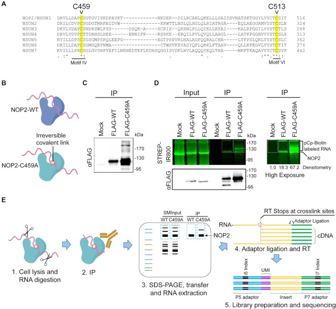
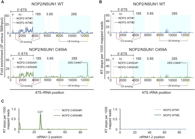
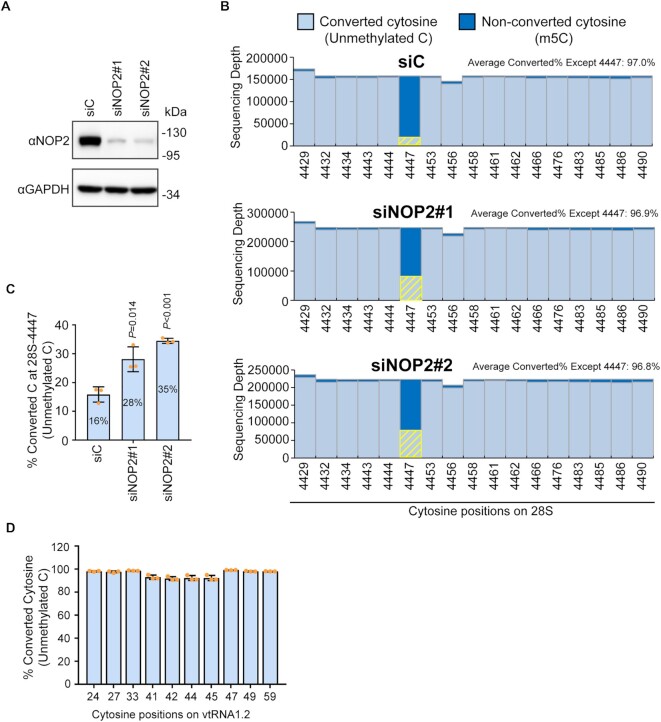
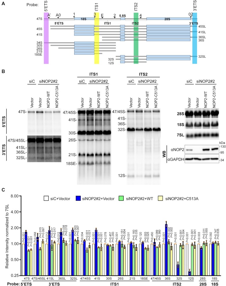
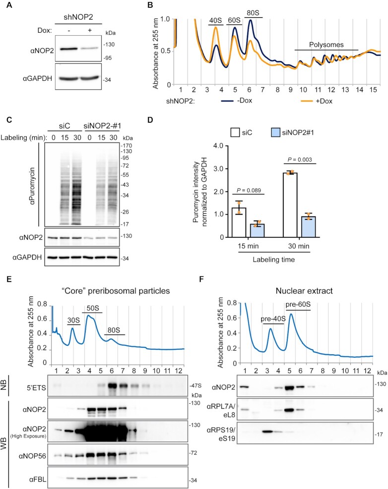
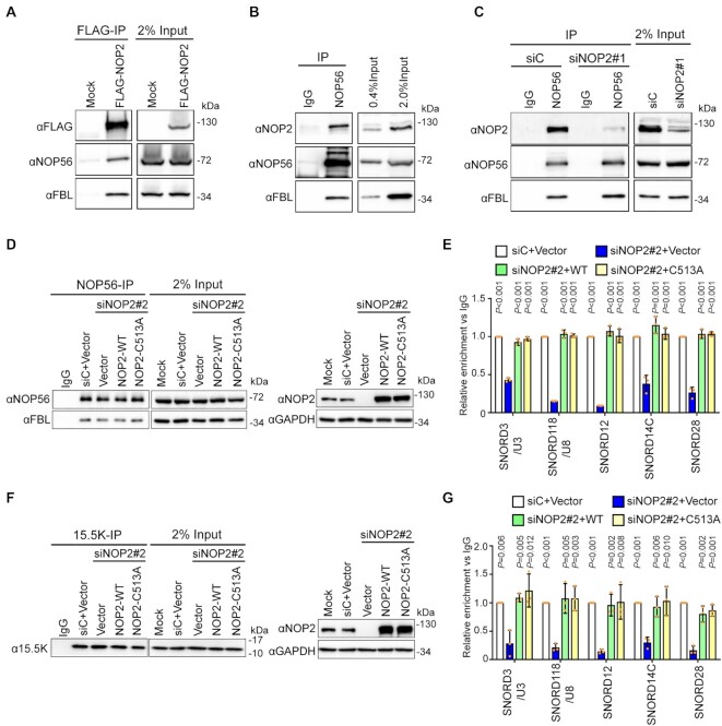
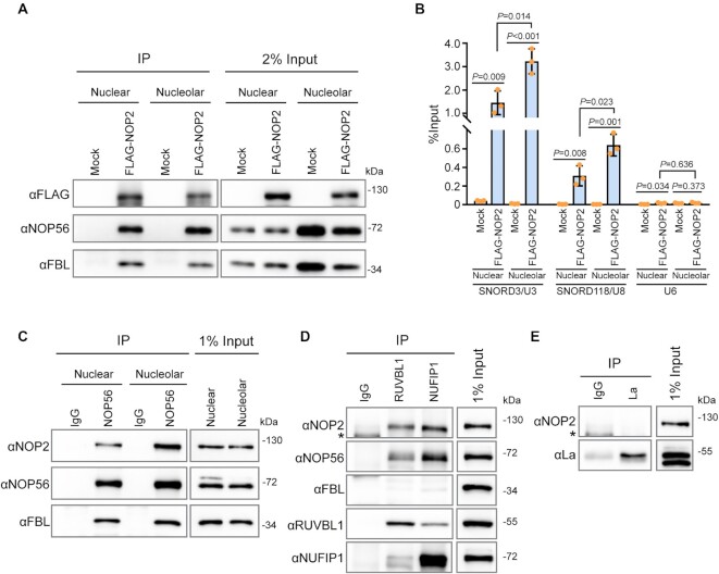
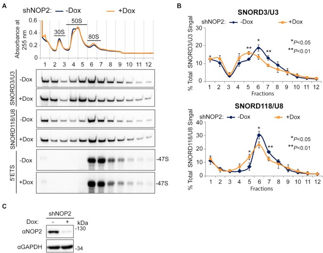
5-Methylcytosine (m5C) is a base modification broadly found on various RNAs in the human transcriptome. In eukaryotes, m5C is catalyzed by enzymes of the NSUN family composed of seven human members (NSUN1-7). NOP2/NSUN1 has been primarily characterized in budding yeast as an essential ribosome biogenesis factor required for the deposition of m5C on the 25S ribosomal RNA (rRNA). Although human NOP2/NSUN1 has been known to be an oncogene overexpressed in several types of cancer, its functions and substrates remain poorly characterized. Here, we used a miCLIP-seq approach to identify human NOP2/NSUN1 RNA substrates. Our analysis revealed that NOP2/NSUN1 catalyzes the deposition of m5C at position 4447 on the 28S rRNA. We also find that NOP2/NSUN1 binds to the 5'ETS region of the pre-rRNA transcript and regulates pre-rRNA processing through non-catalytic complex formation with box C/D snoRNAs. We provide evidence that NOP2/NSUN1 facilitates the recruitment of U3 and U8 snoRNAs to pre-90S ribosomal particles and their stable assembly into snoRNP complexes. Remarkably, expression of both WT and catalytically inactive NOP2/NSUN1 in knockdown background rescues the rRNA processing defects and the stable assembly of box C/D snoRNP complexes, suggesting that NOP2/NSUN1-mediated deposition of m5C on rRNA is not required for ribosome synthesis.


Figures

Figure 1.
Figure 1.
Human NOP2/NSUN1 miCLIP design. (A) Alignment of the protein sequences of human NSUN family members showing conserved cysteine residues (highlighted) required for catalytic activity (C459 and C513). The cysteine mutated for the miCLIP assay is C459 in motif IV. (B) Cartoon depicting that only the NOP2/NUSN1 C459A mutant forms irreversible covalent crosslinks with RNA substrates. (C) FLAG-tagged NOP2/NSUN1 Wildtype (WT) and C459A mutant were expressed in HEK293T cells and immunoprecipitated with a FLAG antibody. Immuno-precipitated proteins were detected by Western Blotting using a FLAG antibody. (D) FLAG-tagged NOP2/NSUN1 WT and C459A mutant expressed in HEK293T cells were immunoprecipitated with a FLAG antibody. Co-precipitated RNA was 3′-end ligated with pCp-biotin for visualization. After SDS-PAGE separation and membrane transfer, the pCp-biotin-labeled RNA was detected with Streptavidin-IR800. Immunoprecipitated NOP2/NSUN1 WT and C459A mutant were detected by immunoblotting with a FLAG antibody. The right panel shows a high exposure of the pCp-biotin-labeled RNA with relative densitometry analysis. (E) Schematic overview of the NOP2/NSUN1 miCLIP-sequencing. HEK293T cells expressing NOP2/NSUN1 WT and C459A mutant were lysed, digested with RNase, immunoprecipitated with FLAG antibody, separated on SDS-PAGE and transferred to membrane. NOP2/NSUN1 WT and C459A mutant-associated RNAs and their respective size-matched inputs (SMInputs) were extracted from the membrane and processed for libraries and Illumina sequencing.
Figure 2.
Figure 2.
Human NOP2/NSUN1 binds to rRNA and snoRNAs. (A–C) HEK293T cells expressing FLAG-tagged NOP2/NSUN1 WT (non-covalently bound to substrate) or C459A mutant (covalently bound to substrate) were subject to CLIP-sequencing analysis (two biological replicates for each sample). CLIP-sequencing reads were aligned to human genome (hg38) and repetitive elements families. CLIP peaks were called by Peakachu. (A) Percentage of miCLIP mapped peak regions in each indicated RNA category with cut-offs of adjusted significance P values < 0.05 and fold change over size-matched input (SMInput) > = 2. (B) Volcano plots of all peaks detected by Peakachu. Each dot represents a peak either enriched in IP (log2 fold change > 0) or SMInput samples (log2 fold change < 0). (C) Number of CLIP peaks with cut-offs of adjusted significant P values < 0.05 and fold change over SMInput ≥2 in each indicated RNA category. C/D box and H/ACA box snoRNA are shown in light and dark blue, respectively. Number of snoRNAs found in CLIP peaks / total number of snoRNAs in the indicated subfamily are shown in parentheses.
Figure 3.
Figure 3.
Human NOP2/NSUN1 binds to the rRNA 5′-ETS region and crosslinks on 28S rRNA at position C4447. (A, B) miCLIP-sequencing data were aligned to human 47S pre-rRNA (NR_046235) and the mapping information was retrieved using Samtools. The location of mature 18S, 5.8S and 28S rRNAs are marked with light blue shadow boxes. A’, A0 and 1 cleavage sites on 5′-ETS are marked with gray lines. Red bars indicate the location of U3 snoRNA binding sites (101). The orange bar indicates the location of U8 snoRNA binding site based on sequence homology and accessibility measurements (103). The yellow bar indicates the location of the U8 snoRNA binding site found by RNA duplex mapping (106). (A) Plot of sequencing depth-normalized reads ratio of IP over size-matched input (SMInput) on the 47S rRNA. (B) Plot of reverse transcription (RT) stops on the 47S rRNA normalized by sequencing depth. RT stops were assigned at the start (+1) sites of the Read1 sequence from NOP2/NSUN1 WT and C459A IP samples. (C) Plot of reverse transcription (RT) stops on vtRNA1.2 normalized by sequencing depth. RT stops were assigned at the start (+1) sites of the Read1 sequence from NOP2/NSUN1 WT and C459A IP samples. (B, C) Plotted data is represented for each independent biological replicate for NOP2/NSUN1 WT (blue lines) and C459A mutant (green lines).
Figure 4.
Figure 4.
Human NOP2/NSUN1 methylates the 28S rRNA at position C4447. (A) HCT116 cells were transfected with non-targeting control (siC), NOP2/NSUN1 siRNA#1 or siRNA#2. 72h later, NOP2/NSUN1 depletion efficiency was determined by western blot. (B) A fraction of cells from (A) were harvested for nuclear RNA extraction. Nuclear RNA was treated with bisulfite salt and the region surrounding C4447 was amplified by RT-PCR for sequencing on the Illumina Mi-Seq platform. Non-converted (protected by 5mC modification) and converted cytosines (lacking m5C modification) near C4447 were plotted. The converted cytosines at position 4447 are highlighted by yellow bars. (C) Percentage of converted cytosine (C) at position 4447 on 28S rRNA. (D) Percentage of converted cytosine on vtRNA1.2. Total RNA from HCT116 cells was treated with bisulfite salt and vtRNA1.2 was amplified by RT-PCR for sequencing on the Illumina Mi-Seq platform. The data are shown as the mean of three independent biological replicates ± standard deviation (SD). Statistical significance between NOP2/NSUN1 depleted samples and non-targeting siRNA control samples was calculated using a two-tailed independent Student's t-test.
Figure 5.
Figure 5.
Human NOP2/NSUN1 is required for efficient pre-rRNA processing. (A) Schematic overview of human precursor rRNA processing and location of probes used for northern blot indicated in colored areas (lavender, yellow, green and blue). (B) HCT116 cells expressing empty vector (Vector), siRNA resistant NOP2/NSUN1 WT, or the C513A catalytically inactive mutant were transfected with non-targeting control (siC) or NOP2 siRNA #2. After 72 h, total RNA was separated on formaldehyde denaturing agarose gel and analyzed by Northern blot using 5′ETS, 3′ETS, ITS-1, ITS-2, 18S, 28S and 7SL probes. A fraction of cells was collected to determine endogenous NOP2/NSUN1 depletion and ectopic expression of NOP2/NSUN1 WT or C513A by Western blot using a NOP2 antibody. (C) Densitometry quantification of each rRNA precursor from (B) normalized to 7SL RNA. The data are presented as the mean of three independent biological replicates ± standard deviation (SD). P-values comparing empty vector with NOP2/NSUN1 knockdown (siNOP2#2 + Vector) were calculated using a two-tailed independent student t-test.
Figure 6.
Figure 6.
Human NOP2/NUSN1 is required for 60S ribosomal subunit biogenesis. (A) HCT116 cells were infected with doxycycline-inducible NOP2 shRNA expressing lentivirus. After puromycin selection, cells were induced with 200 ng/ml doxycycline (Dox+) for four days. Non-induced (Dox–) cells were used as control. NOP2/NSUN1 depletion efficiency was determined by western blot. (B) A fraction of cells from (A) was analyzed by polysome profiling using total cell lysates. Dox+ and Dox– indicate doxycycline-induced and non-induced control, respectively. (C) HCT116 cells were transfected with non-targeting control (siC) or NOP2 siRNA. 72 h after transfection, cells were treated with 5 μg/ml puromycin for the indicated time. Puromycylation of nascent peptides and NOP2/NSUN1 depletion efficiency, was determined by Western blot using puromycin and NOP2 antibodies. (D) Densitometry quantification of the puromycin signal from each lane from (C) was normalized to the corresponding GAPDH signal. The data is presented as the mean of two independent biological replicates ± standard deviation (SD). Statistical significance between NOP2 depleted samples and non-targeting control samples was calculated using a two-tailed independent Student's t-test. (E) High molecular weight complexes containing pre-rRNAs and tightly associated ribosome assembly factors, referred to as ‘core’ preribosomal particles isolated from HCT116 nuclear extracts under high-salt conditions were separated on sucrose gradient ultra-centrifugation followed by fractionation. RNA and proteins were isolated from each fraction and analyzed by western blotting (WB) or northern blot (NB) with the indicated antibodies or probe, respectively. (F) Nuclear extract from HCT116 cells was separated on sucrose gradient ultra-centrifugation followed by fractionation. Proteins were extracted from each fraction and analyzed by western blot with the indicated antibodies.
Figure 7.
Figure 7.
NOP2/NSUN1 is required to maintain the integrity of C/D box snoRNPs. (A) HEK293T expressing empty vector (Mock) or FLAG-tagged NOP2/NUSN1 WT were lysed and immunoprecipitated (IP) with an anti-FLAG antibody. Associated proteins were analyzed by Western blot with the indicated antibodies. Mock transfected cells were used as negative control. (B) NOP56 was immunoprecipitated with an anti-NOP56 antibody from HEK293T cell lysate. Associated proteins were analyzed by Western blot with the indicated antibodies. Normal rabbit IgG was used as a negative immunoprecipitation control. (C) HEK293T cells were transfected with non-targeting control (siC) or NOP2 siRNA. 72 h later, cells were lysed and immunoprecipitated (IP) with an anti-NOP56 antibody. Associated proteins were analyzed by Western blot with the indicated antibodies. Normal rabbit IgG was used as a negative immunoprecipitation control. (D–G) HCT116 cells expressing empty vector (Vector), siRNA resistant NOP2/NSUN1 WT or the C513A catalytically inactive mutant were transfected with non-targeting control (siC) or NOP2 siRNA #2. After 72 h, cells were lysed and immunoprecipitated (IP) with anti-NOP56 (D, E) or anti-15.5K (F, G) antibody. A fraction of associated proteins was analyzed by western blot to control for immunoprecipitation and knockdown efficiency (D, F). The remaining immunoprecipitation fraction was processed for RNA extraction followed by RT-qPCR using SNORD3/U3, SNORD118/U8, SNORD12, SNORD14C, and SNORD28 specific primers (E, G). Relative enrichment over IgG control is represented. Data are presented as mean of three independent biological replicates ± standard deviation (SD). Statistical significance values relative to empty vector NOP2/NSUN1 knockdown (siNOP2 + Vector) were calculated using a two-tailed independent Student's t-test.
Figure 8.
Figure 8.
NOP2/NSUN1 interacts with nuclear and nucleolar snoRNPs. (A) Nuclear and nucleolar extracts from HEK293T cells expressing empty vector (Mock) or FLAG-tagged NOP2/NUSN1 WT were immunoprecipitated (IP) with an anti-FLAG antibody. A fraction of the IP sample was analyzed by western blot. (B) RNA from the remaining IP sample in (A) was extracted and analyzed by RT-qPCR with SNORD3/U3, SNORD118/U8 and U6 specific primers. (C) Nuclear and nucleolar extracts from HCT116 cells were immunoprecipitated with an anti-NOP56 antibody. Associated proteins were analyzed by western blot. Normal rabbit IgG was used as negative immunoprecipitation control. (D, E) HCT116 cells were lysed and immunoprecipitated with anti-RUVBL1, anti-NUFIP1 (D), or anti-La (E) antibodies. Associated proteins were analyzed by western blot. Normal rabbit IgG was used as negative immunoprecipitation control. Asterisk (*) indicates non-specific bands.
Figure 9.
Figure 9.
Depletion of NOP2/NSUN1 reduces SNORD3/U3 and SNORD118/U8 co-sedimentation with 47S rRNA-containing pre-ribosomal particles. (A) HCT116 cells expressing doxycycline-inducible NOP2 shRNA were induced with 200 ng/ml doxycycline (Dox+) for 6 days. Non-induced (Dox–) cells were used as control. High molecular weight complexes containing pre-rRNAs and tightly associated ribosome assembly factors isolated from HCT116 nuclear extracts under high-salt conditions were separated on sucrose gradient ultra-centrifugation followed by fractionation. RNA was isolated from each fraction and analyzed by Northern blot using SNORD3/U3, SNORD118/U8 and 5′ETS probes. (B) Densitometry quantification of SNORD3/U3 and SNORD118/U8 signal from (A) normalized to the respective total signal from all fractions. Data are presented as the mean of three independent biological replicates ± standard deviation (SD). Statistical significance between NOP2 depleted samples (Dox+) and non-induced control samples (Dox–) was calculated using a two-tailed independent Student's t-test. (C) A small fraction of cells from (A) were collected to determine NOP2/NSUN1 depletion efficiency using western blot with the indicated antibodies.
Figure 10.
Figure 10.
The RNA methyltransferase activity of NOP2/NSUN1 is not required to maintain cell proliferation. (A) HCT116 WT or p53 –/– cells were transfected with non-targeting control (siC), NOP2 siRNA #1 or #2. 72h later, a fraction of the cells was harvested for western blot analysis with specific antibodies as indicated. (B) Densitometry quantification of each protein from (A) normalized to GAPDH. Data are presented as the mean of three independent biological replicates ± standard deviation (SD). Statistical significance between NOP2 depleted samples and non-targeting control samples was calculated using a two-tailed independent Student's t-test. A fraction of WT (C) and p53 –/– (D) HCT116 cells from (A) were monitored for proliferation daily using an MTT assay. Relative cell counts were normalized to non-targeting control (siC) on day 3. Data are presented as the mean of three independent biological replicates ± standard deviation (SD). Statistical significance between NOP2 depleted samples and non-targeting control samples was calculated using a two-tailed independent Student's t-test. *P < 0.05 and **P < 0.01: NOP2 siRNA#1 vs siC; #P < 0.05 and ##P < 0.01: NOP2 siRNA#2 versus siC. (E) HCT116 cells expressing empty vector (Vector), siRNA resistant NOP2/NSUN1 WT or C513A mutant were transfected with non-targeting control (siC) or NOP2 siRNA #2. After 72 h, a fraction of cells was collected for western blot analysis with specific antibodies as indicated. (F) A small fraction of cells from (E) was monitored daily for proliferation using an MTT assay. Relative cell counts were normalized to empty vector with non-targeting control (siC + Vector) on day 3. Data are presented as the mean of three independent biological replicates ± standard deviation (SD). Statistical significance values relative to empty vector + NOP2/NSUN1 knockdown (siNOP2#2 + Vector) were calculated using two-tailed independent Student's t-test. *P < 0.05 and **P < 0.01: versus siC; #P < 0.05, ##P < 0.01 and ###P < 0.001: versus siNOP2#2 + WT; +P < 0.05, ++P < 0.01 and +++P < 0.001: versus siNOP2#2 + C513A.

References

    1. Boccaletto P., Machnicka M.A., Purta E., Piątkowski P., Bagiński B., Wirecki T.K., de Crécy-Lagard V., Ross R., Limbach P.A., Kotter A.et al. .. MODOMICS: a database of RNA modification pathways. 2017 update. Nucleic Acids Res. 2017; 46:D303–D307. - PMC - PubMed
    1. Liu N., Zhou K.I., Parisien M., Dai Q., Diatchenko L., Pan T.. N6-methyladenosine alters RNA structure to regulate binding of a low-complexity protein. Nucleic Acids Res. 2017; 45:6051–6063. - PMC - PubMed
    1. Bohnsack K.E., Höbartner C., Bohnsack M.T.. Eukaryotic 5-methylcytosine (m5C) RNA methyltransferases: mechanisms, cellular functions, and links to disease. Genes (Basel). 2019; 10:102. - PMC - PubMed
    1. Louloupi A., Ntini E., Conrad T., Ørom U.A.V.. Transient N-6-Methyladenosine transcriptome sequencing reveals a regulatory role of m6A in splicing efficiency. Cell Rep. 2018; 23:3429–3437. - PubMed
    1. Yang X., Yang Y., Sun B.-F., Chen Y.-S., Xu J.-W., Lai W.-Y., Li A., Wang X., Bhattarai D.P., Xiao W.et al. .. 5-methylcytosine promotes mRNA export — NSUN2 as the methyltransferase and ALYREF as an m5C reader. Cell Res. 2017; 27:606–625. - PMC - PubMed

Publication types

MeSH terms