. 2022 Oct 4;119(40):e2205757119.
doi: 10.1073/pnas.2205757119. Epub 2022 Sep 26.

Modulation of receptor-like transmembrane kinase 1 nuclear localization by DA1 peptidases in Arabidopsis

Affiliations

Modulation of receptor-like transmembrane kinase 1 nuclear localization by DA1 peptidases in Arabidopsis

Benguo Gu et al. Proc Natl Acad Sci U S A. .

Abstract

The cleavage of intracellular domains of receptor-like kinases (RLKs) has an important functional role in the transduction of signals from the cell surface to the nucleus in many organisms. However, the peptidases that catalyze protein cleavage during signal transduction remain poorly understood despite their crucial roles in diverse signaling processes. Here, we report in the flowering plant Arabidopsis thaliana that members of the DA1 family of ubiquitin-regulated Zn metallopeptidases cleave the cytoplasmic kinase domain of transmembrane kinase 1 (TMK1), releasing it for nuclear localization where it represses auxin-responsive cell growth during apical hook formation by phosphorylation and stabilization of the transcriptional repressors IAA32 and IAA34. Mutations in DA1 family members exhibited reduced apical hook formation, and DA1 family-mediated cleavage of TMK1 was promoted by auxin treatment. Expression of the DA1 family-generated intracellular kinase domain of TMK1 by an auxin-responsive promoter fully restored apical hook formation in a tmk1 mutant, establishing the function of DA1 family peptidase activities in TMK1-mediated differential cell growth and apical hook formation. DA1 family peptidase activity therefore modulates TMK1 kinase activity between a membrane location where it stimulates acid cell growth and initiates an auxin-dependent kinase cascade controlling cell proliferation in lateral roots and a nuclear localization where it represses auxin-mediated gene expression and growth.

Keywords: Arabidopsis; DA1 peptidase; TMK1 receptor kinase; auxin signaling; protein cleavage.


Conflict of interest statement

The authors declare no competing interest.

Figures
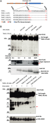
Fig. 1.
Fig. 1.
TMK1 is cleaved by DA1 family peptidases. (A) Diagram and amino acid sequences of TMK1 encoding the intracellular kinase domain (blue box) adjacent to the transmembrane domain (red box). The extracellular region with leucine rich repeats (LRR) is shown as orange boxes. A putative DA1 cleavage site from amino acids 520–537 is underlined. Dashes represent deleted amino acids in the different mutant TMK1 proteins used to assess cleavage. Red text represents amino acids found in the BB cleavage site and the swapped BB cleavage site. (B) Immunoblot of transiently expressed TMK1-3FLAG proteins together with 3HA-DA1 family members in Arabidopsis da1-1 dar1 mesophyll protoplasts. 3HA-DA1pep is a mutant in the peptidase active site. An approximate 50 kDa TMK1-3FLAG cleaved product was observed (red arrow). A separate gel using the same sample with a longer exposure shows DAR1-mediated cleavage. The Lower Panel shows 3HA-DA1, -DAR1, and -DAR2 expression levels. Cleavage levels are the % total FLAG protein in the cleaved band, shown by a red arrow. (C) Immunoblot of transiently expressed TMK1-3FLAG mutant proteins described in panel A together with 3HA-DAR2 in Arabidopsis mesophyll protoplasts. Deletions of the region containing the putative DA1 cleavage site abolished cleavage, while adding the BB cleavage site to the deleted TMK1 protein recovered cleavage (red arrow). The Lower Panel shows 3HA-DAR2 expression levels.
Fig. 2.
Fig. 2.
DAR2 cleavage of TMK1-EGFP relocates it from the plasma membrane to the nucleus of Arabidopsis root protoplasts. (AH) Confocal images of dar2-1 root protoplasts transformed with TMK1-EGFP, histone HT2B-mCherry, and cleavage mutants with or without DAR2. HT2B-mCherry marks the nucleus. Colocation of TMK1-EGFP and HT2B-mCherry shows as yellow fluorescence. (Scale bars: 5 µm.) (I) Ratios of nuclear-localized/total protoplast EGFP in different TMK1-EGFP cleavage mutants. n = 20 protoplasts from each of three independent experiments. All were compared to TMK-EGFP levels without 3HA-DAR2, and significant differences are indicated as *P < 0.05.
Fig. 3.
Fig. 3.
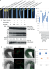
TMK1 cleavage by DA1 family peptidases is induced by auxin to form the apical hook. (A) Complementation of tmk1-1 apical hook angles by TMK1 cleavage mutants and TMK1C expressed by different promoters. (B) Quantitative analyses of apical hook angles in tmk1-1 transformants. n = 25 apical hooks of five independent single locus transformants were measured. *P < 0.05. All measurements were compared to tmk1-1. (C) Immunoblot of TMK1-3FLAG and 3HA-DAR2 transfected dar2 mesophyll protoplasts treated with different levels of IAA. The red arrow indicates cleaved TMK1-FLAG protein. The Lower Panel shows 3HA-DAR2 expression levels. The band above the cleavage product is not related to cleavage activities, *P < 0.05. (D) DR5::NLS-2xEGFP expression levels report auxin distribution across the apical hook of Col-0 and the tmk1-1 mutant. The arcs represent the areas analyzed for EGFP fluorescence. (Scale bar: 100 µm.) (E) Quantitative analyses of EGFP fluorescence on the concave and convex regions of the apical hook in Col-0 and the tmk1-1 mutant. n = 15 hypocotyls. Fluorescence levels were compared to those in the concave region of Col-0.
Fig. 4.
Fig. 4.
Apical hook angle of DA1 family loss-of-function mutants. (A) Apical hook angles of DA1 family peptidases mutants. (B) Analysis of apical hook angles of DA1 family peptidases mutants compared with tmk1-1. n = 15 hypocotyls. Significant differences are indicated as *P < 0.05. (C) qRT-PCR analyses of expression levels of DA1 family peptidases in dark grown seedlings. Expression levels were normalized to DA1 expression levels at 24 h growth in the dark. (D) GUS histochemical analyses of dark-grown transgenic seedlings expressing GUS from DAR1 and DAR2 promoters. The Lower Panels show full length hypocotyls. (Scale bar: 0.5 mm.)

References

    1. Chen M.-K., Hung M.-C., Proteolytic cleavage, trafficking, and functions of nuclear receptor tyrosine kinases. FEBS J. 282, 3693–3721 (2015). - PMC - PubMed
    1. Merilahti J. A. M., Elenius K., Gamma-secretase-dependent signaling of receptor tyrosine kinases. Oncogene 38, 151–163 (2019). - PMC - PubMed
    1. Wolfe M. S., Structure and function of the γ-secretase complex. Biochemistry 58, 2953–2966 (2019). - PMC - PubMed
    1. Smolarkiewicz M., et al. , Gamma-secretase subunits associate in intracellular membrane compartments in Arabidopsis thaliana. J. Exp. Bot. 65, 3015–3027 (2014). - PMC - PubMed
    1. Adamiec M., Ciesielska M., Zalaś P., Luciński R., Arabidopsis thaliana intramembrane proteases. Acta Physiol. Plant. 39, 146 (2017).

Publication types

MeSH terms

LinkOut - more resources