. 2023 Jan 3;22(1):112-122.
doi: 10.1158/1535-7163.MCT-21-0991.

Neoadjuvant Radiation Therapy and Surgery Improves Metastasis-Free Survival over Surgery Alone in a Primary Mouse Model of Soft Tissue Sarcoma

Affiliations

Neoadjuvant Radiation Therapy and Surgery Improves Metastasis-Free Survival over Surgery Alone in a Primary Mouse Model of Soft Tissue Sarcoma

Rutulkumar Patel et al. Mol Cancer Ther. .

Abstract

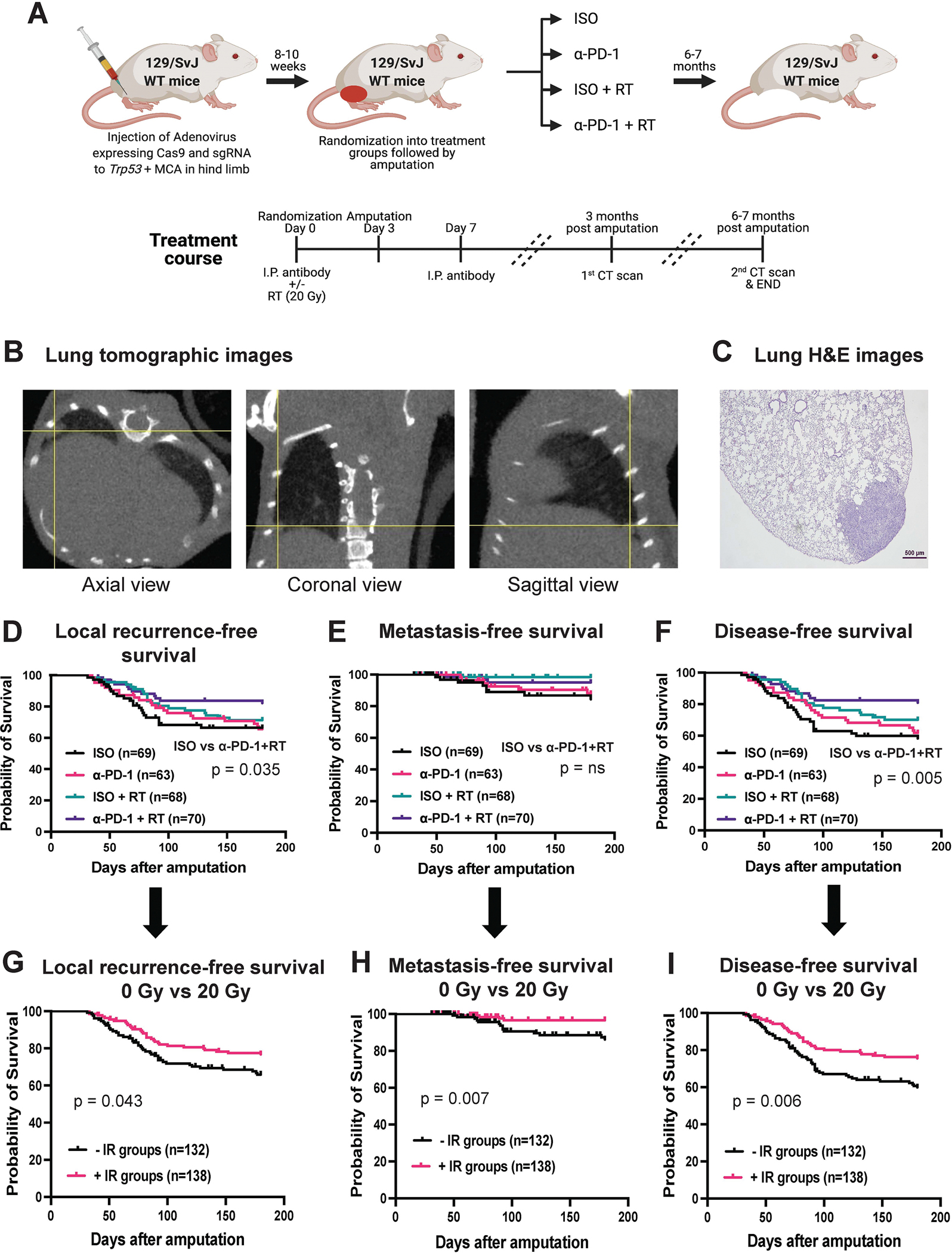
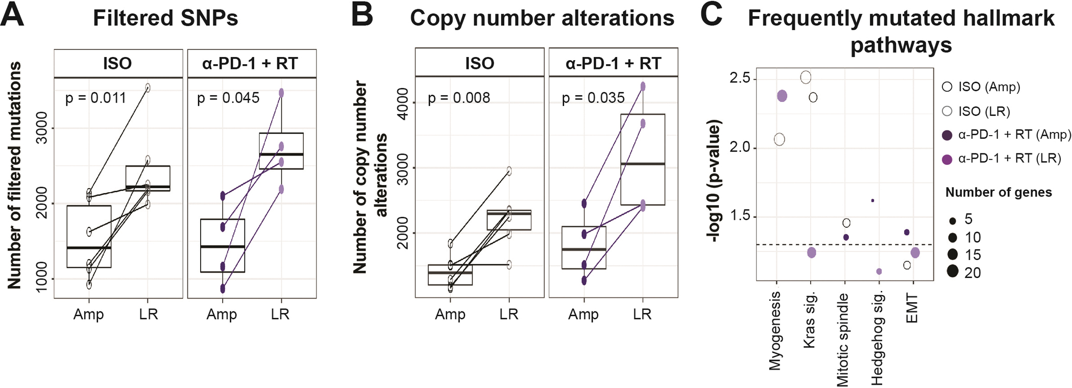
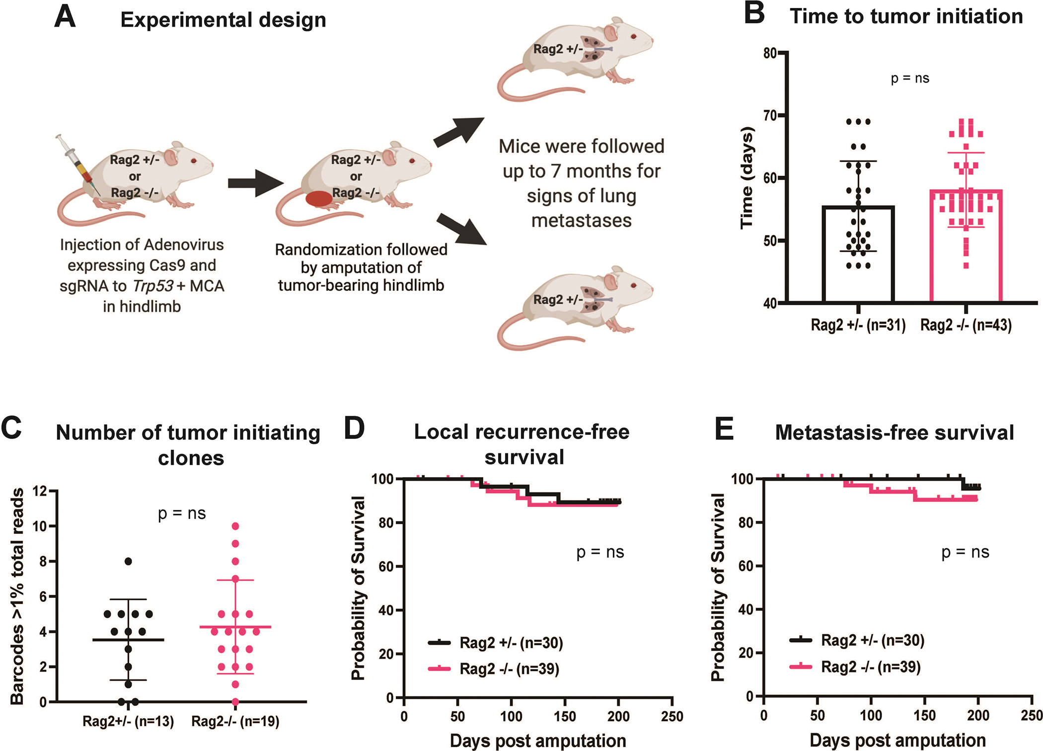
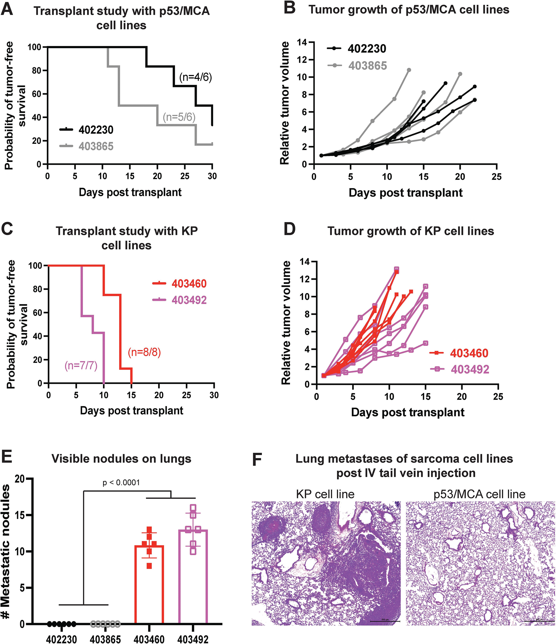
This study aims to investigate whether adding neoadjuvant radiotherapy (RT), anti-programmed cell death protein-1 (PD-1) antibody (anti-PD-1), or RT + anti-PD-1 to surgical resection improves disease-free survival for mice with soft tissue sarcomas (STS). We generated a high mutational load primary mouse model of STS by intramuscular injection of adenovirus expressing Cas9 and guide RNA targeting Trp53 and intramuscular injection of 3-methylcholanthrene (MCA) into the gastrocnemius muscle of wild-type mice (p53/MCA model). We randomized tumor-bearing mice to receive isotype control or anti-PD-1 antibody with or without radiotherapy (20 Gy), followed by hind limb amputation. We used micro-CT to detect lung metastases with high spatial resolution, which was confirmed by histology. We investigated whether sarcoma metastasis was regulated by immunosurveillance by lymphocytes or tumor cell-intrinsic mechanisms. Compared with surgery with isotype control antibody, the combination of anti-PD-1, radiotherapy, and surgery improved local recurrence-free survival (P = 0.035) and disease-free survival (P = 0.005), but not metastasis-free survival. Mice treated with radiotherapy, but not anti-PD-1, showed significantly improved local recurrence-free survival and metastasis-free survival over surgery alone (P = 0.043 and P = 0.007, respectively). The overall metastasis rate was low (∼12%) in the p53/MCA sarcoma model, which limited the power to detect further improvement in metastasis-free survival with addition of anti-PD-1 therapy. Tail vein injections of sarcoma cells into immunocompetent mice suggested that impaired metastasis was due to inability of sarcoma cells to grow in the lungs rather than a consequence of immunosurveillance. In conclusion, neoadjuvant radiotherapy improves metastasis-free survival after surgery in a primary model of STS.


Conflict of interest statement

Conflict of Interest: D.G.K. and Y.M.M. are recipients of a Stand Up To Cancer (SU2C) MSD Catalyst Grant studying pembrolizumab and radiation therapy in sarcoma patients. D.G.K. has received research funding from XRAD Therapeutics, Eli Lilly & Co., Bristol Myers Squibb, Varian Medical Systems, and Merck. D.G.K. owns stock and is on the scientific advisory board of Lumicell, Inc., which is commercializing intraoperative imaging technology. D.G.K. is a cofounder of XRAD Therapeutics, which is developing radiosensitizers. The other authors declare no conflicts of interest.

Figures

Figure 1:
Figure 1:
Long-term metastasis study performed using an autochthonous p53/MCA mouse model of soft-tissue sarcoma (STS) in immunocompetent 129/SvJae wild-type mice induced by CRISPR/Cas9 technology. (A) Schematic representation of experimental design showing the use of CRISPR/Cas9 technology to induce STS followed by randomization of mice into four treatment groups, i.e., isotype control antibody (ISO), anti-PD-1 antibody (α-PD-1), ISO + 20 Gy (ISO + RT), or α-PD-1 + 20 Gy (α-PD-1 + RT). (B) Representative tomographic images of lung with yellow cross-hairs centered on lung metastasis in axial, coronal, and sagittal views. (C) H&E-stained slide showing histological verification of STS metastasis corresponding with lung nodule on micro-CT in panel B. Kaplan-Meier graphs show (D) local recurrence-free survival, (E) metastasis-free survival, and (F) disease-free survival of mice after amputation of tumor-bearing hindlimb for the four treatment groups. Kaplan-Meier graphs represents (G) local recurrence-free survival, (H) metastasis-free survival, and (I) disease-free survival of mice of mice received 0 Gy RT with isotype or α-PD-1 antibody (-IR groups) vs. 20 Gy RT with isotype or α-PD-1 antibody (+IR groups). n = number of mice. p-values in Kaplan-Meier graphs were determined by Log-rank (Mantel-Cox) test. ns = non-significant.
Figure 2:
Figure 2:
Whole exome sequencing (WES) analysis of amputated versus locally recurrent sarcomas. Boxplots represent (A) Filtered SNPs (excluding silent, RNA, intronic, and unknown mutations), and (B) Copy number alterations (including gains and losses) by treatment status and tumor type. (C) Top frequently mutated hallmark pathways determined by hypergeometric test. WES included 4–6 sarcoma samples for each tumor type and treatment group. Amp – amputated tumor, LR – locally recurrent tumor, EMT – epithelial-mesenchymal transition. p-values in boxplots were determined by paired t-tests.
Figure 3:
Figure 3:
Long-term metastasis study performed using an autochthonous p53/MCA mouse model of STS in immuno-proficient (Rag2+/−) vs. immuno-deficient (Rag2−/−) mice. (A) Schematic representation of tumor induction in Rag2 mice using CRISPR/Cas9 technology followed by amputation of tumor-bearing hindlimb. (B) Bar graph represents a time to tumor initiation in Rag2+/− vs. Rag2−/− mice. (C) Bar graph shows the number of tumor-initiating clones determined by CRISPR barcoding in STS arising from Rag2+/− vs. Rag2−/− mice. Kaplan-Meier graphs represent (D) local recurrence-free survival and (E) metastasis-free survival of mice post amputation of the tumor-bearing hindlimb. n = number of mice; data plotted are means ± SD; p-values were determined by student’s t-test in bar graph and Log-rank (Mantel-Cox) test in Kaplan-Meier graph. ns = non-significant.
Figure 4:
Figure 4:
Metastatic potential of sarcoma cell lines measured by intravenous injection of sarcoma cells into immunocompetent 129/SvJae mice. In vivo transplant study shows (A) rate of tumor initiation and (B) rate of tumor growth post intramuscular injection of p53/MCA sarcoma cell lines generated using CRISPR/Cas9 technology. In vivo transplant study shows (C) rate of tumor initiation and (D) rate of tumor growth post intramuscular injection of KP sarcoma cell lines generated using Cre-LoxP technology. (E) Bar graph represents the number of visible nodules detected in the lungs of mice that received either p53/MCA or KP sarcoma cells intravenously via tail vein injection. (F) Representative histological images of lung metastases of sarcoma cell lines post intravenous tail vein injection of either p53/MCA or KP cell lines. n = number of mice; data plotted are means ± SD; p-values were determined by student’s t-test in bar graph.

References

    1. Jo VY, Doyle LA. Refinements in Sarcoma Classification in the Current 2013 World Health Organization Classification of Tumours of Soft Tissue and Bone. Surg Oncol Clin N Am 2016;25(4):621–43 doi 10.1016/j.soc.2016.05.001. - DOI - PubMed
    1. Wang D, Zhang Q, Eisenberg BL, Kane JM, Li XA, Lucas D, et al. Significant Reduction of Late Toxicities in Patients With Extremity Sarcoma Treated With Image-Guided Radiation Therapy to a Reduced Target Volume: Results of Radiation Therapy Oncology Group RTOG-0630 Trial. Journal of clinical oncology : official journal of the American Society of Clinical Oncology 2015;33(20):2231–8 doi 10.1200/JCO.2014.58.5828. - DOI - PMC - PubMed
    1. O’Sullivan B, Davis AM, Turcotte R, Bell R, Catton C, Chabot P, et al. Preoperative versus postoperative radiotherapy in soft-tissue sarcoma of the limbs: a randomised trial. Lancet 2002;359(9325):2235–41 doi 10.1016/S0140-6736(02)09292-9. - DOI - PubMed
    1. Borden EC, Baker LH, Bell RS, Bramwell V, Demetri GD, Eisenberg BL, et al. Soft tissue sarcomas of adults: state of the translational science. Clin Cancer Res 2003;9(6):1941–56. - PubMed
    1. Borghaei H, Paz-Ares L, Horn L, Spigel DR, Steins M, Ready NE, et al. Nivolumab versus Docetaxel in Advanced Nonsquamous Non-Small-Cell Lung Cancer. The New England journal of medicine 2015;373(17):1627–39 doi 10.1056/NEJMoa1507643. - DOI - PMC - PubMed

Publication types

Substances