. 2023 Jan 3;22(1):123-134.
doi: 10.1158/1535-7163.MCT-22-0310.

Sensitization to Ionizing Radiation by MEK Inhibition Is Dependent on SNAI2 in Fusion-Negative Rhabdomyosarcoma

Affiliations

Sensitization to Ionizing Radiation by MEK Inhibition Is Dependent on SNAI2 in Fusion-Negative Rhabdomyosarcoma

Nicole R Hensch et al. Mol Cancer Ther. .

Abstract

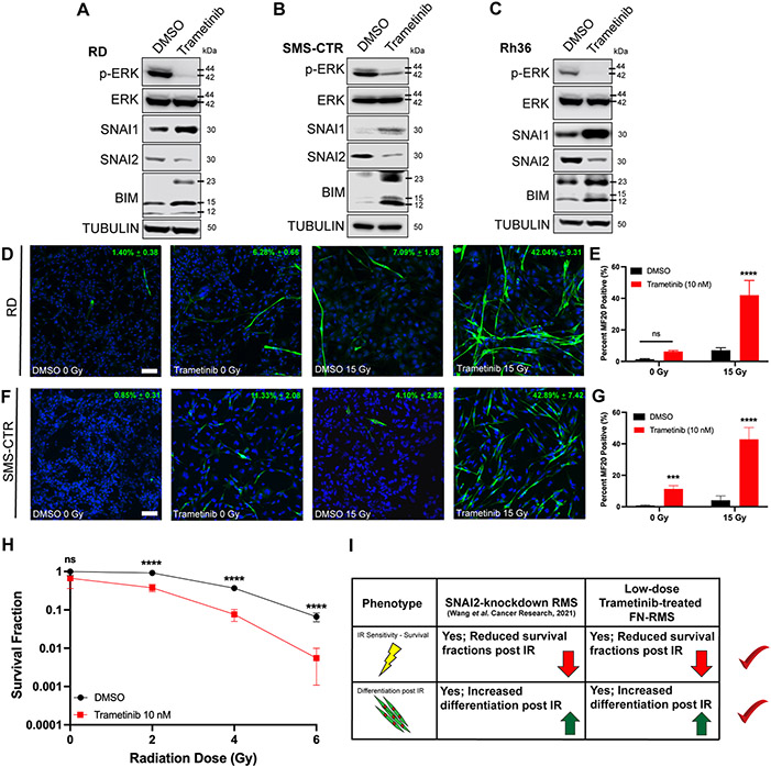
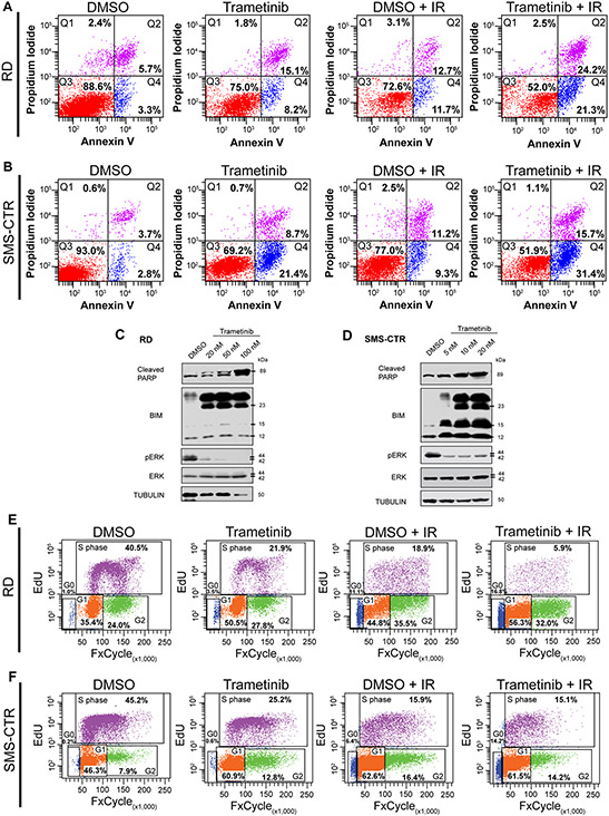
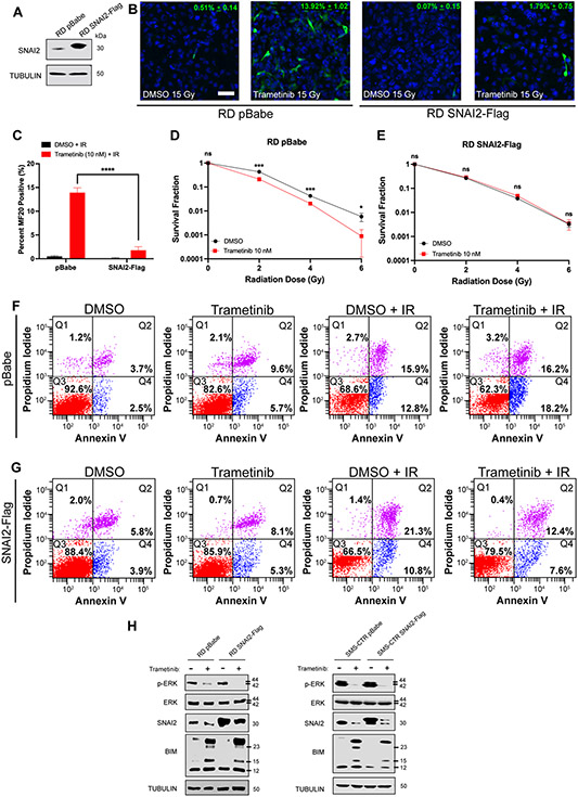
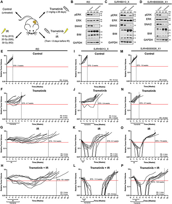
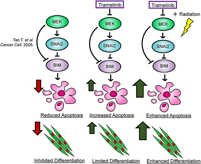
In fusion-negative rhabdomyosarcoma (FN-RMS), a pediatric malignancy with skeletal muscle characteristics, >90% of high-risk patients have mutations that activate the RAS/MEK signaling pathway. We recently discovered that SNAI2, in addition to blocking myogenic differentiation downstream of MEK signaling in FN-RMS, represses proapoptotic BIM expression to protect RMS tumors from ionizing radiation (IR). As clinically relevant concentrations of the MEK inhibitor trametinib elicit poor responses in preclinical xenograft models, we investigated the utility of low-dose trametinib in combination with IR for the treatment of RAS-mutant FN-RMS. We hypothesized that trametinib would sensitize FN-RMS to IR through its downregulation of SNAI2 expression. While we observed little to no difference in myogenic differentiation or cell survival with trametinib treatment alone, robust differentiation and reduced survival were observed after IR. In addition, IR-induced apoptosis was significantly increased in FN-RMS cells treated concurrently with trametinib, as was increased BIM expression. SNAI2's role in these processes was established using overexpression rescue experiments, where overexpression of SNAI2 prevented IR-induced myogenic differentiation and apoptosis. Moreover, combining MEK inhibitor with IR resulted in complete tumor regression and a 2- to 4-week delay in event-free survival (EFS) in preclinical xenograft and patient-derived xenograft models. Our findings demonstrate that the combination of MEK inhibition and IR results in robust differentiation and apoptosis, due to the reduction of SNAI2, which leads to extended EFS in FN-RMS. SNAI2 thus is a potential biomarker of IR insensitivity and target for future therapies to sensitize aggressive sarcomas to IR.


Conflict of interest statement

Conflicts of Interest: The authors declare no potential conflicts of interest.

Figures

Figure 1.
Figure 1.. Low-dose MEK inhibitor reduces SNAI2 expression and in combination with IR induces robust myogenic differentiation and loss of cell survival in FN-RMS.
A, B, C. Protein expression in DMSO- and trametinib-treated FN-RMS cell lines (RD, SMS-CTR, and Rh36). D. Representative confocal microscopy images of RD cells treated with DMSO or trametinib, in non-IR and IR conditions, immunostained with myogenic differentiated myosin (MF20 antibody). Scale bar = 100 μm. E. Quantification of average MF20-positive cells in either non-IR or IR conditions in DMSO- and trametinib-treated RD cells. Error bars represent ±1 SD. ns = not significant, ****p<0.0001 by a two-way ANOVA with a posthoc Sidak’s multiple comparison test. F. Representative confocal microscopy images of SMS-CTR cells treated with DMSO or trametinib, in non-IR and IR conditions, immunostained with myogenic differentiated myosin (MF20 antibody). Scale bar = 100 μm. G. Quantification of average MF20-positive cells in either non-IR or IR conditions in DMSO- and trametinib-treated SMS-CTR cells. Error bars represent ±1 SD. ns = not, ***p < 0.001, ****p < 0.0001 by a two-way ANOVA with a posthoc Sidak’s multiple comparison test. H. Survival fractions of DMSO- and trametinib-treated RD cells were assessed at increasing IR dose exposures. Statistical differences were observed at 2, 4, and 6 Gy. Error bars represent ±1 SD. ns = not significant, ****p < 0.0001 by a Student’s t-test. I. Overview of similar phenotypic characteristics of SNAI2-knockdown RMS cells and low-dose trametinib-treated FN-RMS cells.
Figure 2.
Figure 2.. Combining low-dose MEK inhibition with IR results in increased apoptosis and a G1 cell cycle arrest in FN-RMS.
A. Flow cytometry plots showing propidium iodide vs. annexin V staining of DMSO- and trametinib-treated RD cells after non-IR and IR conditions. Q4 represents cells undergoing early apoptosis, whereas Q2 represents cells undergoing late apoptosis. Q3 represents live cells not undergoing apoptosis. B. Flow cytometry plots showing propidium iodide vs. annexin V staining of DMSO- and trametinib-treated SMS-CTR cells after non-IR and IR conditions. C, D. Cleaved PARP, BIM, pERK, and total ERK protein expression immunoblots in DMSO- and trametinib-treated RD (C) and SMS-CTR (D) cells 96hpIR. Increased trametinib doses were assessed in both cell lines. E. Flow cytometry plots of EdU vs. DAPI staining in DMSO- and trametinib-treated RD cells in non-IR and IR conditions. F. Flow cytometry plots of EdU vs. DAPI staining in DMSO- and trametinib-treated SMS-CTR cells in non-IR and IR conditions.
Figure 3.
Figure 3.. SNAI2 downstream of MEK signaling protects FN-RMS from IR-induced myogenic differentiation and apoptosis.
A. SNAI2 protein expression in RD pBabe and SNAI2-Flag overexpressing cell lines. B. Representative confocal microscopy images of RD pBabe and SNAI2-Flag cells treated with DMSO or trametinib after IR exposure, immunostained with myogenic differentiated myosin MF20 antibody. Scale bar = 100 μm. C. Quantification of average MF20-positive cells in DMSO- and trametinib-treated RD pBabe and SNAI2-Flag cells after IR exposure. Error bars represent ±1 SD. ns = not significant, ****p<0.0001 by a two-way ANOVA with a posthoc Sidak’s multiple comparison test. D, E. Survival fractions of DMSO- and trametinib-treated RD pBabe and SNAI2-Flag cells were assessed at increasing IR dose exposures. Statistical differences were observed at 2, 4, and 6 Gy in RD pBabe, but NOT RD SNAI2-Flag. Error bars represent ±1 SD. ns = not significant, *p < 0.05, ***p < 0.001 by a Student’s t-test. F. Flow cytometry plots showing propidium iodide vs. annexin V staining of DMSO- and trametinib-treated RD pBabe cells after non-IR and IR conditions. G. Flow cytometry plots showing propidium iodide vs. annexin V staining of DMSO- and trametinib-treated RD SNAI2-Flag cells after non-IR and IR conditions. H. Western blots showing pERK, total ERK, and BIM protein expression of RD pBabe, RD SNAI2-Flag, SMS-CTR pBabe, and SMS-CTR SNAI2-Flag cells after DMSO and 10 nM trametinib treatment.
Figure 4.
Figure 4.. MEK inhibitor treatment results in loss of SNAI2 and increases sensitivity to IR, resulting in increased event-free survival in preclinical models.
A. Illustration of in vivo experimental treatment set up. B–D. Western blots showing pERK, total ERK, SNAI2, and BIM protein expression in two representative control tumors and trametinib-treated RD (B), SJRHB013_X (C), and SJRHB000026_X1 (D) tumors. E–H. Relative tumor volume (RTV) of RD tumors receiving control (E), trametinib (F), IR (G), or combination trametinib + IR (H) treatment. Red line depicts 0.5 RTV; vertical dashed line represents the average EFS for each treatment arm. PD: progressive disease, PR: partial response. I–L. Relative tumor volume (RTV) of SJRHB013_X tumors receiving control (I), trametinib (J), IR (K), or combination trametinib + IR (L) treatment. Red line depicts 0.5 RTV; vertical dashed line represents the average EFS for each treatment arm. PD: progressive disease, PR: partial response. CR: complete response. M–P. Relative tumor volume (RTV) of SJRHB000026_X1 tumors receiving control (M), trametinib (N), IR (O), or combination trametinib + IR (P) treatment. Red line depicts 0.5 RTV; vertical dashed line represents the average EFS for each treatment arm. PD: progressive disease, PR: partial response. CR: complete response.
Figure 5.
Figure 5.. Proposed mechanism of inhibiting the MEK/SNAI2 pathway to sensitize FN-RMS to IR-induced apoptosis and myogenic differentiation.

References

    1. Chen X, Stewart E, Shelat AA, Qu C, Bahrami A, Hatley M, et al. Targeting oxidative stress in embryonal rhabdomyosarcoma. Cancer Cell 2013;24(6):710–24 doi 10.1016/j.ccr.2013.11.002. - DOI - PMC - PubMed
    1. Seki M, Nishimura R, Yoshida K, Shimamura T, Shiraishi Y, Sato Y, et al. Integrated genetic and epigenetic analysis defines novel molecular subgroups in rhabdomyosarcoma. Nat Commun 2015;6:7557 doi 10.1038/ncomms8557. - DOI - PMC - PubMed
    1. Shern JF, Chen L, Chmielecki J, Wei JS, Patidar R, Rosenberg M, et al. Comprehensive genomic analysis of rhabdomyosarcoma reveals a landscape of alterations affecting a common genetic axis in fusion-positive and fusion-negative tumors. Cancer discovery 2014;4(2):216–31 doi 10.1158/2159-8290.CD-13-0639. - DOI - PMC - PubMed
    1. Shern JF, Selfe J, Izquierdo E, Patidar R, Chou HC, Song YK, et al. Genomic Classification and Clinical Outcome in Rhabdomyosarcoma: A Report From an International Consortium. J Clin Oncol 2021:JCO2003060 doi 10.1200/JCO.20.03060. - DOI - PMC - PubMed
    1. Smith MA, Altekruse SF, Adamson PC, Reaman GH, Seibel NL. Declining childhood and adolescent cancer mortality. Cancer 2014;120(16):2497–506 doi 10.1002/cncr.28748. - DOI - PMC - PubMed

Publication types

Substances