. 2022 Nov 1;209(9):1746-1759.
doi: 10.4049/jimmunol.2200227. Epub 2022 Sep 26.

α1-Antitrypsin Binds to the Glucocorticoid Receptor with Anti-Inflammatory and Antimycobacterial Significance in Macrophages

Affiliations

α1-Antitrypsin Binds to the Glucocorticoid Receptor with Anti-Inflammatory and Antimycobacterial Significance in Macrophages

Xiyuan Bai et al. J Immunol. .

Abstract

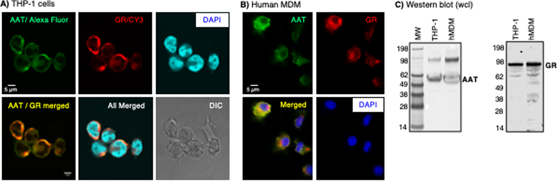
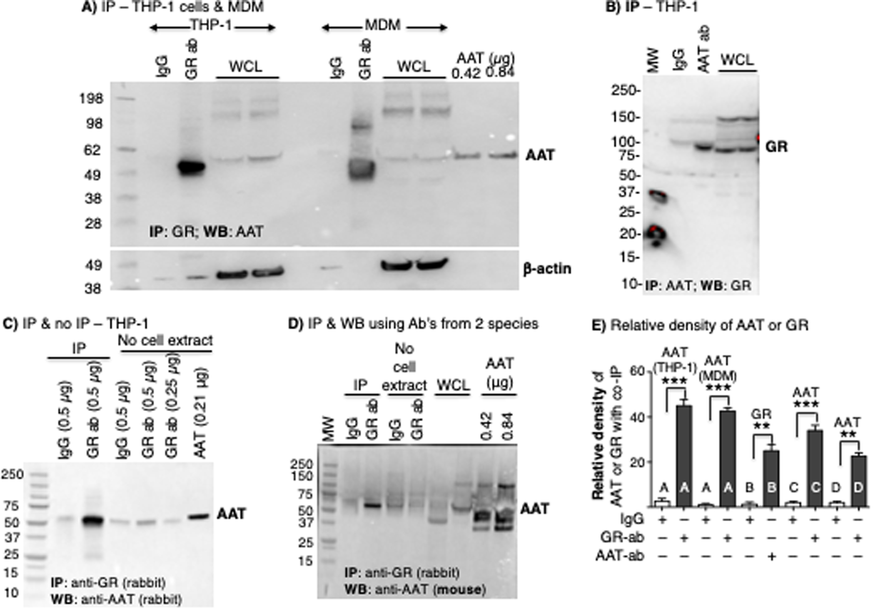
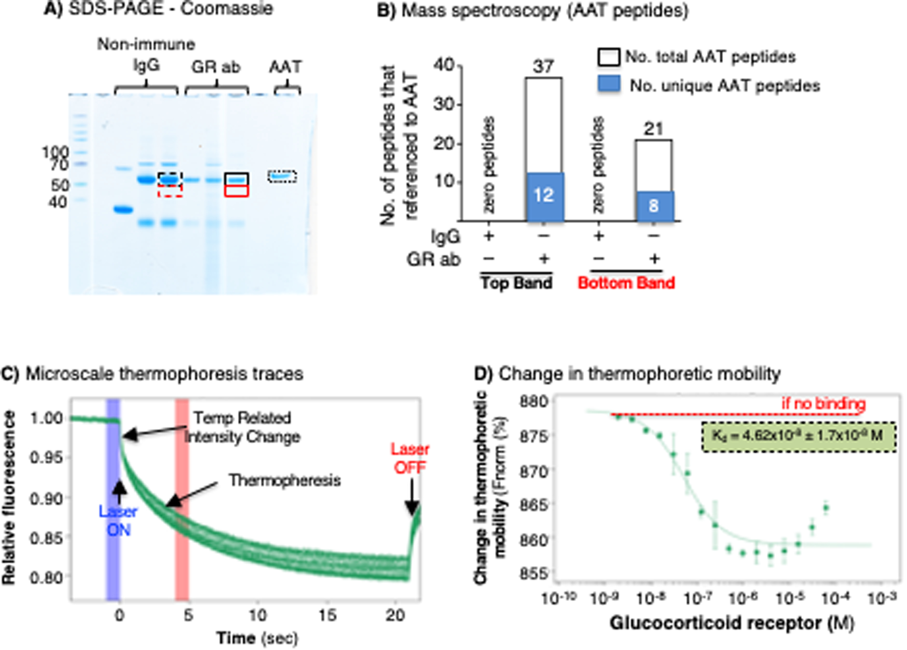
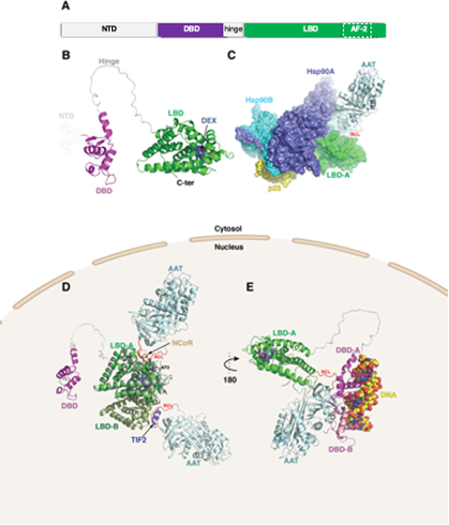
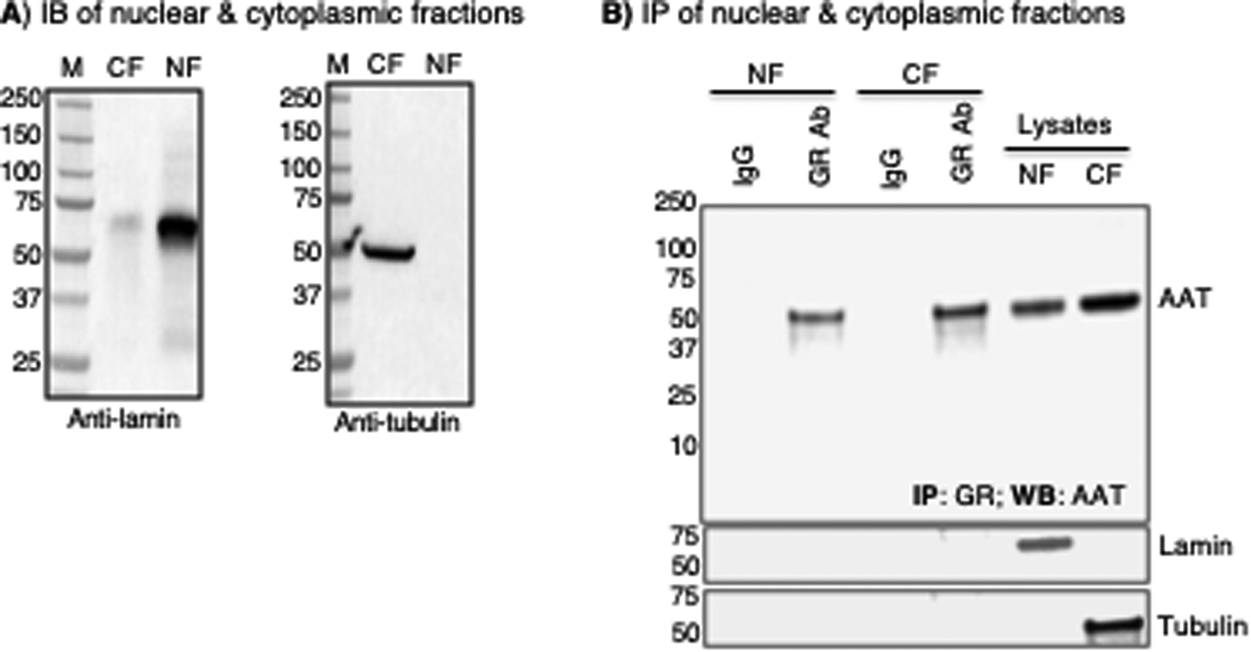
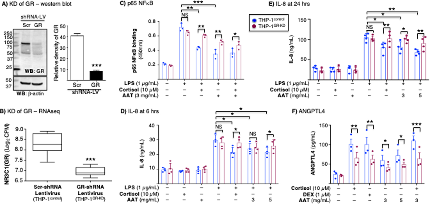
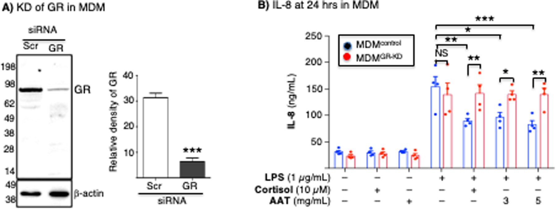
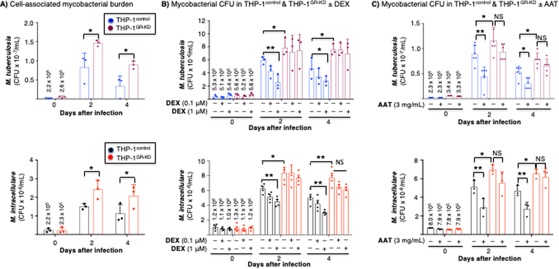
α1-Antitrypsin (AAT), a serine protease inhibitor, is the third most abundant protein in plasma. Although the best-known function of AAT is irreversible inhibition of elastase, AAT is an acute-phase reactant and is increasingly recognized to have a panoply of other functions, including as an anti-inflammatory mediator and a host-protective molecule against various pathogens. Although a canonical receptor for AAT has not been identified, AAT can be internalized into the cytoplasm and is known to affect gene regulation. Because AAT has anti-inflammatory properties, we examined whether AAT binds the cytoplasmic glucocorticoid receptor (GR) in human macrophages. We report the finding that AAT binds to GR using several approaches, including coimmunoprecipitation, mass spectrometry, and microscale thermophoresis. We also performed in silico molecular modeling and found that binding between AAT and GR has a plausible stereochemical basis. The significance of this interaction in macrophages is evinced by AAT inhibition of LPS-induced NF-κB activation and IL-8 production as well as AAT induction of angiopoietin-like 4 protein, which are, in part, dependent on GR. Furthermore, this AAT-GR interaction contributes to a host-protective role against mycobacteria in macrophages. In summary, this study identifies a new mechanism for the gene regulation, anti-inflammatory, and host-defense properties of AAT.


Conflict of interest statement

Conflict of interest

ANG holds equity in Psammiad Therapeutics. All other authors declare that they have no conflicts of interest with the contents of this article.

Figures

Figure 1.
Figure 1.. Immunofluorescence staining for glucocorticoid receptor (GR) and alpha-1-antitrypsin (AAT).
(A) Human THP-1 differentiated macrophages and (B) human monocyte-derived macrophages (MDM) were immunostained using anti-AAT and anti-GR antibodies conjugated to the fluorochromes Alexa Fluor Plus 488 and Cy3, respectively. THP-1 macrophages were imaged with a confocal microscope. Human monocyte-derived macrophages (MDM) were imaged with a fluorescent microscope. The scale bars represent 5 μm. (C) Immunoblotting for AAT and GR in human macrophages. Images shown are representative of three independent experiments.
Figure 2.
Figure 2.. Alpha-1-antitrypsin (AAT) co-immunoprecipitates with glucocorticoid receptor (GR).
(A) THP-1 macrophage and human monocyte-derived macrophage (MDM) lysates were immunoprecipitated (IP’d) with anti-GR polyclonal antibody, followed by immunoblotting of the IP’d fraction to detect AAT and β-actin. (B) THP-1 macrophage lysates were IP’d with anti-AAT polyclonal antibody, followed by immunoblotting of the IP’d fraction for GR. (C) Comparing the relative amount of IgG and anti-GR antibody alone (“No cell extract”) used in the immunoprecipitation (IP) experiments, with THP-1 macrophage lysates IP’d with non-immune IgG or anti-GR, followed by immunoblotting for AAT. (D) THP-1 macrophage lysates were IP’d with anti-GR polyclonal antibodies followed by immunoblotting of the IP’d fraction with a mouse anti-AAT monoclonal antibody. (E) Densitometry of the co-IP protein using non-immune IgG, anti-GR antibody, or anti-AAT antibody for immunoblots represented in Figures A-D (letters A to D shown in the graph correspond to the IP’d bands in Figures A-D). The proteins listed above each bar is the protein detected in the immunoblot. Images in A-D are representative of at least three independent experiments. Data shown in E are the mean ± SD of three independent experiments. AAT=alpha-1-antitrypsin; ab=antibody; GR=glucocorticoid receptor; MDM=monocyte-derived macrophages; WB=western blot; WCL=whole cell lysates. Lanes labeled “No cell extract” contains only IgG or anti-GR antibody alone with no IP performed.
Figure 3.
Figure 3.. Immunoprecipitation-mass spectrometry and microscale thermophoresis demonstrate AAT–GR interaction.
(A) THP-1 macrophages were immunoprecipitated (IP’d) with non-immune IgG or anti-GR antibody, the IP’d fractions were separated by SDS-PAGE, and the gel was stained with Coomassie blue. Stained bands in the gel that ran similarly to AAT (dash and solid black line boxes) were excised along with an area just beneath the aforementioned band (dash and solid red line boxes) and subjected to mass spectrometry. (B) Graphical and numerical representation of the number of total and unique peptides that referenced to the AAT protein in samples IP’d with control IgG (zero AAT peptides for either the top or bottom bands) or with anti-GR antibody (37 total AAT peptides with 12 unique AAT peptides in the top band, and 21 total AAT peptides with 8 unique to AAT in the bottom band). Data shown are representative of two independent experiments. (C) Microscale thermophoresis time-course tracings following the mixing of fluorescent-tagged AAT with 16 different GR concentrations and subjected to a thermogradient. (D) The presence of a “change in thermophoretic mobility” of the fluorescent-tagged AAT with varying GR concentrations demonstrates that the two molecules interact in vitro. Data shown in (D) are the mean ± SD of two independent experiments. AAT=alpha-1-antitrypsin; ab=antibody; GR=glucocorticoid receptor.
Figure 4.
Figure 4.. Molecular modeling of alpha-1-antitrypsin (AAT)–glucocorticoid receptor (GR) complex.
(A) GR is organized into three major domains: an intrinsically disordered N-terminal activation function-1 domain (NTD), a DNA binding domain (DBD), and a C-terminal ligand binding domain (LBD). (B) Structural modeling of the GR protein reveals the presence of the LBD (green) and DBD (magenta), linked by an unstructured hinge region (grey). The intrinsically disordered NTD was not modeled and is depicted by dotted lines. Dexamethasone bound to the LBD is shown as steel blue spheres. (C) AlphaFold2 docking simulations of LBD and AAT (PDB ID 3NE4) superimposed on the GR-Hsp90-p23 co-chaperone complex (PDB ID 7KRJ) show AAT interacting with the LBD via its RCL. (D) The same AAT–GR complex expanded to the LBD dimer (monomers labeled LBD-A and LBD-B). Superimposed are the nuclear coregulator proteins nuclear receptor corepressor (NCoR; derived from PDB ID 3H52) and transcriptional intermediary factor-2 (TIF2; derived from PDB ID 1M2Z) bound to the AF-2 site (broken rectangle) are shown as brown and slate-blue cartoons, respectively. (E) The second model using ClusPro docking shows the RCL of AAT interacting with the LBD of GR. In addition, the DBD is shown complexed with DNA (space-filling atoms), modeled using the GR DNA Binding Domain monomer – TSLP nGRE Complex (magenta; PDB ID 5HN5). Also shown (pink) is a second DBD formed by D-loop-mediated dimerization (PDB ID 5E69), having some degree of steric overlap with AAT. RCL (red)=reactive center loop.
Figure 5.
Figure 5.. The alpha-1-antitrypsin (AAT)–glucocorticoid receptor (GR) complex is found in both the nucleus and cytoplasm.
(A) Pre-immunoprecipitation immunoblot (IB) experiment to detect lamin and tubulin to confirm specific isolation of nuclear and cytoplasmic fractions, respectively. (B) Immunoprecipitation (IP) of the nuclear and cytoplasmic fractions for GR and immunoblot of immunoprecipitated lysates with an anti-AAT antibody. The same membrane was then immunoblotted for lamin and tubulin (two lower panels). All data shown are representative of three independent experiments. CF=cytoplasmic fraction, NF=nuclear fraction
Figure 6.
Figure 6.. Glucocorticoid or AAT inhibition of NFκB activation, inhibition of IL-8 production, and induction of angiopoietin-like 4 is glucocorticoid receptor-dependent.
(A) Western blot of whole-cell lysates of THP-1control and THP-1GR-KD macrophages for GR and β-actin. Densitometry of the GR band on immunoblot normalized for β-actin (***p<0.001 compared to Scr-shRNA lentivirus). The immunoblot and densitometry shown are representative and the mean of three independent experiments, respectively. (B) RNA sequencing (RNAseq) for the GR gene (NR3C1) transcript of the THP-1control and THP-1GR-KD cells using shRNA-lentivirus technology (***p<0.001 compared to Scr-shRNA lentivirus). (C) THP-1control and THP-1GR-KD macrophages were left untreated or pre-treated with cortisol, AAT, or both at the indicated concentrations for 30 minutes, and after stimulation with lipopolysaccharide (LPS) for 6 hours, p65-NFκB binding assay to its consensus oligonucleotide was performed. THP-1control and THP-1GR-KD macrophages were left untreated or pre-treated with cortisol, AAT, or both at the indicated concentrations for 30 minutes, and after stimulation with LPS for (D) 6 hours or (E) 24 hours, the supernatants were assayed for IL-8 by ELISA. (F) Glucocorticoid (cortisol or dexamethasone) or AAT induction of ANGPTL4 in THP-1control and THP-1GR-KD macrophages. Experiments in (C) / (F) and (D) / (E) are the mean ± SEM of three and four independent experiments, respectively, with each experiment done in duplicates. *p<0.05, **p<0.01, ***p<0.001. Blue bars=control shRNA (THP-1control), red bars=GR shRNA (THP-1GR-KD). NS=not significant, KD=knockdown, LV=lentivirus, Scr=scrambled.
Figure 7.
Figure 7.. AAT inhibition of IL-8 production is glucocorticoid receptor-dependent in MDM.
(A) MDM differentiated from the PBMC from a PiMM individual were transfected with scrambled siRNA (control) or GR siRNA. Cell lysates from MDMcontrol or MDMGR-KD were separated by SDS-PAGE and immunoblotted for GR. Densitometry of the GR band on the immunoblot (mean density of two independent experiments). (B) MDMcontrol and MDMGR-KD were left untreated or pre-treated with cortisol (10 μM) or AAT (3 mg/mL) for 30 minutes, followed by stimulation with LPS for 24 hours, and supernatant quantified for IL-8 by ELISA. The immunoblot and densitometry analysis of GR are representative and the mean of three independent experiments, respectively. ELISA for IL-8 is the mean of three independent experiments. *p<0.05, **p<0.01, ***p<0.001. Blue bars=control siRNA (MDMcontrol), red bars=GR siRNA (MDMGR-KD). NS=not significant, KD=knockdown, control=scrambled.
Figure 8.
Figure 8.. Mycobacterial infection of THP-1control and THP-1GR-KD macrophages in the absence or presence of alpha-1-antitrypsin (AAT) or glucocorticoid.
(A) THP-1control and THP-1GR-KD macrophages were infected with M. tuberculosis H37Rv or M. intracellulare for 1 hour, 2 and 4 days. The cells were washed, and intracellular mycobacteria quantified. (B) THP-1control and THP-1GR-KD macrophages were left untreated or pre-treated with dexamethasone (0.1 or 1 μM) for 60 minutes, followed by M. tuberculosis (top panel) or M. intracellulare (bottom panel) infection for 1 hour, 2 days, and 4 days. The cells were washed and intracellular mycobacteria quantified. (C) THP-1control and THP-1GR-KD macrophages were left untreated or pre-treated with 3 mg/mL AAT for 30 minutes, infected with M. tuberculosis (top panel) or M. intracellulare (bottom panel) for 1 hour, 2 and 4 days, the cells washed, and intracellular mycobacteria quantified. Experiments in (A) and (B) are the mean ± SEM of three independent experiments, and in (C), the mean ± SEM of six independent experiments, with each experiment done in duplicates. *p<0.05, **p<0.01. The numbers shown above the bars at t=0 in (B) and (C) are the mean cell-associated CFU at 1 hour after infection. Blue bars=control shRNA (THP-1control), red bars=GR shRNA (THP-1GR-KD). NS=not significant.
Figure 9.
Figure 9.. Glucocorticoid induces expression of a pattern-recognition receptor.
(A) THP-1control and THP-1GR-KD macrophages were left untreated or pre-treated with dexamethasone (1 μM) for 24 hours, followed by immunoblotting of the whole-cell lysates for TLR2 (top). The same membrane was stripped and immunblotted for β-actin (bottom). Data shown in representative of three independent experiments. (B) Relative densitometric measurement of TLR2 protein normalized for β-actin. Data shown are the mean density (arbitrary units) of three independent experiments. *p<0.05, **p<0.01, ***p<0.001. p values for the THP-1GR-KD cells are in comparison to their corresponding THP-1control cells.
Figure 10.
Figure 10.. Illustration of the key findings and hypotheses of the AAT–GR interactions.
(A) Summarizing the key findings, alpha-1-antitrypsin–glucocorticoid receptor (AAT–GR) interaction inhibits nuclear factor-kappa B (NFκB) activation and interleukin-8 (IL-8) production, induces angiopoietin-like 4 (ANGPTL4), and enhances macrophage control of mycobacteria. (B) Several hypotheses of how AAT–GR interaction may affect cellular function: (1) AAT may shuttle GR between the nuclear and cytoplasmic compartments; (2) AAT may facilitate disassembly of GR–chaperone complexes; (3) AAT may stabilize transcriptional complexes of GR–transcription factor (TF, orange structure) or locally modulate the activity of proteases.

References

    1. de Serres F, and Blanco I. 2014. Role of alpha-1 antitrypsin in human health and disease. J Intern Med 276: 311–335. - PubMed
    1. Parr DG, Guest PG, Reynolds JH, Dowson LJ, and Stockley RA. 2007. Prevalence and impact of bronchiectasis in alpha1-antitrypsin deficiency. Am J Respir Crit Care Med 176: 1215–1221. - PubMed
    1. Strnad P, McElvaney NG, and Lomas DA. 2020. Alpha(1)-Antitrypsin Deficiency. N Engl J Med 382: 1443–1455. - PubMed
    1. Bai X, Bai A, Honda JR, Eichstaedt C, Musheyev A, Feng Z, Huitt G, Harbeck R, Kosmider B, Sandhaus RA, and Chan ED. 2019. Alpha-1-antitrypsin enhances primary human macrophage immunity against non-tuberculous mycobacteria,. Front Immunol 10: 1417. - PMC - PubMed
    1. Bryan CL, Beard KS, Pott GB, Rahkola J, Gardner EM, Janoff EN, and Shapiro L. 2010. HIV infection is associated with reduced serum alpha-1-antitrypsin concentrations. Clin Invest Med 33: E384–E389. - PubMed

Publication types

MeSH terms