Review
. 2023 Feb;48(2):172-186.
doi: 10.1016/j.tibs.2022.08.010. Epub 2022 Sep 23.

Structural view of G protein-coupled receptor signaling in the retinal rod outer segment

Affiliations
Review

Structural view of G protein-coupled receptor signaling in the retinal rod outer segment

Sahil Gulati et al. Trends Biochem Sci. 2023 Feb.

Abstract

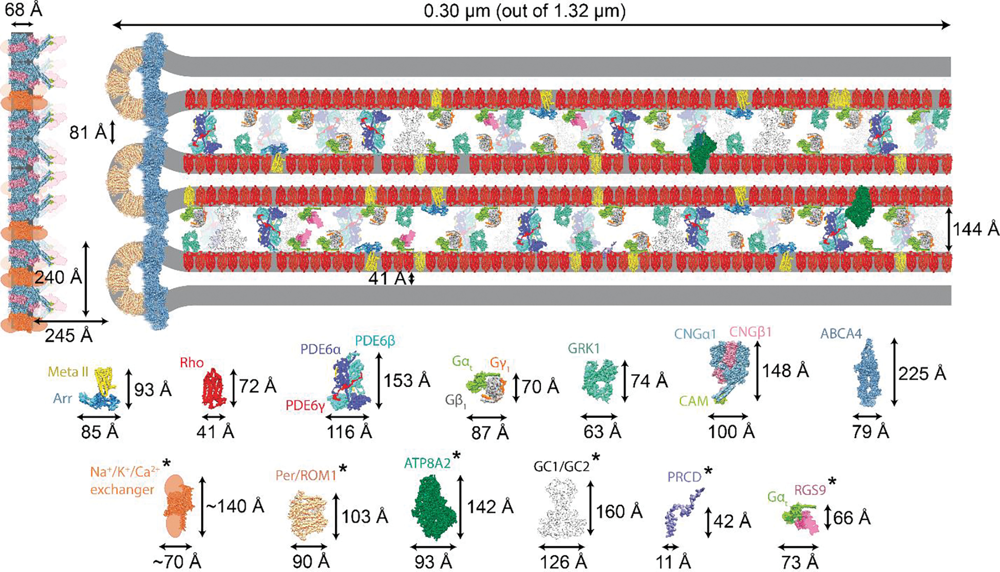
Visual phototransduction is the most extensively studied G protein-coupled receptor (GPCR) signaling pathway because of its quantifiable stimulus, non-redundancy of genes, and immense importance in vision. We summarize recent discoveries that have advanced our understanding of rod outer segment (ROS) morphology and the pathological basis of retinal diseases. We have combined recently published cryo-electron tomography (cryo-ET) data on the ROS with structural knowledge on individual proteins to define the precise spatial limitations under which phototransduction occurs. Although hypothetical, the reconstruction of the rod phototransduction system highlights the potential roles of phosphodiesterase 6 (PDE6) and guanylate cyclases (GCs) in maintaining the spacing between ROS discs, suggesting a plausible mechanism by which intrinsic optical signals are generated in the retina.

Keywords: G protein-coupled receptor (GPCR); GPCR signaling; phototransduction; rhodopsin; rod cell; rod outer segment (ROS).


Conflict of interest statement

Declaration of interests The authors declare no conflicts of interest.

Figures

Figure 1.
Figure 1.. The structure of the rod photoreceptor cell and visual phototransduction.
(A) Schematic diagram of a murine rod photoreceptor showing the major components. In mice, the rod outer segment (ROS) is comprised of ~800 bilayered disc membrane stacks [11] enclosed by a plasma membrane, and contains the components necessary for phototransduction. In WT mice, the average density of discs is 34 discs/μm for the ROS, the average ROS length is 23.8 ± 1.0 μm, and the ROS contains 810 ± 33 discs. The average diameter of the ROS in retinal sections of WT mice is 1.32 ± 0.12 μm [11]. Thus, the total calculated volumes of disc membranes and the ROS cytoplasm in the retina is ~114 × 10−6 ml and 64 × 10−6 ml, respectively [32]. (B) Distances between murine ROS membranes obtained by cryo-ET tomograms [32]. The cryo-ET structure enabled us to precisely measure the distances between ROS components, leading to an increased understanding of the spatial limits within which ROS proteins function. (C) Illustration showing the major steps involved in visual phototransduction. The light-induced conversion of rhodopsin from ground state to signaling state metarhodopsin II (Meta II) results in the activation of Gt. Upon activation, the GTP-bound α-subunit of Gt (Gtα-GTP) binds and activates PDE6, which lowers cGMP levels through hydrolysis, resulting in ion channel closure. Thus, reduction in the cation flux leads to rod cell hyperpolarization in response to a light stimulus, and decreased release of the neurotransmitter glutamate at the synapse.
Figure 2.
Figure 2.. Spatial organization of the key components of visual phototransduction in the ROS.
(A) Sub-section of the ROS depicting the size and abundance of visual phototransduction proteins relative to rhodopsin [67] (Table 1). Distances between the ROS membrane components were obtained from cryo-EM tomograms [30] and are drawn to scale. Individual protein components and their dimensions are displayed below the figure and in Table 1. The asterisks represent modelled protein structures or structures with modeled domains. The longest protein is ABCA4, and its localization is restricted to the rim of the discs. The central part of the disc is occupied primarily by rhodopsin. GCs and PDE6 are the only two proteins that can span the ROS interdiscal space. The separation of plasma membrane from the disc membranes is maintained by a yet unknown mechanism(s). Meta II: metarhodopsin II; Arr: arrestin; Rho: rhodopsin; PDE6: phosphodiesterase 6; GRK1: GPCR kinase 1; CNG1: cGMP-gated channel; ABCA4: Retinal-specific ATP-binding cassette transporter; PRPH2/ROM1: peripherin-ROM1; ATP8A2: ATPase phospholipid transporting 8A2; GC1 and GC2: guanylate cyclases 1 and 2; PRCD: Photoreceptor Disc Component; RGS9: Regulator of G Protein Signaling 9.
Figure 3.
Figure 3.. Rhodopsin complexes that form during the phototransduction sequence.
Structures of metarhodopsin II (Meta II) bound with (from left to right) Gt, GRK1, and arrestin. (A) The complex between photoactivated rhodopsin and the G protein of the visual system, called transducin (Gt). The interaction extends to a 1042 Å2 interface, which involves helix 3 (TM3), intracellular loop 2 (ICL2), TM5, TM6, and helix 8 (H8) of rhodopsin, and helix 5, loops a4-b6, b2-b3 and aN-b1 of Gtα (for structural details see [40]). (B) The complex between photoactivated rhodopsin and GRK1. Dynamic interaction with receptor-bound GRK1 allows formation of a unique intermediate that produces multiple phosphorylation sites in the receptor tail. Changes in rhodopsin involve TM helices 5 and 6, which adopt outward conformations relative to the transmembrane core; TM6 is rotated by about 20°, and H8 is shifted in the complex (for structural details see [45]). (C) The structure of a fusion protein between photoactivated rhodopsin and arrestin. Whether the fusion of the proteins exerts any structural constraints remains to be determined.
Figure 4.
Figure 4.. Major lipid composition of the ROS.
(A) Fatty acid composition of phospholipids from human ROS membranes. Values are expressed as mol % of total fatty acids. At least 70% of the amino phospholipids (i.e., phosphatidylethanolamine and phosphatidylserine) are localized to the outer (cytoplasmic) leaflet of the disc membrane bilayer, while most of the choline phospholipids (i.e., phosphatidylcholine and sphingomyelin) were presumed (by difference) to be localized to the inner (lumenal) membrane leaflet. About 73–87% of the phosphatidylethanolamine and 77–88% of the phosphatidylserine were distributed in the outer monolayer of the disc membrane, while calculations indicated that 65–100% of the phosphatidylcholine resides in the inner monolayer [20]. The most striking feature of ROS lipid composition is the unusually high content of long-chain polyunsaturated acids, which generally comprise 50–60 mol % of the membrane fatty acids. Saturated fatty acids are esterified primarily at the sn1-position, while polyunsaturated fatty acids (PUFAs) are found primarily at the sn2-position of the glycerol moiety. (B) Intradiscal view of ROS showing the localization of major proteins on the surface (cross-section is represented in Figure 2). Tight packing between rhodopsin dimers (pink color) leaves a limited space for lipid binding. Gtα (green); Gtβ (gray); Gtγ (orange); PDE6α (blue); PDE6β (purple); PDE6γ (red). (C) Schematic diagram depicting the lipid composition surrounding a rhodopsin molecule. The relative concentrations of individual lipids are obtained from [20]. The difference between the extracellular and the cytoplasmic total flat surface areas is a result of the number of phospholipids residing at each side and the different packing forces. PC, phosphatidylcholine; PE, phosphatidylethanolamine; PS, phosphatidylserine; CHOL, cholesterol.
Figure 5.
Figure 5.. Hypothetical structural model of PDE6-guided change in ROS morphology.
(A) The cryo-EM structure of the PDE6αβ2γ tetramer, displaying PDE6α (teal), PDE6β (pink) and two molecules of PDE6γ (light gray) [77]. The PDE6αβ heterodimer attains a pseudo-twofold symmetry, where the three domains of PDE6α and PDE6β are organized in a head-to-head arrangement with dimensions of 154 Å X 115 Å X 74 Å. Each of the PDE6α and PDE6β subunits contain two regulatory N-terminal GAF domains (GAF-A and GAF-B) and a C-terminal catalytic domain. The PDE6αβ2γ complex has a 34Å-long N-terminal pony-tail domain (Pt motif). The Pt motif is among the most flexible regions within the PDE6αβ2γ complex and could provide structural stability to the PDE6αβ heterodimer by providing an additional interaction interface of ~895 Å2 between the PDE6α and PDE6β subunits. Due to its hydrophobic properties, this region could provide tethering into the ROS membranes. (B) Light-activation of the dark-adapted ROS leads to conformational rearrangements in the N-terminal GAF (red and purple domains) and C-terminal catalytic domains (green) of the PDE6 heterodimer, resulting in elongation of the PDE6 heterodimer. One side of the PDE6 is tethered to membranes by the isoprenylated moieties on each of the catalytic subunits. The N-terminal side contains the Pt motif, which could intercalate into the adjacent disc membrane, spanning the entire space between discs. (C) The extent of potential conformational changes and ROS extension that occurs during PDE6 activation is illustrated. These structural changes result in the elongation of the PDE6 heterodimer, culminating in the elongation of the ROS.

References

    1. Rodieck RW (1998) The first steps in seeing, Sinauer Associates.
    1. Luo DG et al. (2008) How vision begins: an odyssey. Proc Natl Acad Sci U S A 105 (29), 9855–62. - PMC - PubMed
    1. Polans A et al. (1996) Turned on by Ca2+! The physiology and pathology of Ca(2+)-binding proteins in the retina. Trends Neurosci 19 (12), 547–54. - PubMed
    1. Arshavsky VY and Wensel TG (2013) Timing is everything: GTPase regulation in phototransduction. Invest Ophthalmol Vis Sci 54 (12), 7725–33. - PMC - PubMed
    1. Kawamura S and Tachibanaki S (2021) Molecular bases of rod and cone differences. Prog Retin Eye Res, 101040. - PubMed

Publication types

MeSH terms

Substances