. 2022 Nov;18(11):1184-1195.
doi: 10.1038/s41589-022-01116-1. Epub 2022 Sep 26.

Discovery and molecular basis of subtype-selective cyclophilin inhibitors

Affiliations

Discovery and molecular basis of subtype-selective cyclophilin inhibitors

Alexander A Peterson et al. Nat Chem Biol. 2022 Nov.

Abstract

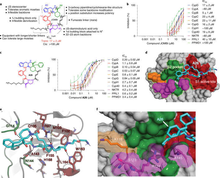
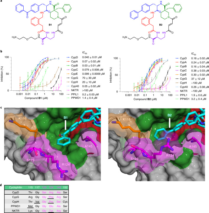
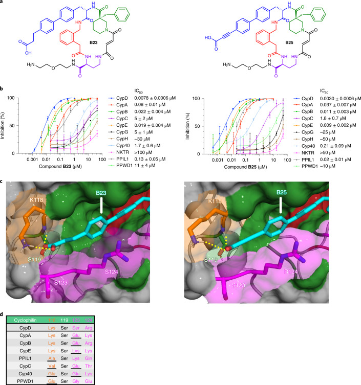
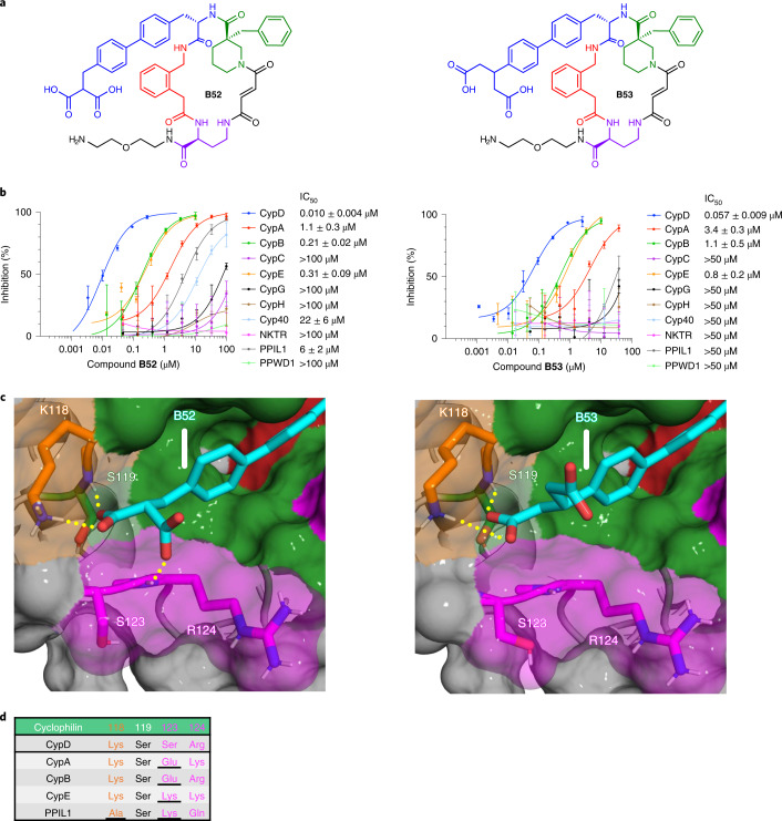
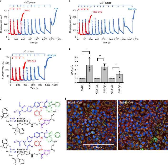
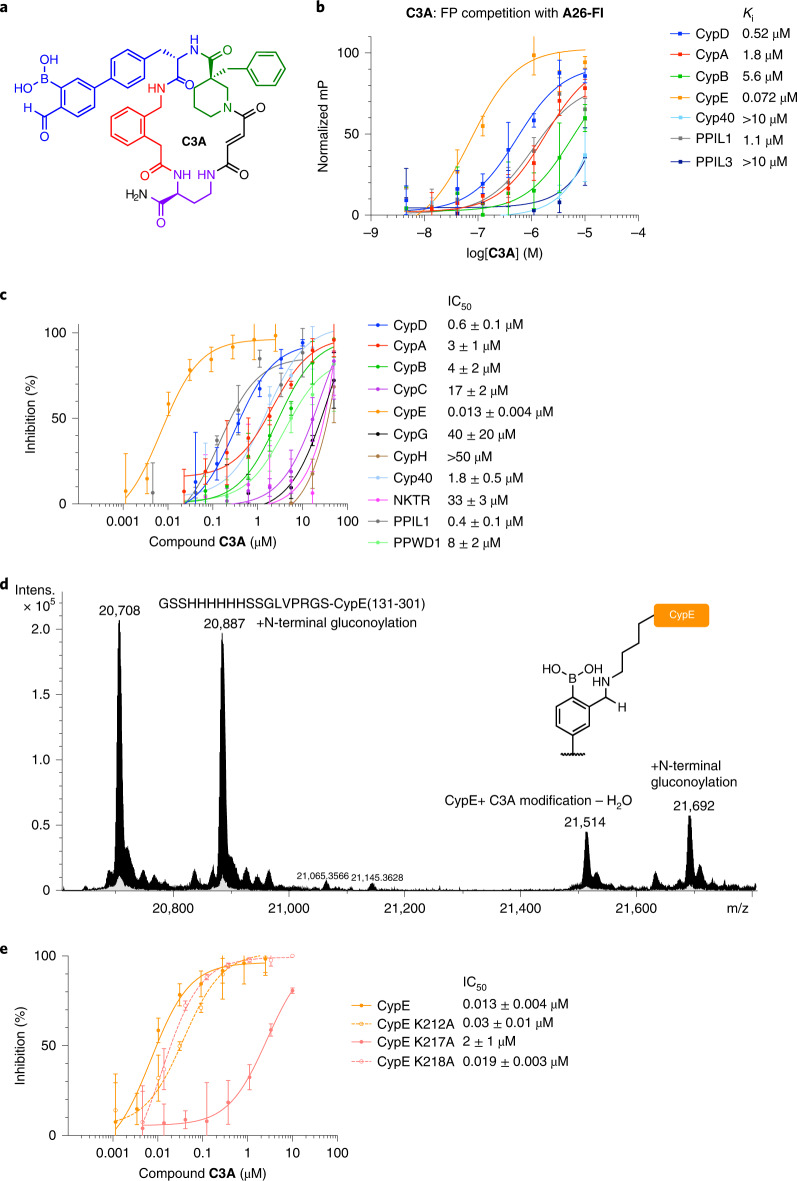
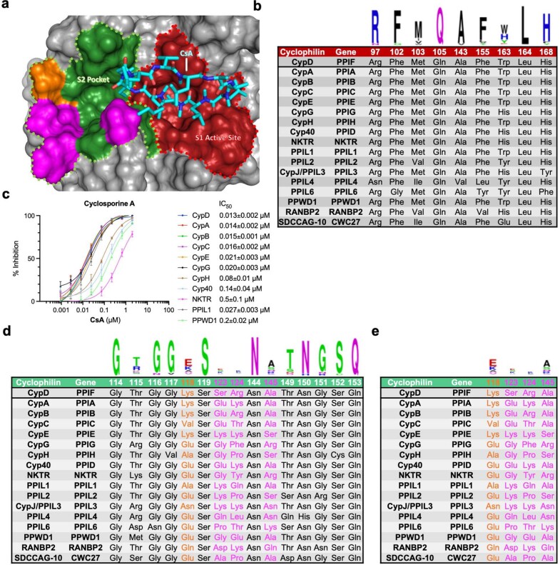
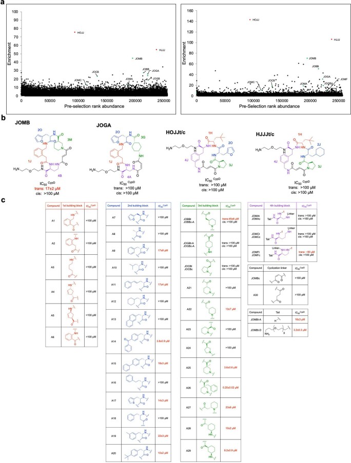
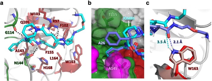
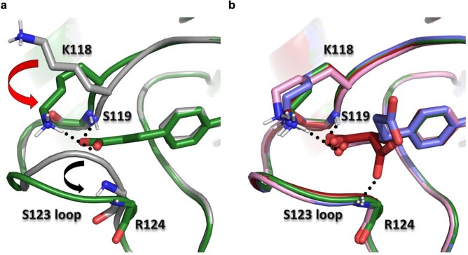
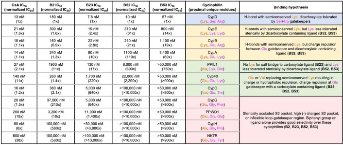
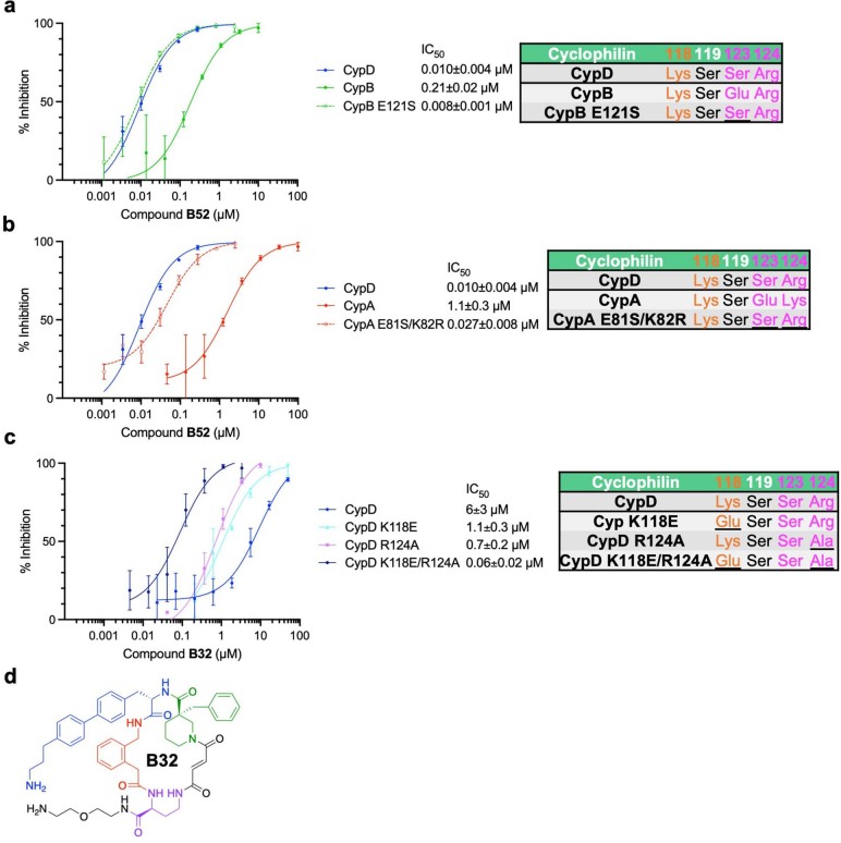
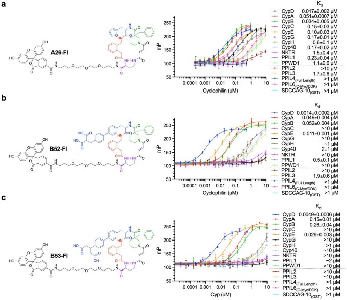
Although cyclophilins are attractive targets for probing biology and therapeutic intervention, no subtype-selective cyclophilin inhibitors have been described. We discovered novel cyclophilin inhibitors from the in vitro selection of a DNA-templated library of 256,000 drug-like macrocycles for cyclophilin D (CypD) affinity. Iterated macrocycle engineering guided by ten X-ray co-crystal structures yielded potent and selective inhibitors (half maximal inhibitory concentration (IC50) = 10 nM) that bind the active site of CypD and also make novel interactions with non-conserved residues in the S2 pocket, an adjacent exo-site. The resulting macrocycles inhibit CypD activity with 21- to >10,000-fold selectivity over other cyclophilins and inhibit mitochondrial permeability transition pore opening in isolated mitochondria. We further exploited S2 pocket interactions to develop the first cyclophilin E (CypE)-selective inhibitor, which forms a reversible covalent bond with a CypE S2 pocket lysine, and exhibits 30- to >4,000-fold selectivity over other cyclophilins. These findings reveal a strategy to generate isoform-selective small-molecule cyclophilin modulators, advancing their suitability as targets for biological investigation and therapeutic development.


Conflict of interest statement

A.A.P. and D.R.L. are co-inventors on patent applications that are based on this work. D.R.L. is a consultant and co-founder of Exo Therapeutics, a company that uses DNA-encoded libraries and exo-site inhibition.

Figures

Fig. 1
Fig. 1. Selection of a DNA-templated library of 256,000 macrocycles for CypD affinity reveals novel cyclophilin inhibitors.
a, Generalized trends in inhibition potency of CypD prolyl-isomerase activity from cyclophilin inhibition profiles of library hits and A-series macrocycles. b, JOMBt, showing weak and promiscuous cyclophilin inhibition of prolyl-isomerase activity. c, Compound A26 showing improved CypD potency but promiscuous inhibition. d, X-ray co-crystal structure of compound A26 (cyan) bound to CypD (PDB ID 7TGT, 1.06 Å resolution), shown as a space-filling model. A26 has a dual-binding mode involving the active site (red) and S2 pocket (green) of CypD. e, Active site binding interactions with A26. The phenyl group provides the primary hydrophobic interactions with F102, M103, A143, F155, L164, and H168. Black dashes show predicted hydrogen bonding interactions with R97, Q105, G114, N144, and W163 and the backbone of the A26. f, S2 pocket binding pose of the furan of A26, exhibiting a shallow interaction that does not engage non-conserved residues K118 (orange), S123 (magenta), and R124 (magenta) on the far side of the pocket. IC50 values reflect mean ± s.e.m. of three technical replicates. Data points and error bars reflect mean ± s.d. of individual assays at one dose. Source data
Fig. 2
Fig. 2. Compounds with large S2 pocket binding groups show reduced potency against cyclophilins with sterically occluded S2 pockets.
a, Structure of B1, B2. b, Cyclophilin prolyl-isomerase inhibition screens for B1 and B2. c, Co-crystal structures of B1 (PDB ID 7TGU, 1.21 Å resolution) or B2 (PDB ID 7TGV, 1.46 Å resolution) bound to CypD, viewing the S2 pocket. Active site binding is identical to that of A26 (Fig. 1e). d, Residues within the S2 pocket of cyclophilins inhibited less potently by B1 and B2, with important residues underlined. The benzophenone or biphenyl group of B1 or B2, respectively, fills the S2 pocket more completely, resulting in selectivity over cyclophilins with more sterically occluded or inflexible S2 pockets. CypD by contrast contains a relatively un-occluded S2 pocket and flexible R124 residue. IC50 values reflect mean ± s.e.m. of three technical replicates. Data points and error bars reflect mean ± s.d. of individual assays at one dose. Source data
Fig. 3
Fig. 3. Carboxylate-containing biphenyl derivatives of B2 offer enhanced CypD selectivity.
a, Structure of B23 and B25. b, Cyclophilin prolyl-isomerase inhibition screens for B23 and B25. c, Co-crystal structures of B23 (PDB ID 7TH7, 1.18 Å resolution) and B25 (PDB ID 7THC, 1.57 Å resolution) bound to CypD, viewing the S2 pocket. Active site binding is identical to that of A26 (Fig. 1e). Yellow dashes indicate predicted hydrogen bonds. d, List of residues on the far side of the S2 pocket of cyclophilins that are proximal to the ligand carboxylates, with important deleterious interactions underlined. The biphenyl group with 3-carbon carboxylates, B23 and B25, achieve strong selectivity over cyclophilins without a residue homologous to CypD K118. B23 and B25 show similar inhibition potencies for CypD and other cyclophilins that contain a Lys residue homologous to CypD K118, but slightly attenuated depending on the identity of residue 123. IC50 values reflect mean ± s.e.m. of three technical replicates. Data points and error bars reflect mean ± s.d. of individual assays at one dose. Source data
Fig. 4
Fig. 4. Biphenyl dicarboxylates achieve strong CypD selectivity.
a, Structure of B52 and B53. b, Cyclophilin prolyl-isomerase inhibition screens for B52 and B53. c, Co-crystal structures of B52 (PDB ID 7THD, 1.16 Å resolution) and B53 (PDB ID 7THF, 1.10 Å resolution) bound to CypD, viewing the S2 pocket. Active site binding is identical to A26 (Fig. 1e). Yellow dashes indicate predicted hydrogen bonds. d, List of residues on the far side of the S2 pocket of cyclophilins that are proximal to the ligand carboxylates. Both compounds retain CypD potency similar to that of mono-carboxylate B23, while enhancing selectivity over CypA, CypB, CypE, and PPIL1. The malonic and glutaric acids of B52 and B53, respectively, position the carboxylate in a similar pose as B23 (Fig. 3c), while presenting a second carboxylate to the S123 residue. B52 forms a predicted hydrogen bond with the peptide backbone of S123–R124. R124 is pushed out of the S2 pocket, consistent with other macrocycles containing large S2-binding groups such as B1. B52 and B53 achieve selectivity over CypA and CypB through charge repulsion with a glutamate at the analogous 123 position, while creating a steric clash with PPIL1 and the lysine of CypE at this same position. IC50 values reflect mean ± s.d. of four independent replicates (each comprising three technical replicates). Graphs show a representative single independent replicate (independent replicate 1 is shown, containing three technical replicates) with data points and error bars reflecting mean ± s.d. of individual assays at one dose. Further independent replicates are shown in Supplementary Fig. 18b,c. Source data
Fig. 5
Fig. 5. Cy5-conjugated cyclophilin D inhibitors delay calcium induced opening of the mPTP in isolated mouse liver mitochondria and enter human cells as ester prodrugs.
The calcium retention capacity of mitochondria was determined in isolated mouse liver mitochondria (0.5 µg mL−1) in response to pulses of 60 µM CaCl2 in the presence of the indicated CypD inhibitors (or inactive enantiomers). Concentrations used were 2 µM CsA, 10 µM B52-Cy5, 10 µM *B52-Cy5, 20 µM B53-Cy5, 20 µM *B53-Cy5. Mitochondrial uptake of extramitochondrial Ca2+ was assessed by monitoring the fluorescence of Calcium green 5n, depicted in arbitrary units (AU). The rapid increase in fluorescence after several pulses of Ca2+ are taken up corresponds to mitochondrial Ca2+ release through mPTP opening. ac, Traces are shown from a representative experiment, with all assays performed on the same mitochondrial preparation and day. d, Quantitation of calcium retention capacity (CRC) reported as the ratio of the number of Ca2+ pulses required to induce mPTP opening in the listed condition relative to DMSO control conditions on the same mitochondrial preparation and day. e, Structures of Cy5-conjugated CypD-selective inhibitors and prodrugs. f, Fluorescence microscopy of HeLa cells co-incubated with ester prodrugs B52-Et-Cy5 and B53-Et-Cy5 (red), co-stained with mitochondrial (green) and nuclear (blue) dyes (Mitotracker Green and Hoechst 33342, respectively). Both prodrugs show good plasma membrane permeability and co-localization with Mitotracker Green, quantified in Extended Data Fig. 8a–i. Calcium retention data are from three independent experiments/mitochondrial isolations. Data bars and error bars represent mean ± s.d. DMSO versus CsA, P = 0.0135; *B52-Cy5 versus B52-Cy5, P = 0.0011; *B53-Cy5 versus B53-Cy5, P = 0.0382. *P < 0.05, **P < 0.005 by one-sided Student’s t-test. Microscopy images are a representative image of three technical replicates. Scale bars, 200 µm. Source data
Fig. 6
Fig. 6. Aryl-carbonyl boronic acid C3A achieves selective inhibition of CypE.
a, Structure of C3A. b, C3A fluorescence polarization (FP) competition with A26-Fl against cyclophilins with S2 pocket lysines. c, Cyclophilin prolyl-isomerase inhibition screen of C3A, showing potency and selectivity for CypE. d, Mass spectroscopy trace of CypE incubated with C3A and reduced with sodium cyanoborohydride. C3A shows an adduct consistent with CypE + amine–H2O (+806 Da), the result of iminoboronate formation followed by reductive amination. The mass of CypE is 20,708 Da, The CypE preparation also included N-terminal gluconoylation. e, Prolyl-isomerase inhibition by C3A against CypE S2 pocket lysine to alanine mutants. For the FP assay, the y-axis is normalized to internal control wells containing A26-Fl only (100%) and A26-Fl with cyclophilin (0%). Values reflect mean of three technical replicates and error bars reflect s.d. of individual assays at one dose. For the prolyl-isomerase assay in c and the CypE wild-type dose–response curve in e, IC50 values reflect mean ± s.d. of four independent replicates (each comprising three technical replicates). Graph shows a representative single independent replicate (Independent replicate 3 is shown, containing three technical replicates) with data points and error bars reflecting mean ± s.d. of individual assays at one dose. Further independent replicates are shown in Supplementary Fig. 28b. For the prolyl-isomerase assay in e, IC50 values for the CypE mutants reflect mean ± s.e.m. of three technical replicates, while data points and error bars reflect mean ± s.d. of individual assays at one dose. Source data
Extended Data Fig. 1
Extended Data Fig. 1. Exclusive active site binding results in promiscuous cyclophilin inhibition.
a, Co-crystal structure of CsA bound to CypD (PDB ID 2Z6W). CsA (cyan) binds to the active site (red) of CypD, a binding mode that does not engage more diversified residues that lie in the S2 pocket (green), including the primary gatekeepers (magenta) and the semiconserved K118 residue (orange). b, Active site (S1 pocket) residues with WebLogo plots, showing high conservation between the 17 cyclophilin isoforms with corresponding gene and protein identifier. c, Isomerization of the reporter peptide Suc-AAPF-AMC was measured with 5 nM cyclophilin and varying concentrations of CsA. CsA shows potent, but promiscuous inhibitory activity against all prolyl isomerase-active cyclophilins. Selective inhibition could rather be achieved through interactions with S2 pocket residues of cyclophilin subtypes (d). e, Cyclophilin S2 pocket residues that are the primary diversification sites within the active site-S2 pocket groove with WebLogo plots. Typical cyclophilin inhibitors such as CsA do not engage in interactions with these residues. All residue numbering is in reference to CypD. IC50 values reflect mean ± s.e.m. of three technical replicates. Data points and error bars reflect mean ± s.d. of individual assays at one dose. Source data
Extended Data Fig. 2
Extended Data Fig. 2. Overview of macrocycle library hits from the CypD binding selection and JOMBt structure-activity relationships.
a, Two replicates of this selection yielded consistent enrichment of the JO** family of macrocycles. Library hits re-synthesized in DNA-free form resulted in novel CypD inhibitors JOMBt and JOBBt, the starting points of CypD inhibitors developed in this study. b, Structure JOMB, JOGA, and H*JJ macrocycles that enriched well in the DNA-templated macrocycle library selection. JOMB in the trans isomer (JOMBt) exhibits weak prolyl isomerase inhibition. c, Library hits and derivatives of JOMBt and their corresponding prolyl isomerase inhibition IC50 values for CypD. Each listed compound has the JOMBt structure except for the drawn building block substitution. Note, library encoded macrocycles that denote an ‘-A’ nomenclature (JOGBt-A, JOGBc-A, JOBBc-A) include the same primary amide tail shown in JOMBt-A. Values and error bars reflect mean ± s.e.m. of three technical replicates. Source data
Extended Data Fig. 3
Extended Data Fig. 3. X-ray co-crystal structure of JOMBt:CypD compared to A26:CypD.
a, Active site binding pose of JOMBt (PDB ID 7TGS, 1.75 Å resolution) showing almost identical interactions with CypD as A26 (Fig. 1d, e). Black dashes indicate predicted hydrogen bonds. Notable differences between the binding mode of JOMBt and A26 include b, An extra hydrophobic contact between the benzyl group of A26 (purple) and T115 (green) is absent with JOMBt (cyan). c, An extra hydrogen bond between A26 (purple) and W163 (salmon or red) that JOMBt (cyan) lacks. The W163–JOMBt distance is 3.5 Å (cyan dashes), compared to 2.1 Å for W163–A26 (purple dashes). Van der Waals surfaces of T115 and A145 (magenta) are shown in the structure with A26.
Extended Data Fig. 4
Extended Data Fig. 4. Carboxylate-containing S2-binding moieties induce K118 side-chain and S123 backbone migration.
a, Superposition of co-crystal structures of CypD with B2 (gray) and B23 (green). The side-chain of K118 typically is oriented away from the S2 pocket, as shown in co-crystal structures that do not contain carboxylate ligands, such as that of B2. A properly placed carboxylate group (B23) migrates K118’s side-chain into the S2 pocket (red arrow), forming a salt bridge along with a hydrogen bond with S119’s peptide backbone (black dashes). Also shown is the S123 loop migration that occurs when carboxylate-containing biphenyl groups bind deep into the S2 pocket (black arrow). b, Co-crystal structure superposition of CypD with B23 (green), B25 (pink), B52 (red) and B53 (purple). All properly placed carboxylate-containing ligands induce the same K118 and S123 conformational change. Dicarboxylates such as B52 and B53 also present the second carboxylate towards the S123 gatekeeper residue, the former exhibiting a hydrogen bond (black dashes) with the S123–R124 peptide backbone.
Extended Data Fig. 5
Extended Data Fig. 5. Analysis of CypD-selective prolyl isomerase inhibition based on S2 pocket containing residues.
IC50 values for each cyclophilin are accompanied by fold difference normalized to IC50CypD. Residues listed next to each cyclophilin are the important proximal residues near the carboxylate-containing ligands. CsA does not bind the S2 pocket and shows almost no cyclophilin isoform selectivity, while B2’s large biphenyl group exhibits selectivity over cyclophilins with sterically occluded S2 pockets. Further selectivity over CypC, Cyp40, and PPIL1 is achieved through interactions between CypD’s K118 residue and carboxylate-containing ligands such as B23. Installation of dicarboxylates in B52 and B53 result in CypD selectivity by presenting a second carboxylate near the analogous S123 position on CypD. Potency for CsA, B2, and B23 values reflect mean of three technical replicates. Potency for B52 and B53 values reflect mean of four independent replicates.
Extended Data Fig. 6
Extended Data Fig. 6. Inhibition potency is dependent on favorable interactions with S2 pocket residues.
Dose response curves for B52 against a, CypB E121S mutant and b, CypA E81S/K82R double mutant, with residue tables for important dicarboxylate proximal residues. Mutated residues are underlined. B52 inhibits both CypB and CypA mutants that have the appropriate ‘CypD’ gatekeeper residues at similar potency values compared to wild-type CypD. c, Dose response curves for B32 against wild-type CypD and CypD mutants shown in the table. Two mutations to remove positively charged residues K118 and R124 restore inhibition potency of amine derivative B32. d, Structure of B32. For CypD, CypA, and CypB wild-type IC50 data with B52, values reflect mean ± s.d. of four independent replicates (each comprising three technical replicates). Graphs show a representative single independent replicate (Independent replicate 1 is shown, containing three technical replicates) with data points and error bars reflecting mean ± s.d. of individual assays at one dose. Further independent replicates are shown in Supplementary Fig. 18b-c. All other IC50 values reflect mean ± s.e.m. of three technical replicates, with data points and error bars reflecting mean ± s.d. of individual assays at one dose. Source data
Extended Data Fig. 7
Extended Data Fig. 7. Cyclophilin binding profiles using fluorescence polarization of fluorescein-labeled macrocycles.
Each cyclophilin was titrated against 0.5 nM fluorescein-labeled macrocycle a, A26-Fl; b, B52-Fl; or c, B53-Fl. Trends for selectivity follow those observed in the prolyl-isomerase assay. Cyclophilins in the legend below the dashed line are either prolyl-isomerase inactive or require much higher concentrations to observe prolyl isomerization in vitro. Kd values and error bars reflect mean ± s.e.m. of three technical replicates. Data points and error bars reflect mean ± s.d. of individual assays at one dose. Source data
Extended Data Fig. 8
Extended Data Fig. 8. Quantification of mitochondrial localization in HeLa cells by fluorescence microscopy of Cy5-conjugated compounds.
HeLa cells were treated with Cy5-conjugated compounds and analyzed for a, total identifiable Cy5 spots; b, Cy5 spots per well; c, mean fluorescence intensity of identified Cy5 spots; d, sum of fluorescence intensity of identified Cy5 spots; e, mean Cy5 fluorescence intensity per cell; f, sum of Cy5 fluorescence in all measured cells; g, percent of Cy5 spots that overlap >70% with Mitotracker Green co-stain; and h, fluorescence intensity of Cy5 spots that overlap >70% with Mitotracker Green. i, values of data shown in a-h. Values and error bars reflect mean ± s.d. of three technical replicates. Source data
Extended Data Fig. 9
Extended Data Fig. 9. Hydrolysis of ester prodrug CypD inhibitors.
Compounds were evaluated for their ability to be hydrolyzed from di-ester to mono-ester, or to di-acid CypD inhibitors. Each reaction was analyzed by LC-MS, and ion abundances for each are shown as a percent of the total sum. These were conducted under conditions of: a, Tris-HCl buffer only; b, 250 nM carboxylesterase 1 (CES1); c, 250 nM carboxylesterase 2 (CES2); d, incubated with A549 cells for 48 h and intracellular fraction isolated; e, incubated with HeLa cells for 48 h and intracellular fraction isolated; f, incubated with HEK293T cells for 48 h and intracellular fraction isolated; g, incubated with MEFs for 48 h and intracellular fraction isolated; h, incubated with HepG2 cells for 36 h and intracellular fraction isolated. Esters show good stability in buffer and are only cleaved under esterase conditions, or intracellularly, with B52-Et-Cy5 showing the most rapidly hydrolyzed esters. Values and error bars reflect mean ± s.d. of three technical replicates. Source data
Extended Data Fig. 10
Extended Data Fig. 10. Mass spectroscopy analysis of compounds co-incubated with CypE.
a, Mass spectroscopy analysis of lysine covalent modification of CypE with C3A, C5A, and C6A. b, Mass spectroscopy analysis of lysine covalent modification after treatment with NaCNBH3 to trap the iminoboronate as a reduced lysine-linked secondary amine, highlighting relevant m/z regions. C3A and C5A show covalent modification (+779 Da and +806 Da, respectively) of CypE after reductive amination with NaCNBH3.

References

    1. Davis TL, et al. Structural and biochemical characterization of the human cyclophilin family of peptidyl-prolyl isomerases. PLoS Biol. 2010;8:e1000439. - PMC - PubMed
    1. Chen J, et al. Biochemical features of recombinant human cyclophilin J. Anticancer Res. 2016;36:1175–1180. - PubMed
    1. Dunyak BM, Gestwicki JE. Peptidyl-proline isomerases (PPIases): targets for natural products and natural product-inspired compounds. J. Med. Chem. 2016;59:9622–9644. - PMC - PubMed
    1. Austin, C. et al. Prolyl Isomerases—Old Proteins as New Therapeutic Targets (Selcia Ltd, 2015).
    1. Izzo V, Bravo-San Pedro JM, Sica V, Kroemer G, Galluzzi L. Mitochondrial permeability transition: new findings and persisting uncertainties. Trends Cell Biol. 2016;26:655–667. - PubMed

Publication types