. 2022 Nov;39(11):2699-2707.
doi: 10.1007/s11095-022-03398-5. Epub 2022 Sep 26.

Formulation of Lipid-Free Polymeric Mesoscale Nanoparticles Encapsulating mRNA

Affiliations

Formulation of Lipid-Free Polymeric Mesoscale Nanoparticles Encapsulating mRNA

Rachel Skelton et al. Pharm Res. 2022 Nov.

Abstract

Introduction: Nanoparticle-mediated gene therapy has found substantial clinical impact, primarily focused on lipid-based nanoparticles. In comparison with lipid nanoparticles, polymeric particles may have certain advantages such as increased biocompatibility and controlled release. Our prior studies have found that polymeric mesoscale nanoparticles exhibited specific targeting to the renal proximal tubules. Thus, in this study, we sought to identify formulation parameters that allow for development of polymeric mesoscale nanoparticles encapsulating functional mRNA for delivery into tubular epithelial cells.

Methods: We evaluated particle uptake in vitro prior to exploring formulation parameters related to introduction of a primary mixture of polymer in acetonitrile and hydrophilic mRNA in water. Finally, we evaluated their functionality in a renal tubular epithelial cell line.

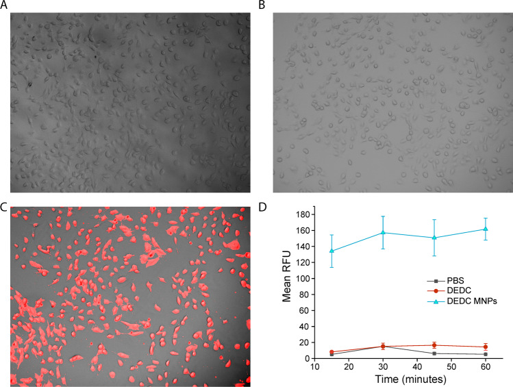
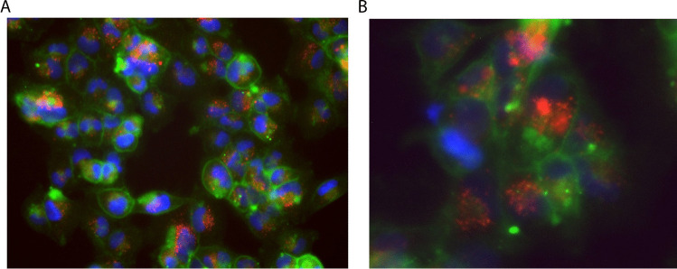
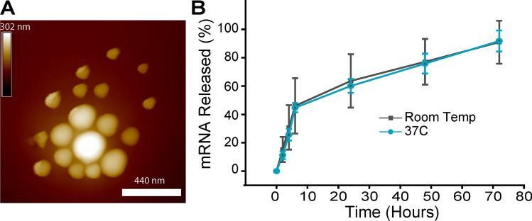
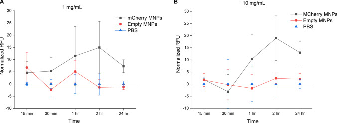
Results: We found that MNPs are endocytosed within 15 min and that the mesoscale nanoparticle formulation procedure was generally robust to introduction of a primary mixture and encapsulation of mRNA. These particles exhibited substantial uptake in renal cells in vitro and rapid (< 1 h) expression of a model mCherry fluorescent protein.

Conclusion: We anticipate these findings having potential in the delivery of specific gene therapies for renal disorders and cancer.

Keywords: gene delivery; kidney disease; mRNA; polymeric nanoparticles; translation.


Conflict of interest statement

RMW is a scientific advisor with equity interest in Goldilocks Therapeutics, Inc.

Figures

Fig. 1
Fig. 1
Uptake of fluorescent dye-loaded MNPs in 786-O cells. (A) Transmitted light and Cy5 fluorescence of 786-O cells incubated with PBS for 15 min imaged at 100X. (B) Transmitted light and Cy5 fluorescence of 786-O cells incubated with 1 µg/mL DEDC dye alone for 15 min imaged at 100X. (C) Transmitted light and Cy5 fluorescence of 786-O cells incubated with 1 mg/mL DEDC MNPs for 15 min imaged at 100X. (D) Normalized Cy5 signal from 786-O cells after incubation for up to one hour in 15-min increments. Each point represents mean ± standard deviation of six images.
Fig. 2
Fig. 2
Subcellular localization of fluorescent dye-loaded MNPs in 786-O cells. (A) Subcellular localization of DEDC-MNPs in 786-O cells at 200X and (B) 400X. Green: MemBrite cell membrane dye; Blue: Hoechst nuclear dye; Red: Cy5 encapsulated within MNPs.
Fig. 3
Fig. 3
Characterization of mCherry mRNA-MNPs. (A) Atomic force microscopy of a region of mRNA-MNPs. (B) Release of mRNA from MNPs up to 72 h in 10% FBS either at room temperature or at 37°C. Data points represent mean ± standard deviation of 3 replicates.
Fig. 4
Fig. 4
Expression of mCherry mRNA after intracellular delivery by MNPs. (A) 1 mg/mL of mCherry mRNA-MNPs was incubated with 786-O renal tubular epithelial cells for 1, 4, and 24 h. Top row: RFP filter; Bottom row: transmitted light. (B) 10 mg/mL of mCherry mRNA-MNPs were incubated the same as in Panel A. (C) Control experiments of 786-O cells treated with no MNPs or empty MNPs (left, center respectively) for 24 h or imaging of mCherry mRNA MNPs with no cells present (right). Scale bar = 300 µm. (D) Mean relative fluorescence was obtained from each timepoint and concentration treatment of mCherry mRNA MNPs with 786-O cells and compared for 20 ROIs per image. NS = not significant; *** = p < 0.05.
Fig. 5
Fig. 5
Quantification of mCherry expression in 786-O cells via automated image-based cytometry. (A) Normalized relative fluorescence units (RFU) obtained from 4–6 automated images in cells treated with 1 mg/mL mCherry MNPs or Empty MNPs compared to background PBS controls. (B) Normalized relative fluorescence units (RFU) obtained from 4–6 automated images in cells treated with 10 mg/mL mCherry MNPs or Empty MNPs compared to background PBS controls. Each point represents mean ± standard deviation.

References

    1. Chen J, et al. Production and clinical development of nanoparticles for gene delivery. Molecular Therapy-Methods & Clinical Development. 2016;3:16023. doi: 10.1038/mtm.2016.23. - DOI - PMC - PubMed
    1. Hou X, et al. Lipid nanoparticles for mRNA delivery. Nat Rev Mater. 2021;6(12):1078–1094. doi: 10.1038/s41578-021-00358-0. - DOI - PMC - PubMed
    1. Mitchell MJ, et al. Engineering precision nanoparticles for drug delivery. Nat Rev Drug Discovery. 2021;20(2):101–124. doi: 10.1038/s41573-020-0090-8. - DOI - PMC - PubMed
    1. Zhang MM, et al. The growth of siRNA-based therapeutics: Updated clinical studies. Biochem Pharmacol. 2021;189:114432. doi: 10.1016/j.bcp.2021.114432. - DOI - PMC - PubMed
    1. Hu B, et al. Therapeutic siRNA: state of the art. Signal Transduct Target Ther. 2020;5(1):1–25. doi: 10.1038/s41392-020-0207-x. - DOI - PMC - PubMed

LinkOut - more resources