. 2022 Oct;7(10):1661-1672.
doi: 10.1038/s41564-022-01227-4. Epub 2022 Sep 26.

Computational identification of a systemic antibiotic for gram-negative bacteria

Affiliations

Computational identification of a systemic antibiotic for gram-negative bacteria

Ryan D Miller et al. Nat Microbiol. 2022 Oct.

Abstract

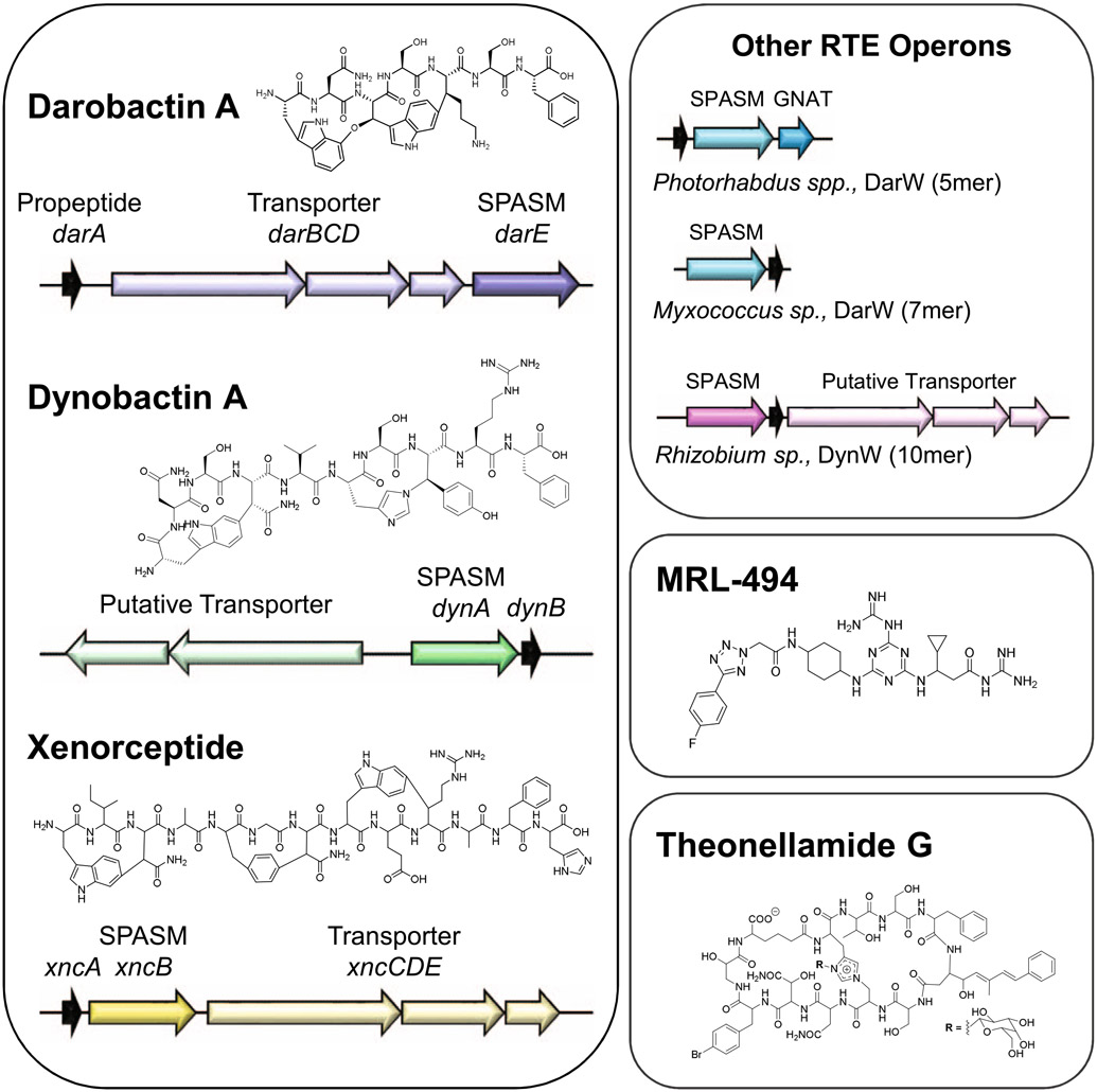
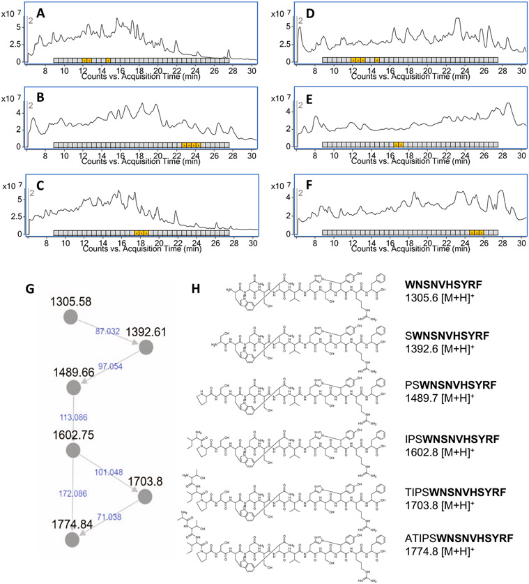
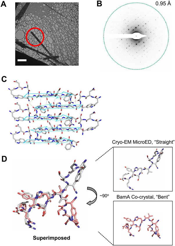
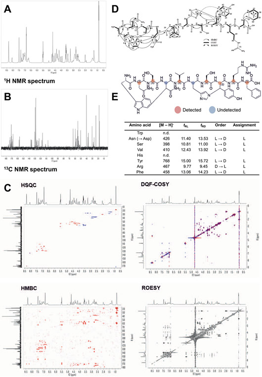
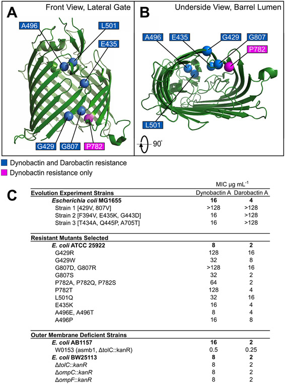
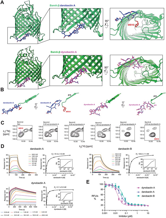
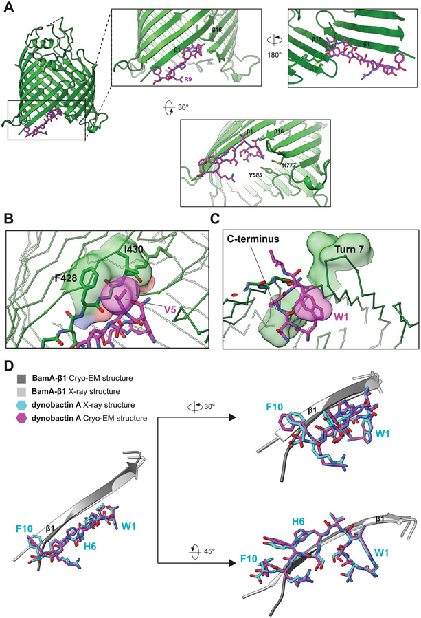
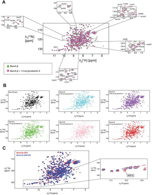
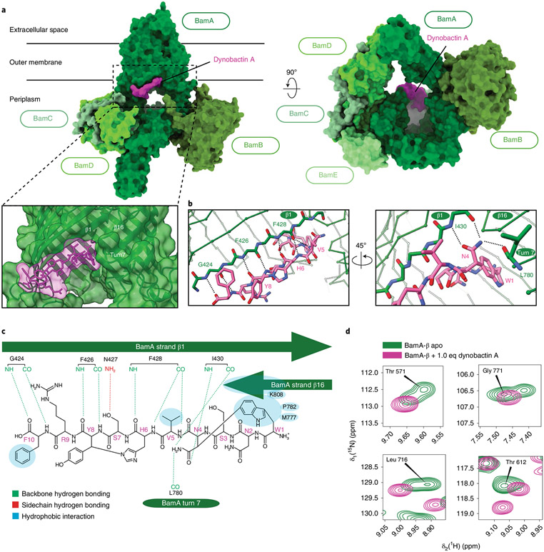
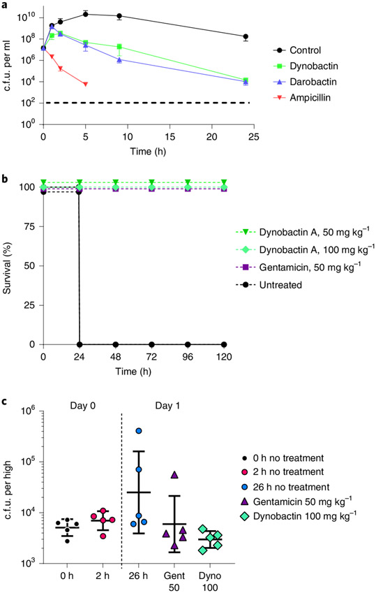
Discovery of antibiotics acting against Gram-negative species is uniquely challenging due to their restrictive penetration barrier. BamA, which inserts proteins into the outer membrane, is an attractive target due to its surface location. Darobactins produced by Photorhabdus, a nematode gut microbiome symbiont, target BamA. We reasoned that a computational search for genes only distantly related to the darobactin operon may lead to novel compounds. Following this clue, we identified dynobactin A, a novel peptide antibiotic from Photorhabdus australis containing two unlinked rings. Dynobactin is structurally unrelated to darobactins, but also targets BamA. Based on a BamA-dynobactin co-crystal structure and a BAM-complex-dynobactin cryo-EM structure, we show that dynobactin binds to the BamA lateral gate, uniquely protruding into its β-barrel lumen. Dynobactin showed efficacy in a mouse systemic Escherichia coli infection. This study demonstrates the utility of computational approaches to antibiotic discovery and suggests that dynobactin is a promising lead for drug development.


Figures

Extended Data Fig. 1 ∣
Extended Data Fig. 1 ∣. Compound and Operon structures.
Shown are darobactin and putatively-related cyclophane RiPP products (dynobactin A, xenorceptide), BamA-targeting synthetic molecule (MRL-494), and theonellamide G which contains an unusual histidine Nε2 to alanine β-carbon linkage, similar to dynobactin histidine Nε2 to tyrosine β-carbon linkage.
Extended Data Fig. 2 ∣
Extended Data Fig. 2 ∣. Rapid identification of target metabolites.
Total ion chromatogram (TIC) and Escherichia coli MG1655 activity assay. Panels A-C correspond to C18 (A), PFP (B), and Phenyl (C) columns applied to a 0.1% (v/v) formic acid in water:acetonitrile gradient condition. Panels (D), (E), and (F) correspond to the same columns applied to a 0.1% (v/v) formic acid in water:methanol condition. Timeslice fractions which inhibit E. coli growth are indicated by highlighted boxes containing a ‘+’ symbol. (G) Summary of networked masses in antimicrobial ion exchange fraction. (H) Structure of biologically-active metabolites purified from Photorhabdus australis with GNPS network relationship to the dominant metabolite (dynobactin A).
Extended Data Fig. 3 ∣
Extended Data Fig. 3 ∣. CryoEM microED structure determination.
MicroED data collection and analysis of 19 independent crystals of dynobactin A yielded structure, further details are elaborated within Methods. (A) Bright-field TEM image of dynobactin crystals (Scale bar: 5 μm). (B) Electron diffraction pattern with resolution ring at 0.95 Å. (C) 2D crystal packing arrangements of dynobactin. The intramolecular hydrogen bonds are shown as dashed lines for the top two molecules. The crystallographic b axis is parallel to the vertical direction of the figure. (D) Dynobactin A shows flexible conformations. Left shows superimposition dynobactin A microED structure (straight) and the dynobactin A structure observed in co-crystal with target BamA (bent). Right panels depict individual structures side-by-side. Separation of the two macrocycle rings in dynobactin A allows for free rotation about 4 bonds, creating an approximate 90° kink in the dynobactin A structure.
Extended Data Fig. 4 ∣
Extended Data Fig. 4 ∣. Dynobactin Secondary Structural Confirmations.
Full NMR assignment available in Supplementary Table 4. (A) 1H NMR spectrum (900 MHz, D2O). (B) 13C NMR spectrum (225 MHz, D2O). (C) 2D NMR spectra recorded in D2O (top left HSQC, top right DQF-COSY, bottom left HMBC, bottom right ROESY). (D) Key 2D NMR correlations in D2O and DMSO-d6. (E) Retention times (tR, min) of FDLA derivatives from dynobactin A Marfey’s analysis.
Extended Data Fig. 5 ∣
Extended Data Fig. 5 ∣. Target identification and resistance mutations.
BamA crystal structure (green) with labeled resistance mutation sites identified in this paper: (A) front view of lateral gate and (B) view of the barrel lumen from the periplasmic side (underside). Sites identified which gave resistance to both compounds are labeled in blue, and a mutation site which gives resistance to only dynobactin A is labeled in magenta. (C) Table listing MICs for bacteria from the previously described darobactin-resistance evolution experiment, the isolated dynobactin-resistant mutants from this study, and other E. coli strains with outer membrane deficiencies (that is porin or efflux knockouts).
Extended Data Fig. 6 ∣
Extended Data Fig. 6 ∣. Unique features of dynobactin A binding.
(A) Comparison of the co-crystal structure of BamA-β with bound darobactin A (PDB:7NRF) and with bound dynobactin A (this work). In the close-up panels, W810 is highlighted in red. (B) Comparison of the orientation of the compound relative to strand β16 of BamA in the two structures. The bulky C-terminal extension of dynobactin A displaces the C-terminus of BamA further into the barrel lumen, with residue W810 becoming flexibly disordered (C) Selected region of a 2D [15N1H]-TROSY spectrum of BamA-β in LDAO micelles upon titration with dynobactin A. Tentative assignment for indole W810 is indicated. (D) Affinity measurements of darobactins and dynobactin A to BamA-β via Surface Plasmon Resonance, sensorgrams and the corresponding steady-state affinity plots show dynobactin A binds one order of magnitude tighter to BamA-barrel than the darobactins. (E) Efficacy of the compounds in inhibiting BAM-mediated folding in native outer membrane vesicles (OMVs) (data are presented as mean values ± SD, n = 2). Fitting of these data resulted in IC50 values of 30 ± 6 nM, 48 ± 8 nM, and 16 ± 2 nM for darobactin A, darobactin B and dynobactin A with a 95% confidence interval, respectively.
Extended Data Fig. 7 ∣
Extended Data Fig. 7 ∣. Cryo-EM and X-ray structure comparison of dynobactin A-bound BamA.
(A) X-ray crystal structure of BamA-β with bound dynobactin A at 2.5 Å resolution. Zoomed-in panels highlight specific residues involved in the interaction. (B) Hydrophobic interaction between V5 of dynobactin A to the side chains of F428 and I430 of BamA in the co-crystal structure. (C) Interaction between W1 of dynobactin A and BamA. (D) Superimposition of the X-ray and cryo-EM structures (β-strand 1 only). The comparison shows a high degree of similarity in the conformation of dynobactin A between the cryo-EM and X-ray structure.
Extended Data Fig. 8 ∣
Extended Data Fig. 8 ∣. Solution NMR spectroscopy of BamA-β interacting with dynobactin A.
(A) 2D [15N,1H]-TROSY spectra of apo BamA-β in LDAO micelles (green) overlaid with BamA-β with 1.0 eq of dynobactin A (magenta). Zoomed-in panels show selected resonances. Tentatively assigned W810 is indicated with a frame on the spectrum. (B) 2D [15N,1H]-TROSY spectra of BamA-β in a titration experiment with increasing concentration of dynobactin A, as indicated, from black to red. (C) NMR spectrum of mutant W810F to confirm the assignment of W810.
Extended Data Fig. 9 ∣
Extended Data Fig. 9 ∣. Time-lapse microscopy of E. coli undergoing dynobactin A treatment.
E. coli MG1655 cells were spotted onto a 1.5% agarose pad containing dynobactin A (8x MIC), the membrane stain FM4-64 10 μg mL−1 (false-colored in magenta), and membrane permeabilization stain Sytox Green 0.5 μM (false-colored in green). Cells were incubated at 37 °C in a thermostatic chamber and imaged every 15 minutes under the microscope. The panels and selected time points were chosen to best represent the population of E. coli MG1655 undergoing dynobactin A treatment. White arrows indicate representative examples of membrane blebbing; orange arrows indicate examples of swelling or cell lysis. Scale bars, 5 μm. This experiment is representative of two biologically-independent experiments performed, each showing similar results.
Fig. 1 ∣
Fig. 1 ∣. Schematic overview of the workflow.
The darobactin operon radical SAM enzyme, DarE, was used to query NCBI BLASTp. BLASTp hits were examined for associations with putative RiPPs in the surrounding genomic neighbourhood using the RiPPER search tool or manual curation, and were applied as further BLASTp queries in such cases. Producers of putatively identified RiPPs were fermented in media to screen for the presence of these RiPPs by biological activity assays or mass spectrometry. Once candidates were identified, these were purified and characterized through both biological assays to determine spectrum of action/cytotoxicity and structural elucidation approaches. A promising compound with low toxicity was then tested for efficacy in animal models of E. coli infection and its mechanism of action identified.
Fig. 2 ∣
Fig. 2 ∣. Phylogenetic tree of rSAM-SPASM enzymes.
a, Neighbour-joining tree generated from ClustΩ multi-sequence alignment of distantly related SPASM enzymes. Putatively identified cyclophane RTEs were plotted alongside major characterized SPASM enzymes and their closest homologues (selected using NCBI BLASTp). RTE clades of note are highlighted. b, Neighbour-joining tree generated from ClustalΩ multi-sequence alignment of known RTEs (XyeB, DarE), close homologues and newly identified clades neighbouring DarE (DynA, DynW and DarW).
Fig. 3 ∣
Fig. 3 ∣. Identification of dynobactin A from P. australis.
a, Lawn bio-assay of 20x concentrated P. australis W34 supernatant. b, Mass spectra of dominant Gram-negative selective metabolite, exact mass (M) 1,304.57. c, Bottom: RP-HPLC chromatogram of XAD16N-extracted and SP-FF ion-exchanged P. australis supernatant. Top: isolated peaks assayed against lawns of E. coli and S. aureus. d, Well from high-throughput crystallization screen containing fluorescent crystals. e, Top: cryo-EM microED-generated 3D crystal structure of dynobactin A molecule. Dashed green box, carbon-carbon bond formed between the W1 C6 and the β-carbon of N4; dashed orange box, nitrogen-carbon linkage between the H6 imidazole Nε2 and the β-carbon of Y8. Bottom: accompanying 2D structure.
Fig. 4 ∣
Fig. 4 ∣. Dynobactin A binds BamA lateral gate.
a, Three-dimensional structure of the BAM complex in DDM detergent micelles with bound dynobactin A, resolved by cryo-EM to a resolution of 3.6 Å. The proteins are shown as surfaces with colours as annotated. b, Close-up view of the dynobactin A binding site in a 2.5 Å crystal structure of dynobactin A bound to BamA-β. Amino acids involved in dynobactin A binding are shown in stick representation and polar contacts are shown as dashed lines: blue, nitrogen; red, oxygen; green and magenta (BamA and dynobactin, respectively), carbon. c, Schematic representation of the interactions between dynobactin A and BamA. d, Selected regions from a 2D [15N,1H]-TROSY spectrum of BamA-β in LDAO detergent micelles in the absence (green) and presence (magenta) of dynobactin A. The four selected amino acids show doublet peaks resolved into a singlet upon dynobactin A addition. Full spectra are shown in Extended Data Fig. 8.
Fig. 5 ∣
Fig. 5 ∣. Efficacy of dynobactin A.
a, Time-dependent killing of E. coli ATCC 25922 by dynobactin, darobactin and ampicillin. Antibiotics were added at 4x their respective MICs. Time points are graphed as the mean c.f.u. ± s.d. The experiment was performed in biological triplicate. Ampicillin-killed E. coli fell below the limit of detection, denoted by a dashed line. b, Mouse septicaemia model, wherein mice were inoculated with a lethal dose of multidrug-resistant E. coli AR350, followed by administration of a single intraperitoneal dose of antibiotics: dynobactin 50 mg kg−1, dynobactin 100 mg kg−1 and gentamicin 50 mg kg−1 at 1 h post infection; an untreated mouse group was included. Four mice were tested per group. c, In a neutropenic thigh model of E. coli AR350 infection, gentamicin (purple triangle) or dynobactin (green diamond) were delivered to groups of mice (n = 5) by intraperitoneal injection at 2 h post infection (red circle). Infection was monitored over 26 h. At 26 h post infection (blue circle), thighs were homogenized, serially diluted and plated in triplicate for c.f.u. Error bars represent c.f.u. mean ± s.d. for each group.

Comment in

References

    1. Brown ED & Wright GD Antibacterial drug discovery in the resistance era. Nature 529, 336–343 (2016). - PubMed
    1. Lewis K The science of antibiotic discovery. Cell 181, 29–45 (2020). - PubMed
    1. Ling LL et al. A new antibiotic kills pathogens without detectable resistance. Nature 517, 455–459 (2015). - PMC - PubMed
    1. Tacconelli E et al. Discovery, research, and development of new antibiotics: the WHO priority list of antibiotic-resistant bacteria and tuberculosis. Lancet Infect. Dis 18, 318–327 (2018). - PubMed
    1. Zgurskaya HI, Rybenkov VV, Krishnamoorthy G & Leus IV Trans-envelope multidrug efflux pumps of Gram-negative bacteria and their synergism with the outer membrane barrier. Res. Microbiol 169, 351–356 (2018). - PMC - PubMed

Publication types

MeSH terms

LinkOut - more resources