. 2022 Nov 15;204(11):e0027322.
doi: 10.1128/jb.00273-22. Epub 2022 Sep 27.

HfaE Is a Component of the Holdfast Anchor Complex That Tethers the Holdfast Adhesin to the Cell Envelope

Affiliations

HfaE Is a Component of the Holdfast Anchor Complex That Tethers the Holdfast Adhesin to the Cell Envelope

Nelson K Chepkwony et al. J Bacteriol. .

Abstract

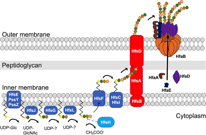
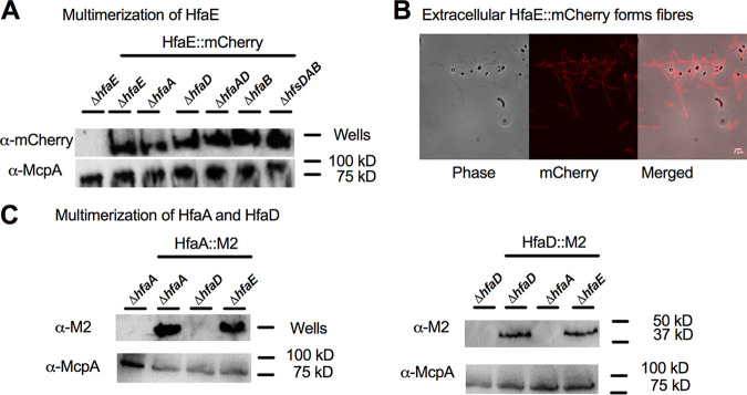
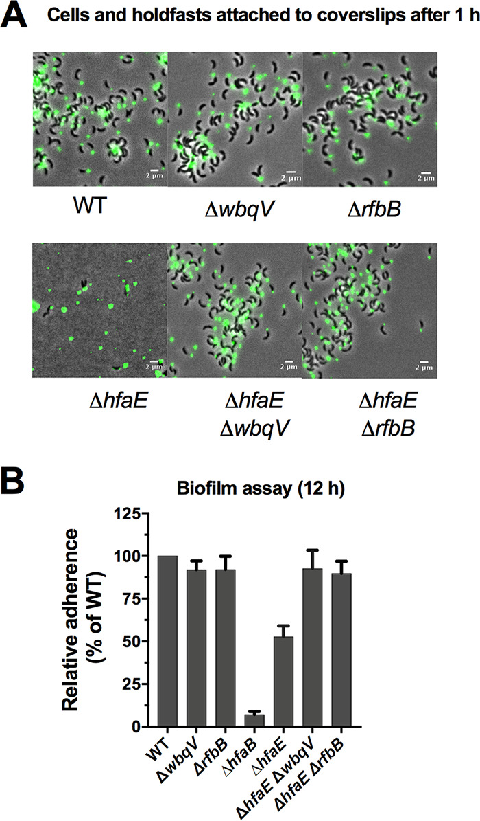
Bacteria use adhesins to colonize different surfaces and form biofilms. The species of the Caulobacterales order use a polar adhesin called holdfast, composed of polysaccharides, proteins, and DNA, to irreversibly adhere to surfaces. In Caulobacter crescentus, a freshwater Caulobacterales species, the holdfast is anchored at the cell pole via the holdfast anchor (Hfa) proteins HfaA, HfaB, and HfaD. HfaA and HfaD colocalize with holdfast and are thought to form amyloid-like fibers that anchor holdfast to the cell envelope. HfaB, a lipoprotein, is required for the translocation of HfaA and HfaD to the cell surface. Deletion of the anchor proteins leads to a severe defect in adherence resulting from holdfast not being properly attached to the cell and shed into the medium. This phenotype is greater in a ΔhfaB mutant than in a ΔhfaA ΔhfaD double mutant, suggesting that HfaB has other functions besides the translocation of HfaA and HfaD. Here, we identify an additional HfaB-dependent holdfast anchoring protein, HfaE, which is predicted to be a secreted protein. HfaE is highly conserved among Caulobacterales species, with no predicted function. In planktonic culture, hfaE mutants produce holdfasts and rosettes similar to those produced by the wild type. However, holdfasts from hfaE mutants bind to the surface but are unable to anchor cells, similarly to other anchor mutants. We showed that fluorescently tagged HfaE colocalizes with holdfast and that HfaE forms an SDS-resistant high-molecular-weight species consistent with amyloid fiber formation. We propose that HfaE is a novel holdfast anchor protein and that HfaE functions to link holdfast material to the cell envelope. IMPORTANCE For surface attachment and biofilm formation, bacteria produce adhesins that are composed of polysaccharides, proteins, and DNA. Species of the Caulobacterales produce a specialized polar adhesin, holdfast, which is required for permanent attachment to surfaces. In this study, we evaluate the role of a newly identified holdfast anchor protein, HfaE, in holdfast anchoring to the cell surface in two different members of the Caulobacterales with drastically different environments. We show that HfaE plays an important role in adhesion and biofilm formation in the Caulobacterales. Our results provide insights into bacterial adhesins and how they interact with the cell envelope and surfaces.

Keywords: Caulobacterales; adhesion; bacterial adhesin; biofilm; biofilms; extracellular polysaccharides; holdfast.


Conflict of interest statement

The authors declare no conflict of interest.

Figures

FIG 1
FIG 1
Model of C. crescentus holdfast synthesis, modification, export, and anchor machinery. Shown is a schematic of holdfast synthesis, modification, secretion, and anchor machinery. Holdfast polysaccharide synthesis is initiated by the glycosyltransferases HfsE, PssY, and PssZ, which transfers activated sugar precursors (UDP-Glc) in the cytoplasm to a lipid carrier. Three glycosyltransferases, HfsJ, HfsG, and HfsL, add different monosaccharide components to the growing lipid-linked repeat unit. The deacetylase HfsH modifies one or more sugar residues within the repeat unit before the flippase HfsF transports the lipid carrier-repeat unit structure into the periplasm. Repeat units are then assembled by the polymerases HfsC and HfsI. The polysaccharide is transported outside the cell through an export complex composed of HfsA, HfsB, and HfsD. The exported polysaccharide is then anchored to the cell wall by the holdfast anchor proteins HfaA, HfaB, HfaD, and HfaE. HfaB is a lipoprotein that is predicted to secrete HfaA and HfaD. The different colors of hexagons represent different sugars in the holdfast polysaccharides.
FIG 2
FIG 2
HfaE is involved in cell adhesion. (A) Genomic organization of the holdfast anchor genes (hfa) in C. crescentus and H. baltica. Genes were identified using a reciprocal best-hit analysis with C. crescentus and H. baltica genomes. In the C. crescentus genome, hfaE is found outside the hfa locus, while in the H. baltica genome, hfaE is found downstream of hfaD. ORFs, open reading frames. (B) Multiple-sequence alignment of HfaE from selected members of the Caulobacterales. Residues are color-coded based on their physicochemical properties using ClustalW (64). Consensus residues are indicated by an asterisk, and amyloid domains are indicated by black lines. B. subvibrioides, Brevundimonas subvibrioides; A. biprosthecum, Asticcacaulis biprosthecum; A. excentricus, Asticcacaulis excentricus; M. maris, Maricaulis maris. (C and D) Quantification of biofilm formation by a crystal violet assay after incubation for 12 h, expressed as the mean percentage of WT crystal violet staining normalized to the OD600. Errors are expressed as the standard errors of the means from three independent biological replicates, each with four technical replicates.
FIG 3
FIG 3
HfaE contributes to holdfast anchoring. (A) Representative images showing merged phase and fluorescence channels of exponentially growing planktonic cultures of C. crescentus (left) and H. baltica (right) strains. Holdfast polysaccharides are labeled with AF488-WGA (green). White arrows indicate shed holdfast, while blue arrows indicate rosettes. (B) Quantification of rosettes formed in C. crescentus (left) and H. baltica (right) strains grown to an OD600 of 0.8. Data are expressed as the mean numbers of rosettes formed. Errors are represented as the standard errors of the means from two biological replicates with five technical replicates each. (C and D) Representative images showing merged phase and fluorescence channels of C. crescentus and H. baltica strains bound to a glass slide. Holdfast is labeled with AF488-WGA (green). Exponentially growing cultures were incubated on glass slides for 1 h (C) or 4 h to 12 h (D) and washed to remove unbound cells before AF488-WGA labeling. (E) Quantification of biofilm formation by a crystal violet assay after incubation for 12 h and 24 h, expressed as the mean percentage of WT crystal violet staining normalized to the OD600. Errors are expressed as the standard errors of the means from three independent biological replicates, each with four technical replicates. The variance in biofilm formation was analyzed using a t test. ****, P < 0.0001; ns, not significant.
FIG 4
FIG 4
Epistasis analysis of hfaE and other hfa genes. (A) Representative images showing merged phase and fluorescence channels of exponentially growing, planktonic C. crescentus (left) and H. baltica (right) strains on agarose pads. Holdfast polysaccharides are labeled with AF488-WGA (green). (B) Quantification of shed holdfasts in C. crescentus (left) and H. baltica (right) strains grown to an OD600 of 0.8. Data are expressed as the mean numbers of shed holdfasts formed. Errors are represented as the standard errors of the means from two biological replicates with five technical replicates each. The variance in the number of shed holdfasts was analyzed using a t test (****, P < 0.0001; **, P < 0.01; ns, not significant). (C) Representative images showing merged phase and fluorescence channels of C. crescentus (left) and H. baltica (right) strains bound to a glass slide. Holdfast polysaccharides are labeled with AF488-WGA (GlcNAc) (green), and holdfast thiols are labeled with the maleimide dye AF594-Mal (thiols) (red). Exponential-phase cultures were incubated on glass slides for 1 h and washed to remove unbound cells before colabeling with AF488-WGA and AF594-Mal.
FIG 5
FIG 5
HfaE colocalizes with the holdfast in C. crescentus. (A and B) Representative images showing merged phase and fluorescence channels of the indicated C. crescentus strains expressing HfaE::mCherry. Holdfasts were labeled with AF488-WGA (green), specific for GlcNAc in holdfast polysaccharides, and the mCherry channel (red) was used to visualize the localization of HfaE::mCherry. Exponential-phase planktonic cultures were used to quantify the percentage of predivisional cells with HfaE::mCherry foci at the cell pole, which is indicated numerically at the bottom of each set of representative images. Data are expressed as the means from two independent biological replicates, along with the standard errors of the means. A total of 5,000 cells were quantified per replicate using MicrobeJ. (C) Time-lapse montages of C. crescentus hfaE::mCherry on soft agarose pads (0.8% agarose). Exponential-phase cultures were placed onto soft agarose pads containing holdfast-specific AF488-WGA (green) and covered with a glass coverslip. The blue arrow indicates a predivisional cell, and the white arrows indicate the polar localization of HfaE::mCherry as well as the newly synthesized holdfast in the swarmer cell. Images were collected every 10 min for 6 h.
FIG 6
FIG 6
Localization of anchor proteins in hfaE mutants. (A) Representative immunofluorescence images showing merged phase and fluorescence channels of C. crescentus strains expressing HfaA-M2 or HfaD-M2. Exponentially growing cells were fixed with formaldehyde. Anti-FLAG (M2) primary antibody and goat anti-rabbit secondary antibody conjugated to IRDye 680 (red fluorescence) were used to visualize the localization of the anchor protein. WT EV is wild-type with empty vector. (B) Representative images showing phase and fluorescence channels of exponentially growing C. crescentus hfaB::mCherry strains. The mCherry fluorescence channel was used to visualize HfaB::mCherry (red). For panels A and B, exponential-phase planktonic cultures were used to quantify the percentage of predivisional cells with fluorescent foci at the cell pole, which is indicated numerically at the bottom of each set of representative images. Data are expressed as the means from two independent biological replicates, along with the standard errors of the means. A total of 1,000 cells were quantified per replicate using MicrobeJ.
FIG 7
FIG 7
HfaE forms high-molecular-weight complexes. (A and C) Western blotting of whole-cell lysates of C. crescentus hfaE::mCherry (A) and C. crescentus hfaA::FLAG and C. crescentus hfaD::FLAG (C) using anti-FLAG tag (α-M2) antibody with goat anti-rabbit HRP-conjugated secondary antibody. Exponentially growing cells grown to an OD600 of 0.6 to 0.8 were normalized to the equivalent of 1 mL of culture at an OD600 of 1.0, and 10 μL of the cell lysate was loaded into each lane. Anti-McpA was used as a loading control. (B) Representative images showing phase, fluorescence, and merged channels of C. crescentus hfaE::mCherry with extracellular HfaE::mCherry protein forming fibers on a glass coverslip.
FIG 8
FIG 8
Mutations in sugar-nucleotide synthesis genes suppress the hfaE mutation. (A) Representative images showing merged phase and fluorescence channels of exponentially growing planktonic C. crescentus strains. Exponentially growing cultures were incubated on glass slides for 1 h and washed to remove unbound cells before labeling with AF488-WGA (GlcNAc) (green). (B) Quantification of biofilm formation by a crystal violet assay after incubation for 12 h, expressed as the mean percentage of WT crystal violet staining normalized to the OD600. Errors are expressed as the standard errors of the means from three independent biological replicates, each with four technical replicates.

Comment in

Similar articles

Cited by

  • What Glues the Glue to the Cell Surface?
    Fiebig A. Fiebig A. J Bacteriol. 2022 Nov 15;204(11):e0038622. doi: 10.1128/jb.00386-22. Epub 2022 Oct 26. J Bacteriol. 2022. PMID: 36286485 Free PMC article.
  • Bacteria in Fluid Flow.
    Padron GC, Shuppara AM, Palalay JS, Sharma A, Sanfilippo JE. Padron GC, et al. J Bacteriol. 2023 Apr 25;205(4):e0040022. doi: 10.1128/jb.00400-22. Epub 2023 Mar 23. J Bacteriol. 2023. PMID: 36951552 Free PMC article. Review.

References

    1. Berne C, Ducret A, Hardy GG, Brun YV. 2015. Adhesins involved in attachment to abiotic surfaces by Gram-negative bacteria. Microbiol Spectr 3(4):MB-0018-2015. 10.1128/microbiolspec.MB-0018-2015. - DOI - PMC - PubMed
    1. Dang H, Lovell CR. 2016. Microbial surface colonization and biofilm development in marine environments. Microbiol Mol Biol Rev 80:91–138. 10.1128/MMBR.00037-15. - DOI - PMC - PubMed
    1. O’Toole GA, Wong GC. 2016. Sensational biofilms: surface sensing in bacteria. Curr Opin Microbiol 30:139–146. 10.1016/j.mib.2016.02.004. - DOI - PMC - PubMed
    1. Lee CK, Vachier J, de Anda J, Zhao K, Baker AE, Bennett RR, Armbruster CR, Lewis KA, Tarnopol RL, Lomba CJ, Hogan DA, Parsek MR, O’Toole GA, Golestanian R, Wong GCL. 2020. Social cooperativity of bacteria during reversible surface attachment in young biofilms: a quantitative comparison of Pseudomonas aeruginosa PA14 and PAO1. mBio 11:e02644-19. 10.1128/mBio.02644-19. - DOI - PMC - PubMed
    1. Smyth CJ, Marron MB, Twohig JM, Smith SG. 1996. Fimbrial adhesins: similarities and variations in structure and biogenesis. FEMS Immunol Med Microbiol 16:127–139. 10.1111/j.1574-695X.1996.tb00129.x. - DOI - PubMed

Publication types

MeSH terms

LinkOut - more resources