. 2023 Mar;306(3):e221795.
doi: 10.1148/radiol.221795. Epub 2022 Sep 27.

SARS-CoV-2 Variants Infection in Relationship to Imaging-based Pneumonia and Clinical Outcomes

Affiliations

SARS-CoV-2 Variants Infection in Relationship to Imaging-based Pneumonia and Clinical Outcomes

Jong Eun Lee et al. Radiology. 2023 Mar.

Abstract

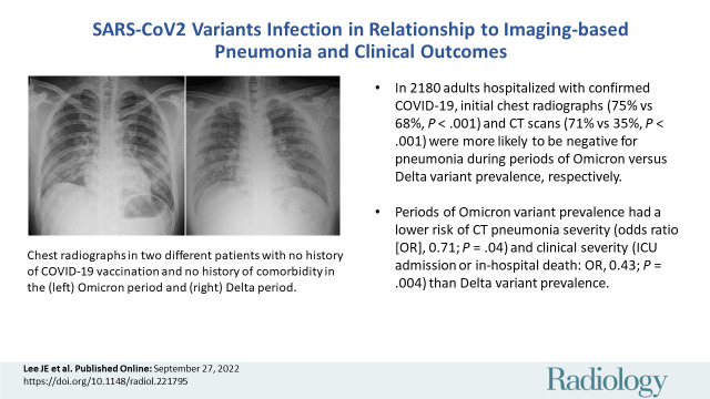
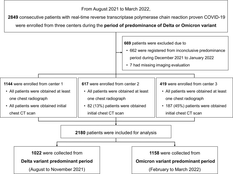
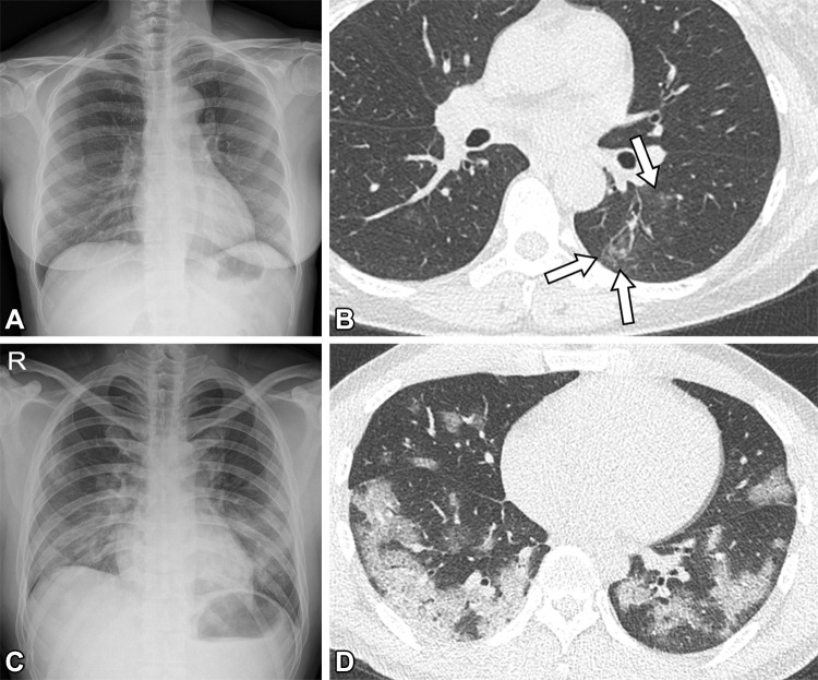
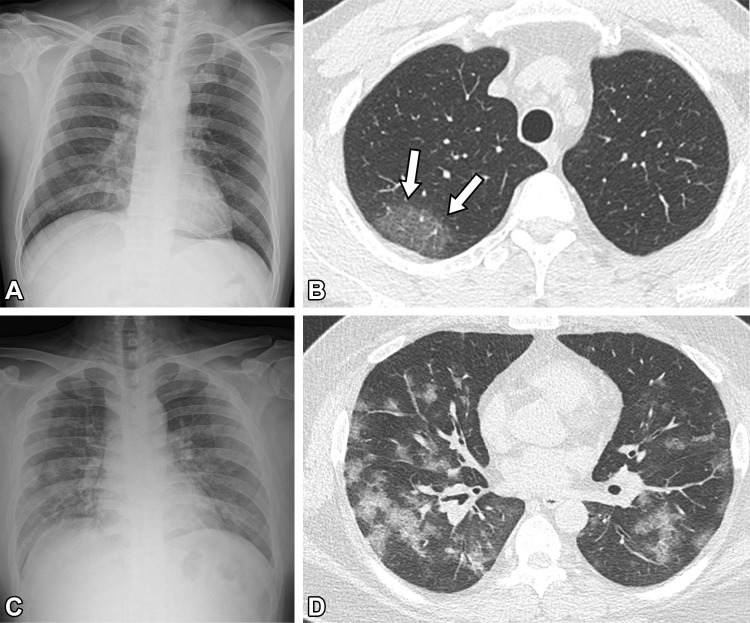
Background Few reports have evaluated the effect of the SARS-CoV-2 variant and vaccination on the clinical and imaging features of COVID-19. Purpose To evaluate and compare the effect of vaccination and variant prevalence on the clinical and imaging features of infections by the SARS-CoV-2. Materials and Methods Consecutive adults hospitalized for confirmed COVID-19 at three centers (two academic medical centers and one community hospital) and registered in a nationwide open data repository for COVID-19 between August 2021 and March 2022 were retrospectively included. All patients had available chest radiographs or CT images. Patients were divided into two groups according to predominant variant type over the study period. Differences between clinical and imaging features were analyzed with use of the Pearson χ2 test, Fisher exact test, or the independent t test. Multivariable logistic regression analyses were used to evaluate the effect of variant predominance and vaccination status on imaging features of pneumonia and clinical severity. Results Of the 2180 patients (mean age, 57 years ± 21; 1171 women), 1022 patients (47%) were treated during the Delta variant predominant period and 1158 (53%) during the Omicron period. The Omicron variant prevalence was associated with lower pneumonia severity based on CT scores (odds ratio [OR], 0.71 [95% CI: 0.51, 0.99; P = .04]) and lower clinical severity based on intensive care unit (ICU) admission or in-hospital death (OR, 0.43 [95% CI: 0.24, 0.77; P = .004]) than the Delta variant prevalence. Vaccination was associated with the lowest odds of severe pneumonia based on CT scores (OR, 0.05 [95% CI: 0.03, 0.13; P < .001]) and clinical severity based on ICU admission or in-hospital death (OR, 0.15 [95% CI: 0.07, 0.31; P < .001]) relative to no vaccination. Conclusion The SARS-CoV-2 Omicron variant prevalence and vaccination were associated with better clinical outcomes and lower severe pneumonia risk relative to Delta variant prevalence. © RSNA, 2022 Supplemental material is available for this article. See also the editorial by Little in this issue.


Conflict of interest statement

Disclosures of conflicts of interest: J.E.L. No relevant relationships. M.H. No relevant relationships. Y.H.K. No relevant relationships. M.J.C. No relevant relationships. B.H.S. No relevant relationships. W.G.J. No relevant relationships. Y.J.J. No relevant relationships.

Figures

None
Graphical abstract
Flow diagram shows patients with real-time reverse transcriptase
polymerase chain reaction–proven COVID-19.
Figure 1:
Flow diagram shows patients with real-time reverse transcriptase polymerase chain reaction–proven COVID-19.
Representative cases show pneumonia extents and patterns with chest
radiographs and CT images during the Omicron period. (A, B) Images in a
52-year-old woman with a breakthrough COVID-19 infection 4 months after a
second dose of the mRNA-1273 vaccine (fully vaccinated) in the Omicron
period. The patient had no history of comorbidity. (A) Chest radiograph
obtained at admission shows no abnormality in both lungs. The chest
radiograph extent of pneumonia was scored as 0 (no evidence of pneumonia).
(B) Axial chest CT image obtained on the same day shows poorly defined
centrilobular nodules in the left lower lobe (arrows). The extent of
pneumonia with CT was scored as 1 (1%–25% involvement). This case was
classified as “atypical” for COVID-19 pneumonia, as per the
RSNA chest CT classification system. (C, D) Images in a 30-year-old man with
no history of COVID-19 vaccination and no history of comorbidity in the
Omicron period. (C) Chest radiograph taken at admission shows patchy
ground-glass opacities in the middle to lower zones of both lungs. The chest
radiograph extent of pneumonia was scored as 2 (>25% involvement).
(D) Axial chest CT image obtained on the same day shows multifocal
ground-glass opacities with a crazy-paving appearance in bilateral lungs.
The extent of pneumonia at CT was scored as 2 (>25% involvement), and
the appearance of COVID-19 pneumonia was classified as
“typical,” according to the RSNA chest CT classification
system.
Figure 2:
Representative cases show pneumonia extents and patterns with chest radiographs and CT images during the Omicron period. (A, B) Images in a 52-year-old woman with a breakthrough COVID-19 infection 4 months after a second dose of the mRNA-1273 vaccine (fully vaccinated) in the Omicron period. The patient had no history of comorbidity. (A) Chest radiograph obtained at admission shows no abnormality in both lungs. The chest radiograph extent of pneumonia was scored as 0 (no evidence of pneumonia). (B) Axial chest CT image obtained on the same day shows poorly defined centrilobular nodules in the left lower lobe (arrows). The extent of pneumonia with CT was scored as 1 (1%–25% involvement). This case was classified as “atypical” for COVID-19 pneumonia, as per the RSNA chest CT classification system. (C, D) Images in a 30-year-old man with no history of COVID-19 vaccination and no history of comorbidity in the Omicron period. (C) Chest radiograph taken at admission shows patchy ground-glass opacities in the middle to lower zones of both lungs. The chest radiograph extent of pneumonia was scored as 2 (>25% involvement). (D) Axial chest CT image obtained on the same day shows multifocal ground-glass opacities with a crazy-paving appearance in bilateral lungs. The extent of pneumonia at CT was scored as 2 (>25% involvement), and the appearance of COVID-19 pneumonia was classified as “typical,” according to the RSNA chest CT classification system.
Representative cases show pneumonia extents and patterns with chest
radiographs and CT images during the Delta period. (A, B) Images in a
54-year-old man 1 month after a first dose of BNT162b2 vaccine (partially
vaccinated) in the Delta period. The patient had no history of comorbidity.
(A) Chest radiograph obtained at admission shows no abnormality in both
lungs. The chest radiograph extent of pneumonia was scored as 0 (no evidence
of pneumonia). (B) Axial chest CT image obtained on the same day shows
unilateral focal ground-glass opacity in the right upper lobe (arrows). The
extent of pneumonia with CT was scored as 1 (1%–25% involvement), and
this case was classified as an "indeterminate" for COVID-19 pneumonia,
according to the RSNA chest CT classification system. (C, D) Images in a
32-year-old man with no history of COVID-19 vaccination and no history of
comorbidity in the Delta period. (C) Chest radiograph at admission shows
patchy ground-glass opacities in the middle to lower zones of both lungs.
The chest radiograph extent of pneumonia was scored as 2 (>25%
involvement). (D) Axial chest CT image obtained on the same day shows
multifocal ground-glass opacities with a crazy-paving appearance in
bilateral lungs. The extent of pneumonia with CT was scored as 2
(>25% involvement), and this case was classified as "typical" for
COVID-19 pneumonia, as per the RSNA chest CT classification
system.
Figure 3:
Representative cases show pneumonia extents and patterns with chest radiographs and CT images during the Delta period. (A, B) Images in a 54-year-old man 1 month after a first dose of BNT162b2 vaccine (partially vaccinated) in the Delta period. The patient had no history of comorbidity. (A) Chest radiograph obtained at admission shows no abnormality in both lungs. The chest radiograph extent of pneumonia was scored as 0 (no evidence of pneumonia). (B) Axial chest CT image obtained on the same day shows unilateral focal ground-glass opacity in the right upper lobe (arrows). The extent of pneumonia with CT was scored as 1 (1%–25% involvement), and this case was classified as an “indeterminate” for COVID-19 pneumonia, according to the RSNA chest CT classification system. (C, D) Images in a 32-year-old man with no history of COVID-19 vaccination and no history of comorbidity in the Delta period. (C) Chest radiograph at admission shows patchy ground-glass opacities in the middle to lower zones of both lungs. The chest radiograph extent of pneumonia was scored as 2 (>25% involvement). (D) Axial chest CT image obtained on the same day shows multifocal ground-glass opacities with a crazy-paving appearance in bilateral lungs. The extent of pneumonia with CT was scored as 2 (>25% involvement), and this case was classified as "typical" for COVID-19 pneumonia, as per the RSNA chest CT classification system.

Comment in

Similar articles

Cited by

References

    1. World Health Organization . Weekly epidemiological update on COVID-19 . https://www.who.int/publications/m/item/weekly-epidemiological-update-on.... Published June 8, 2022. Accessed August 21, 2022 .
    1. World Health Organization . Coronavirus (COVID-19) dashboard . https://covid19.who.int/. Published August 16, 2022. Accessed August 21, 2022 .
    1. Korea Disease Control and Prevention Agency . COVID-19 dashboard, Republic of Korea . http://ncov.mohw.go.kr/en/. Published June 30, 2022. Accessed June 30, 2022 .
    1. Stouten V , Hubin P , Haarhuis F , et al. . Incidence and Risk Factors of COVID-19 Vaccine Breakthrough Infections: A Prospective Cohort Study in Belgium . Viruses 2022. ; 14 ( 4 ): 802 . - PMC - PubMed
    1. Lee JE , Hwang M , Kim YH , et al. . Imaging and Clinical Features of COVID-19 Breakthrough Infections: A Multicenter Study . Radiology 2022. ; 303 ( 3 ): 682 – 692 . - PMC - PubMed

Supplementary concepts