

Does the release of potassium from astrocyte endfeet regulate cerebral blood flow?
- PMID: 3616619
- PMCID: PMC2505270
- DOI: 10.1126/science.3616619
Does the release of potassium from astrocyte endfeet regulate cerebral blood flow?
Abstract
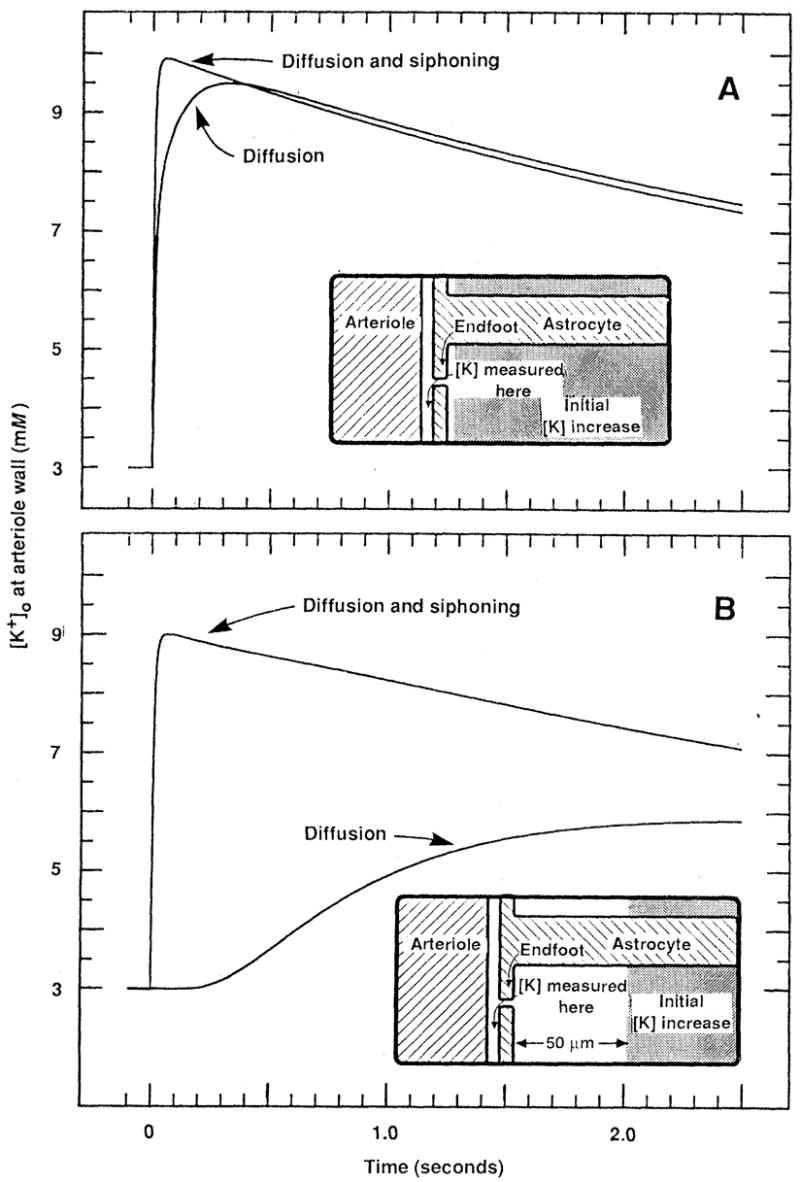
Local increases in neuronal activity within the brain lead to dilation of blood vessels and to increased regional cerebral blood flow. Increases in extracellular potassium concentration are known to dilate cerebral arterioles. Recent studies have suggested that the potassium released by active neurons is transported through astrocytic glial cells and released from their endfeet onto blood vessels. The results of computer simulations of potassium dynamics in the brain indicate that the release of potassium from astrocyte endfeet raises perivascular potassium concentration much more rapidly and to higher levels than does diffusion of potassium through extracellular space, particularly when the site of a potassium increase is some distance from the vessel wall. On the basis of this finding, it is proposed that the release of potassium from astrocyte endfeet plays an important role in regulating regional cerebral blood flow in response to changes in neuronal activity.
Figures
References
-
- Olesen J. Brain. 1971;94:635. - PubMed
-
- Brodersen P, et al. Arch Neural. 1973;28:334.
-
- Fox PT, Raichle ME. J Neurophysiol. 1984;51:1109. - PubMed
-
-
Regional cerebral blood How can. increase by 30 to 35% during normal sensory stimulation in humans [(4); Henriksen L, Paulson OB, Lassen NA. Eur J Nucl Med. 1981;6:487. by 111% during epileptic seizures in humans 3, and by 260% during seizures in experimental animals [ Plum F, Posner JF, Troy B. Arch Neural. 1968;18:1. Such rCBF increases occur rapidly [within seconds Leniger-Follert E. J Cereb Blood Flow Metab. 1984;4:150. although their precise time course is not known
-
Publication types
MeSH terms
Substances
Grants and funding
LinkOut - more resources
Full Text Sources
Other Literature Sources
Medical
