. 2022 Nov 8;7(21):e161244.
doi: 10.1172/jci.insight.161244.

Interleukin-37 protects against acinar cell pyroptosis in acute pancreatitis

Affiliations

Interleukin-37 protects against acinar cell pyroptosis in acute pancreatitis

Nan Ma et al. JCI Insight. .

Abstract

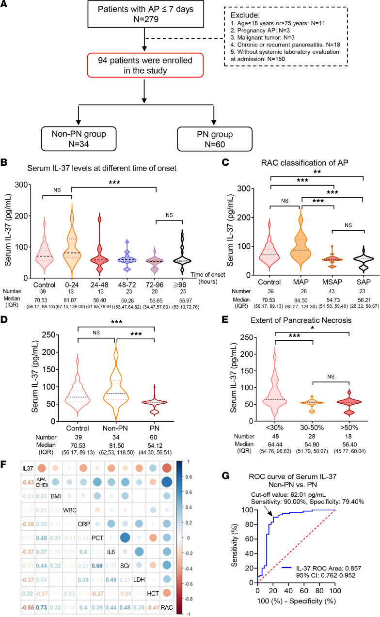
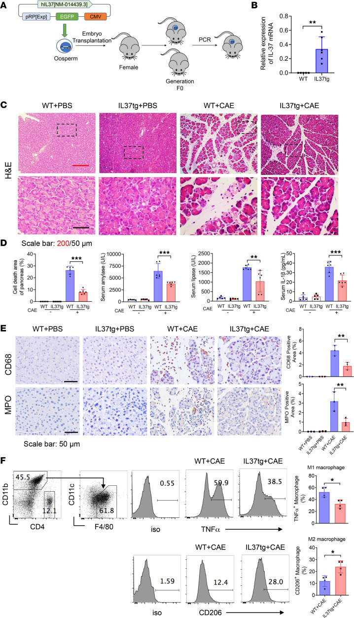
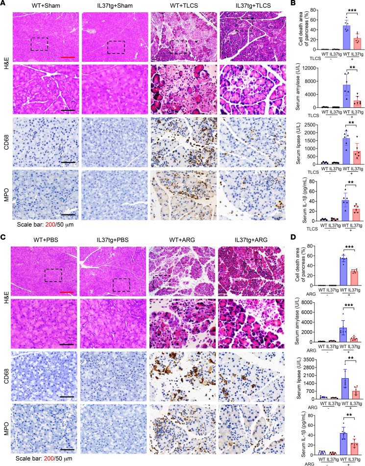
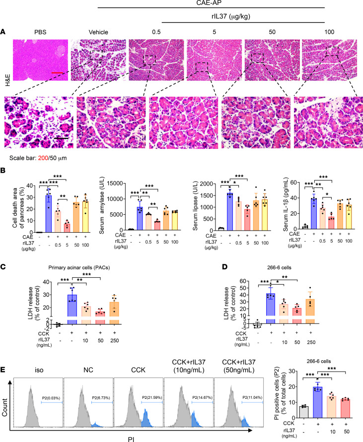
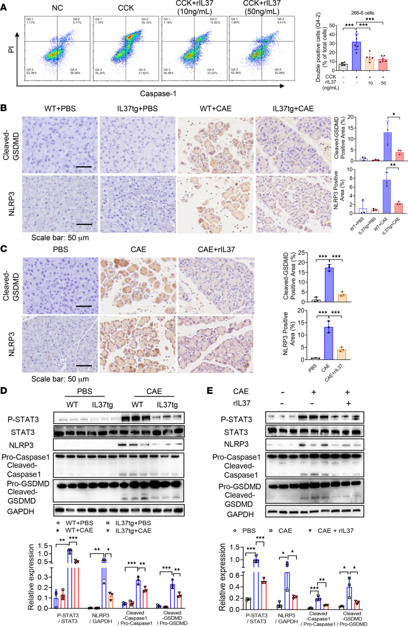
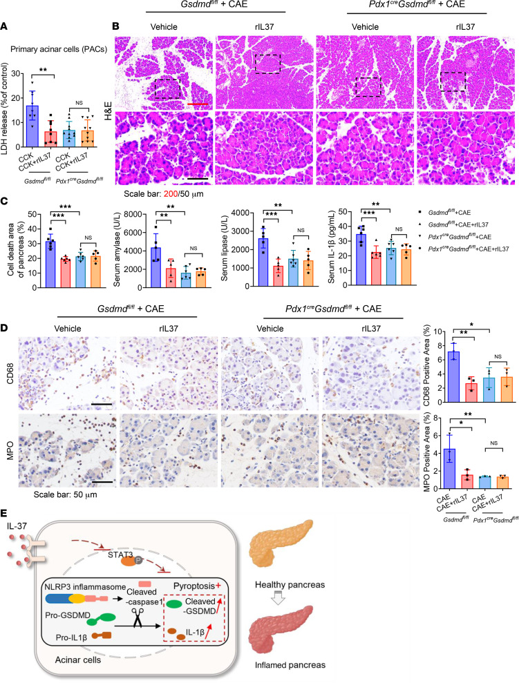
Acute pancreatitis (AP) is a local and/or systemic inflammatory disease that starts with acinar cell injury and necrosis; it has no effective medical treatment and thus remains a life-threatening condition. Interleukin-37 (IL-37), a natural immunomodulator, has demonstrated an antiinflammatory effect; however, the role of IL-37 in AP remains unknown. The serum IL-37 levels of 39 healthy controls and 94 patients with AP were measured. Cholecystokinin was applied to induce pancreatic acinar cell injury in vitro. Classical experimental AP models, such as caerulein, l-arginine, and taurolithocholic acid 3-sulfate disodium salt, were included in the in vivo study. A transgenic mouse model with the IL-37 gene and administration of recombinant IL-37 were used to further investigate the function of IL-37 in AP. Pancreas-specific gasdermin D-knockout (GSDMD-knockout) mice were used to explore the protective mechanism of IL-37. Our results showed that serum IL-37 levels in humans were negatively correlated with the severity of AP. Furthermore, IL-37-transgenic mice and supplementation with recombinant IL-37 could both protect against AP. Mechanistically, IL-37 was able to suppress pyroptosis of injured acinar cells, and specific depletion of GSDMD in the pancreas counteracted the protective effect of IL-37. Our study demonstrates that IL-37 protects against acinar cell pyroptosis in AP.

Keywords: Cytokines; Gastroenterology; Inflammation; Pharmacology.


Figures

Figure 1
Figure 1. Serum IL-37 levels in AP patients.
(A) Flowchart of the patients with AP in the clinical study. (B) Serum IL-37 levels in controls and AP patients at different times of onset. (C) ELISA analysis of serum IL-37 levels in healthy controls and different classification of AP patients (n = 94). (D) ELISA levels of control, non-PN, and PN groups. (E) Serum IL-37 levels in AP patients with different extents of pancreatic necrosis (PN). (F) Heatmap of correlation coefficients between serum IL-37 levels and other clinical indices at admission. (G) Receiver operating characteristic (ROC) curve analysis of serum IL-37 levels for diagnosing PN. Data were analyzed using Kruskal-Wallis test. Data are presented as median (IQR), and statistical significance is denoted as *P < 0.05, **P < 0.01, and ***P < 0.001. APACHE II, Acute Physiology and Chronic Health Evaluation II; CRP, C-reactive protein; HCT, hematocrit; LDH, lactate dehydrogenase; MAP, mild AP; MSAP, moderately severe AP; PCT, procalcitonin; RAC, Revised Atlanta Classification; SAP, severe AP; SCr, serum creatinine; WBC, white blood cells.
Figure 2
Figure 2. Reduced severity of experimental AP in IL37tg mice.
(A) Schematic diagram of the generation of human IL-37 transgenic mice (IL37tg). (BF) WT and IL37tg mice were injected with PBS and caerulein (CAE; 200 μg/kg, 1-hour intervals, 10 times total) to generate control and AP groups, respectively. Mice were harvested at 24 hours after the first injection. (B) Quantitative real-time PCR analysis of IL-37 mRNA expression in pancreatic tissues from IL37tg and WT littermates in normal condition. (C) H&E staining of pancreatic tissues from the indicated groups (n = 5–6 per group). Scale bars: 200 or 50 μm. (D) Percentages of pancreatic cell death area, and serum amylase, lipase, and IL-1β levels at 12 hours. (E) IHC staining of CD68 and MPO in pancreatic tissues, which marked macrophages and neutrophils, respectively (n = 3 per group). Scale bars: 50 μm. (F) Flow cytometry analysis of pancreatic leukocytes from the WT-CAE and IL37tg-CAE groups. Representative flow cytometry gating and the proportion of M1 (CD11b+F4/80+TNF-α+ of CD45.2+) and M2 (CD11b+F4/80+CD206+ of CD45.2+) macrophages (n = 4 per group). Experiments were repeated 3 times. Statistical comparisons were made using 1-way ANOVA or Student’s 2-tailed t test. Data are presented as mean ± SD, and statistical significance is denoted as *P < 0.05, **P < 0.01, and ***P < 0.001.
Figure 3
Figure 3. Protective effects of IL-37 in TLCS- and ARG-induced AP models.
(A and B) Taurolithocholic acid 3-sulfate disodium salt (TLCS; 2.5%, 2 mg/kg) was injected in retrograde into the pancreatic bile duct to induce AP in IL37tg and WT mice. The sham group was used as the control in TLCS-induced experimental AP models. (A) H&E staining of pancreatic tissues from the indicated groups at 24 hours, and representative IHC staining of CD68 and MPO in pancreatic tissues. Scale bars: 200 or 50 μm. (B) Percentages of pancreatic cell death area, and serum amylase, lipase, and IL-1β levels at 24 hours (n = 5–6 per group). (C and D) WT and IL37tg littermates were given i.p. injections of 8% l-arginine (ARG; 3.3 g/kg, hourly, 3 times total) to induce AP, while an equivalent volume of PBS was injected into mice from the control group. (C) H&E staining at 72 hours, and representative IHC staining of CD68 and MPO in pancreatic tissues. Scale bars: 200 or 50 μm. (D) Percentages of pancreatic cell death area, and serum amylase, lipase, and IL-1β levels (n = 5–6 per group). Statistical comparisons were made using 1-way ANOVA. Data are presented as mean ± SD, and statistical significance is denoted as **P < 0.01, and ***P < 0.001.
Figure 4
Figure 4. Recombinant IL-37 protects mice from experimental AP.
(A and B) C57BL/6J WT mice were injected with CAE to induce AP, and after 1 hour, the indicated doses of rIL37 were administered (n = 5–6 per group). Mice injected with PBS were used as normal controls. All mice were sacrificed and harvested after 24 hours. (A) H&E staining of pancreatic tissue from the indicated mice. Scale bars: 200 or 50 μm. (B) Percentages of pancreatic cell death area, and serum amylase, lipase, and IL-1β levels at 12 hours. (C and D) Pancreatic acinar cell line 266-6 or primary acinar cells were treated with CCK, together with gradient doses of rIL37 for 12 or 6 hours, respectively. The levels of LDH release are shown. (E) 266-6 cells were treated with CCK together with rIL37 for 12 hours; then cells were harvested for propidium iodide (PI) staining. Representative flow cytometry histograms and the proportion of PI-positive cells are shown. Experiments were repeated 3 times. Statistical comparisons were made using 1-way ANOVA. Data are presented as mean ± SD, and statistical significance is denoted as *P < 0.05, **P < 0.01, and ***P < 0.001. NC, normal control.
Figure 5
Figure 5. IL-37 suppresses GSDMD-mediated pyroptosis of acinar cells in AP.
(A) 266-6 cells were stimulated with CCK and treated or not treated with rIL37 (10 or 50 ng/mL) for 12 hours. Caspase-1 and PI staining was performed to identify pyroptotic cells. Representative flow cytometry gating and proportion of double-positive cells are shown. (B and C) Pancreatic tissues collected from mice were stained with anti-GSDMD and anti-NLRP3 antibodies. (B) Representative images and quantitative analysis for GSDMD and NLRP3 staining of pancreatic tissues from IL37tg and WT mice with or without CAE-AP (n = 3 per group). (C) Representative images and quantitative analysis for cleaved GSDMD and NLRP3 staining in pancreatic tissues of the AP group and the rIL37-treated group (5 μg/kg) (n = 3 per group). Scale bars: 50 μm. (D and E) Western blot analyses and quantification of the expression of GSDMD (pro- and cleaved), NLRP3, and caspase-1 (pro- and cleaved) in pancreatic tissues from the indicated mice (n = 3 per group). Experiments were repeated 3 times. Statistical comparisons were made using 1-way ANOVA. Data are presented as mean ± SD, and statistical significance is denoted as *P < 0.05, **P < 0.01, and ***P < 0.001.
Figure 6
Figure 6. Specific knockdown of GSDMD in pancreatic acinar cells neutralizes the protective effect of IL-37.
(A) Primary pancreatic acinar cells (PACs) were isolated from Gsdmdfl/fl and Pdx1cre Gsdmdfl/fl (GsdmdΔPan) mice. PACs were treated with CCK with or without rIL37 (50 ng/mL). The LDH levels are shown. (BD) AP was induced in Gsdmdfl/fl and Pdx1cre Gsdmdfl/fl mice with CAE, and rIL37 (5 μg/kg) was administered 1 hour after AP induction. (B) H&E staining of pancreatic tissues. Scale bars: 200 or 50 μm. (C) Percentages of pancreatic cell death area, and serum amylase, lipase, and IL-1β levels at 12 hours (n = 5–6 per group). (D) Representative images and quantitative analysis for IHC staining of CD68 and MPO (n = 3 per group). Scale bars: 50 μm. (E) Schematic diagram showing that IL-37 protects against AP through suppression of the pyroptosis pathway. Statistical comparisons were made using 1-way ANOVA. Data are presented as mean ± SD, and statistical significance is denoted as *P < 0.05, **P < 0.01, and ***P < 0.001.

Similar articles

Cited by

References

    1. Tenner S, et al. American College of Gastroenterology guideline: management of acute pancreatitis. Am J Gastroenterol. 2013;108(9):1400–1415. doi: 10.1038/ajg.2013.218. - DOI - PubMed
    1. NCD Risk Factor Collaboration (NCD-RisC) Worldwide trends in body-mass index, underweight, overweight, and obesity from 1975 to 2016: a pooled analysis of 2416 population-based measurement studies in 128·9 million children, adolescents, and adults. Lancet. 2017;390(10113):2627–2642. doi: 10.1016/S0140-6736(17)32129-3. - DOI - PMC - PubMed
    1. Petrov MS, Yadav D. Global epidemiology and holistic prevention of pancreatitis. Nat Rev Gastroenterol Hepatol. 2019;16(3):175–184. doi: 10.1038/s41575-018-0087-5. - DOI - PMC - PubMed
    1. Schepers NJ, et al. Impact of characteristics of organ failure and infected necrosis on mortality in necrotising pancreatitis. Gut. 2019;68(6):1044–1051. doi: 10.1136/gutjnl-2017-314657. - DOI - PubMed
    1. Van Dijk SM, et al. Acute pancreatitis: recent advances through randomised trials. Gut. 2017;66(11):2024–2032. doi: 10.1136/gutjnl-2016-313595. - DOI - PubMed

Publication types