. 2022 Nov 8;7(21):e161214.
doi: 10.1172/jci.insight.161214.

DIO3 protects against thyrotoxicosis-derived cranio-encephalic and cardiac congenital abnormalities

Affiliations

DIO3 protects against thyrotoxicosis-derived cranio-encephalic and cardiac congenital abnormalities

M Elena Martinez et al. JCI Insight. .

Abstract

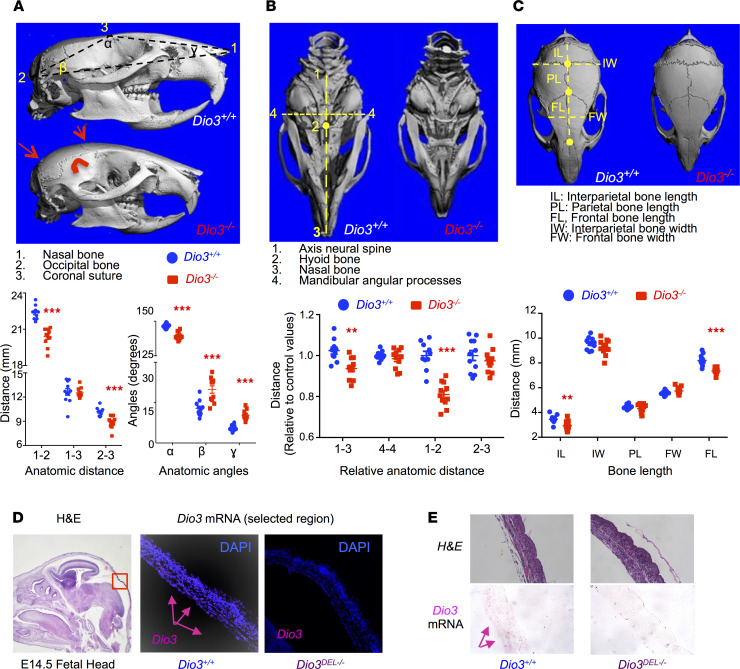
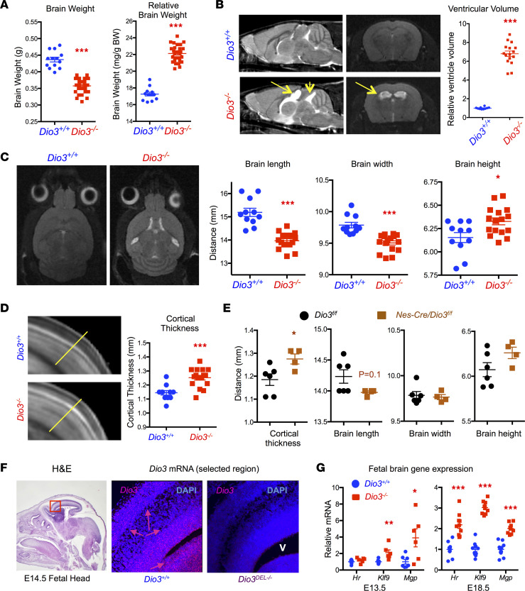
Maternal hyperthyroidism is associated with an increased incidence of congenital abnormalities at birth, but it is not clear which of these defects arise from a transient developmental excess of thyroid hormone and which depend on pregnancy stage, antithyroid drug choice, or unwanted subsequent fetal hypothyroidism. To address this issue, we studied a mouse model of comprehensive developmental thyrotoxicosis secondary to a lack of type 3 deiodinase (DIO3). Dio3-/- mice exhibited reduced neonatal viability on most genetic backgrounds and perinatal lethality on a C57BL/6 background. Dio3-/- mice exhibited severe growth retardation during the neonatal period and cartilage loss. Mice surviving after birth manifested brain and cranial dysmorphisms, severe hydrocephalus, choanal atresia, and cleft palate. These abnormalities were noticeable in C57BL/6J Dio3-/- mice at fetal stages, in addition to a thyrotoxic heart with septal defects and thin ventricular walls. Our findings stress the protecting role of DIO3 during development and support the hypothesis that human congenital abnormalities associated with hyperthyroidism during pregnancy are caused by transient thyrotoxicosis before clinical intervention. Our results also suggest thyroid hormone involvement in the etiology of idiopathic pathologies including cleft palate, choanal atresia, Chiari malformations, Kaschin-Beck disease, and Temple and other cranio-encephalic and heart syndromes.

Keywords: Development; Embryonic development; Endocrinology; Mouse models.


Conflict of interest statement

Conflict of interest: The authors have declared that no conflict of interest exists.

Figures

Figure 1
Figure 1. Neonatal growth and cartilage deficits in Dio3–/– mice.
(A) Neonatal growth in Dio3+/+ and Dio3–/– littermates (n = 26–49 per data point). (B) P5 serum levels of growth hormone (GH) and insulin-like growth factor 1 (IGF-1). (C) Representative photographs taken from skeletal preparations of P11 mice showing reduced cartilage in several anatomic locations. Original magnification, 12.5×, 20×, and 10×. *, **, and *** indicate P < 0.05, 0.01, and 0.001, respectively, as determined by the Student’s 2-tailed t test. Error bars represent the standard error of the mean.
Figure 2
Figure 2. Cranial dysmorphisms in Dio3–/– mice.
(A) Lateral-view microCT images and anatomic parameters of adult mouse crania. (B) Ventral-view microCT images and anatomic parameters of adult mouse crania. (C) Top-view microCT images and anatomic parameters of adult mouse crania. (D and E) Dio3 mRNA expression in E14.5 fetal calvaria (pink arrows) as determined by in situ hybridization (RNAscope). MicroCT images shown represent female littermates of each genotype. Original magnification, 10× (fetal head), 200× (calvaria), 200× (E). **, *** indicate P < 0.01 and 0.001, respectively, as determined by the Student’s 2-tailed t test (n = 8–11 per genotype, including at least 4 males and 4 females).
Figure 3
Figure 3. Craniofacial and palate abnormalities in Dio3–/– mice.
(A) Representative caudo-rostral (light blue arrow) MR slice image of adult male mice (n = 4 Dio3+/+, 4 Dio3–/–) revealing choanal atresia (red arrows) in Dio3–/– mice. (B) Representative MR images of E14.5 and P15 mice showing cleft palate and palate defects (red arrows) in Dio3–/– mice (n = 5 E14.5 Dio3+/+, 4 E14.5 Dio3–/–, 3 P15 Dio3+/+, 3 P15 Dio3–/–). (C) In situ hybridization (RNAscope) demonstrating strong Dio3 mRNA expression in the palates of E14.5 fetuses (pink arrows) (H&E image is the same as in Figure 2 and provided for anatomical reference). Original magnification, 10× (fetal head) and 200× (palate). (D) Expression of T3-regulated genes in E18.5 craniofacial tissue as determined by real-time quantitative PCR (qPCR). **, *** indicate P < 0.01 and 0.001, respectively, as determined by the Student’s 2-tailed t test (n = 11 Dio3+/+, 9 Dio3–/–). OB, olfactory bulb; P, palate; T, tongue.
Figure 4
Figure 4. Brain dysmorphisms and hydrocephalus in Dio3–/– mice.
(A) Brain weight and relative brain weight of adult mice (both sexes are included). (B) Representative rostral-caudal and coronal MR imaging of adult male brains exhibiting severe hydrocephalus (yellow arrows) in Dio3–/– mice and quantification of total ventricular volume (n = 10 Dio3+/+, 16 Dio3–/–). (C) Representative top-view MR images of adult brain and brain dimensions in Dio3+/+ and Dio3–/– littermates (n = 10, 16). (D) Representative MR image and measured line and quantification of brain cortical thickness in adult mice (n = 10, 16). (E) Measurements of brain dimensions and cortical thickness in adult mice with neural tissue-specific DIO3 deficiency (n = 6, 4). (F) In situ hybridization (RNAscope) demonstrating robust Dio3 mRNA expression in E14.5 fetal brain during corticogenesis (pink arrows), especially in the periventricular area and the newly formed outer cortex layer as determined by in situ hybridization (RNAscope) (H&E image is the same as in Figure 2 and provided for anatomical reference). Original magnification, 10× (fetal head) and 200× (brain cortex). (G) Expression of T3-regulated genes in E13.5 and E18.5 brains as determined by real-time qPCR. *, **, *** indicate P < 0.05, 0.01, and 0.001, respectively, as determined by the Student’s 2-tailed t test. V, ventricle.
Figure 5
Figure 5. Fetal heart defects in Dio3–/– fetuses.
(A) Representative MR images (n = 5 Dio3+/+, 4 Dio3–/–) illustrating congenital defects (yellow arrows) in Dio3–/– E14.5 fetal hearts. (B) H&E staining (n = 3, 4) illustrating reduced ventricular wall thickness (yellow lines) in the right ventricle of a Dio3–/– E14.5 fetal heart. (C) Representative MR images (n = 13 Dio3+/+, 6 Dio3+/–, 9 Dio3–/–) illustrating congenital defects (yellow arrows) in Dio3+/– and Dio3–/– E18.5 fetal hearts. Scale bar, 150 microns. ASD and VSD, atrial and ventricular septal defect, respectively; yellow lines indicate ventricular wall thickness; RV, right ventricle; LV, left ventricle; RA, right atria.
Figure 6
Figure 6. Altered gene expression in the heart of Dio3–/– fetuses.
(A) Volcano plot of gene expression in the Dio3–/– E14.5 fetal heart. (B) Heatmap of 364 differentially expressed genes (P < 0.05) in the E14.5 fetal heart (n = 4, values for each gene were normalized to 1 as the mean of all samples). (C) Top upstream regulators (based on P values and activation scores) whose pathways are altered in Dio3–/– E14.5 fetal heart as determined by Ingenuity analyses. Dotted lines indicate the threshold established by the software to indicate substantial activation/inactivation (D) qPCR validation of differentially expressed genes in independent RNA samples isolated from E14.5 fetal hearts (n = 8, 6). (E) Expression of the same genes in E18.5 fetal hearts (n = 11, 8). *** indicates P < 0.001, as determined by the Student’s 2-tailed t test.
Figure 7
Figure 7. Systemic thyrotoxicosis in E18.5 Dio3–/– fetuses.
(A and B) Serum T3 and T4, respectively, in E18.5 fetuses. Dotted lines indicate the sensitivity of the assays as determined by 2 standard deviations above the zeros. (A) n = 11, 11, 13, and 7. (B) n = 8, 12, 18, and 7. (CF) Expression of selected T3-responsive genes in liver, intestine, lung, and placenta of E18.5 fetuses (n = 11 Dio3+/+, 8 Dio3–/–). **, *** indicate P < 0.01 and 0.001, respectively, as determined by 1-way ANOVA and Tukey’s post hoc test (A) or by the Student’s 2-tailed t test.
Figure 8
Figure 8. Palate and cardiac defects and altered fetal growth trajectories.
(A) Representative MR images (n = 5, 10, 4) illustrating craniofacial, palate, and cardiac defects (yellow arrows) in littermate E14.5 fetuses of the 3 genotypes. (B) Correlation of cranial length and weight in E14.5 fetuses and association with congenital defects. Data included E14.5 fetuses of all genotypes and also fetuses generated by wild-type parents. (C) Photograph illustrating growth retardation in a Dio3–/– E10.5 embryos compared to a Dio3+/+ littermate. (D and E) E13.5 and E18.5 weight, respectively, of fetuses of different genotypes and generated by heterozygous crosses or by WT crosses (n = 6–43). (F and G) E13.5 and E18.5 weight, respectively, of placentas from fetuses of different genotypes and by heterozygous crosses or by WT crosses (n = 6–24). **, *** indicate P < 0.01 and 0.001, respectively, as determined by 1-way ANOVA and Tukey’s post hoc test.

Similar articles

Cited by

References

    1. Mulvihill JJ. Craniofacial syndromes: no such thing as a single gene disease. Nat Genet. 1995;9(2):101–103. doi: 10.1038/ng0295-101. - DOI - PubMed
    1. Findley TO, et al. Congenital heart defects and copy number variants associated with neurodevelopmental impairment. Am J Med Genet A. 2022;188(1):13–23. doi: 10.1002/ajmg.a.62484. - DOI - PubMed
    1. Helm BM, et al. Genetic evaluation of inpatient neonatal and infantile congenital heart defects: new findings and review of the literature. Genes (Basel) 2021;12(8):1244. doi: 10.3390/genes12081244. - DOI - PMC - PubMed
    1. Mai CT, et al. National population-based estimates for major birth defects, 2010-2014. Birth Defects Res. 2019;111(18):1420–1435. doi: 10.1002/bdr2.1589. - DOI - PMC - PubMed
    1. Legrand J. Effects of thyroid hormones on central nervous system development. In: Yanai J, ed. Neurobehavioral Teratology. Elsevier; 1984:331–363.

Publication types