. 2022 Sep 27;17(9):e0275163.
doi: 10.1371/journal.pone.0275163. eCollection 2022.

Peripheral Exudative Hemorrhagic Chorioretinopathy with and without treatment-Clinical and multimodal imaging characteristics and prognosis

Affiliations

Peripheral Exudative Hemorrhagic Chorioretinopathy with and without treatment-Clinical and multimodal imaging characteristics and prognosis

Margarita Safir et al. PLoS One. .

Abstract

Purpose: To describe clinical and imaging characteristics of patients with Peripheral Exudative Hemorrhagic Chorioretinopathy (PEHCR), prognosis and treatment response.

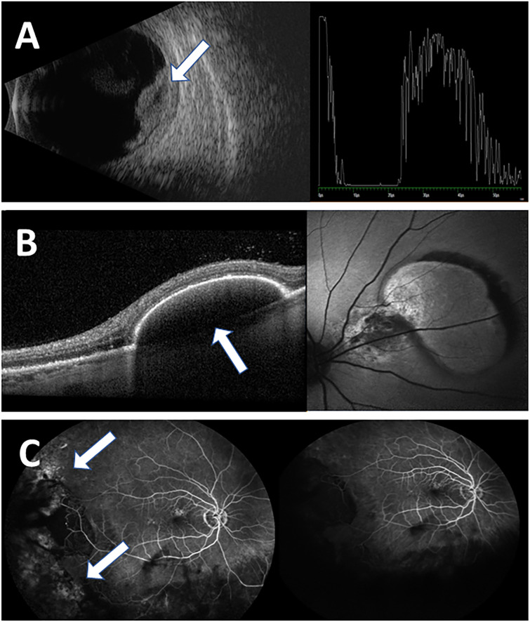
Methods: In this retrospective cohort study medical records of patients diagnosed with PEHCR in a tertiary medical center between 2008 and 2018 were reviewed. Collected data included demographics, medical history, ophthalmologic examination and multi-modal imaging including fundus autofluorescence, optical coherence tomography (OCT), ultrasound (US), fluorescein angiography and indocyanine green angiography when available. Bevacizumab treatment results were analyzed when applied.

Results: 35 eyes of 32 patients were included, with a female predominance (56.25%) and an average age of 79.0±9.87 years at presentation. Most common OCT and US findings were subretinal mass (68.75%), pigment epithelial detachment (30.00%) and atrophic changes (21.86%). Median follow-up period was 18.00 months (range 0-102). Visual acuity (VA) remained stable (39.29%) or improved (25.00%) in most cases available for follow-up. Treatment with intravitreal bevacizumab induced a statistically significant clinical resolution in 88.89% of eyes available for follow-up (8/9 eyes) (p = 0.02).

Conclusions: PEHCR is presented with high clinical variability and generally good prognosis. This is the first publication demonstrating a statistically significant clinical resolution of disease following intravitreal bevacizumab injections.


Conflict of interest statement

The authors have declared that no competing interests exist.

Figures

Fig 1
Fig 1. Clinical subtypes of PEHCR.
Three different subtypes were observed in our cohort (arrows): A, Hemorrhagic; B, Exudative; C combined.
Fig 2
Fig 2. Main imaging findings of PEHCR in our cohort.
The imaging modalities used for evaluation and follow-up of our patients included mostly ultrasound (US) and optical coherence test (OCT), in addition fluorescein angiography (FA) and Indocyanine Green Angiography (ICGA) in several patients. A, US image, depicting a choroidal mass (B-scan right side) with a high internal reflectivity (A-mode left side, arrow); B, The most common finding at presentation on OCT was PED (30%) (arrow) C, FA imaging reveals mostly hyperflourescent lesions (arrows) (55.56%), with no evidence of CNV.
Fig 3
Fig 3. Clinical appearance and OCT findings before and after anti-VEGF treatment in a patient with macula threatening PEHCR.
Color fundus photography of the same eye A before and B after 10 anti-VEGF intravitreal injections. A demonstrating a large elevated hemorrhagic lesion at the superior arcade consistent with hemorrhagic PEHCR (white arrow) old fibrotic lesion (yellow arrow), B resolution of the bleeding, clinical shrinkage of the lesion and residual atrophic changes (white arrow). C+D, OCT of the same eye at the level of the aforementioned lesion before and after treatment: C demonstrating a large hemorrhagic PED (white arrow) with no subretinal fluid. D After treatment the PED shrunk in size with a residual subretinal hyper-reflective layer consistent with scaring. No subretinal fluid is seen.

References

    1. Reese A.B. and Jones I.S. (1961) Hematomas Under the Retinal Pigment Epithelium. Trans Am Ophthalmol Soc, 1961. 59: p. 43–79. ; PMCID: PMC1316398. - PMC - PubMed
    1. Sliva V.B. and J Brockhurst R. (1976) Hemorrhagic detachment of the peripheral retinal pigment epithelium. Arch Ophthalmol, 1976. 94(8): p. 1295–1300. doi: 10.1001/archopht.1976.03910040167009 - DOI - PubMed
    1. Kingham J.D (1978) Hemorrhagic detachment of the peripheral retinal pigment epithelium. Ann Ophthalmol, 1978. 10(2): p. 175–8. . - PubMed
    1. Bloome M.A. and Ruiz R.S. (1978) Massive spontaneous subretinal hemorrhage. Am J Ophthalmol, 1978. 86(5): p. 630–7. doi: 10.1016/0002-9394(78)90181-2 - DOI - PubMed
    1. Mantel I., Uffer S., and Zografos L. (2009) Peripheral exudative hemorrhagic chorioretinopathy: a clinical, angiographic, and histologic study. Am J Ophthalmol, 2009. 148(6): p. 932–8 e1. doi: 10.1016/j.ajo.2009.06.032 - DOI - PubMed