Review
. 2023 Feb 1;29(3):513-520.
doi: 10.1158/1078-0432.CCR-21-1258.

NASH and Hepatocellular Carcinoma: Immunology and Immunotherapy

Affiliations
Review

NASH and Hepatocellular Carcinoma: Immunology and Immunotherapy

Matthias Pinter et al. Clin Cancer Res. .

Abstract

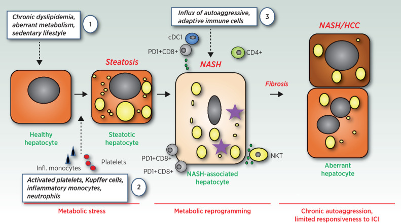
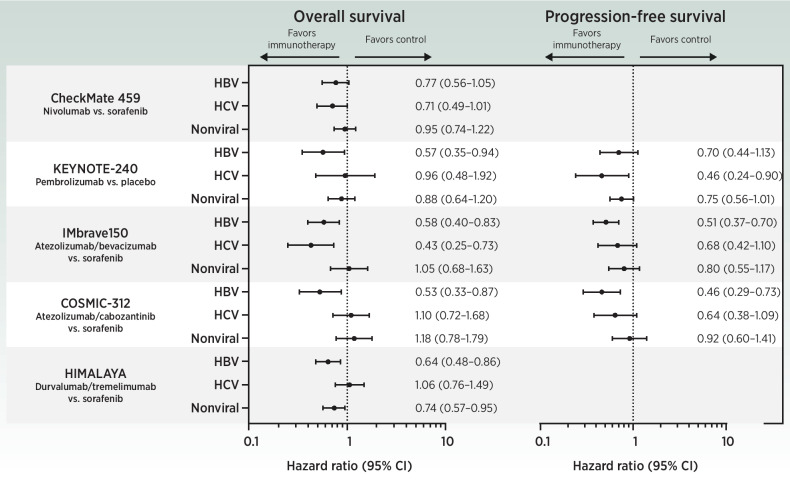
The last 10 years have revolutionized our basic understanding of nonalcoholic fatty liver disease and consequent liver cancer. It has become clear that several innate and adaptive immune cells play an important role in initiating, maintaining, or exacerbating nonalcoholic steatohepatitis (NASH)-a disease that has been recently defined as autoaggressive. Despite improved disease management aimed at reducing the progression of fibrosis, NASH is set to become a leading cause for hepatocellular carcinoma (HCC). Preliminary data from preclinical studies suggest that immunotherapy efficacy may be reduced in NASH-related HCC compared with viral HCC; however, conclusive evidence supporting clinical translation of these findings is lacking. Comprehensive clinical and immunologic phenotyping of mechanisms linking NASH progression with carcinogenesis and therapeutic resistance is key to prevent progression to cirrhosis, improve monitoring and stratification of NASH according to predicted cancer risk, and ultimately increase survival of patients with NASH-HCC. In this review, we summarize the state of the art in the field of NASH and NASH-HCC with focus on immunobiology. We discuss preclinical and clinical findings underpinning NASH as an immunologically distinct pro-tumorigenic disease entity, and explore areas of potential therapeutic vulnerabilities in NASH-associated HCC.


Figures

Figure 1. On the role of adaptive and innate immune cells in NASH and NASH-HCC transition (1). Chronic dyslipedimia and sedentary lifestyle cause aberrant heapticyte metabolism, increased ER- and mitochondrial stress. This over time leads to a metabolic catastrophy in hepatocytes – (2) driving first local inflammation by Kupffer cells, hepatocyte damage and influx of platelets, neutrophils and inflammatory monocytes. Over time different adaptive immune cells (e.g., CD8+PD-1+ T cells) as well as innate immune cells (e.g., CXCR1+ cDCs) infiltrate the liver and support the development of autoaggressive CD8+ T cells (3). This type of inflammation, either triggered by CD8+PD-1+ T cell or NKT-cell secreted cytokines (e.g., LIGHT) or cell–cell contact with hepatocytes causes a downregulation of the metabolic machinery in hepatocytes and a consequent increase of lipid accumulation, lipid toxicity and further liver damage. NASH as well as the autoaggressive T cells further develop and induce a fibrotic responses as well as an inflammatory hepatic environment that in the context of NASH-HCC does not well respond to ICI.
Figure 1.
On the role of adaptive and innate immune cells in NASH and NASH-HCC transition (1). Chronic dyslipidemia and sedentary lifestyle cause aberrant hepatocyte metabolism, increased ER-, and mitochondrial stress. This over time leads to a metabolic catastrophe in hepatocytes – (2) driving first local inflammation by Kupffer cells; hepatocyte damage; and influx of platelets, neutrophils, and inflammatory monocytes. Over time different adaptive immune cells (e.g., CD8+PD-1+ T cells) as well as innate immune cells (e.g., CXCR1+ cDCs) infiltrate the liver and support the development of autoaggressive CD8+ T cells (3). This type of inflammation, either triggered by CD8+PD-1+ T cell or NKT-cell secreted cytokines (e.g., LIGHT) or cell–cell contact with hepatocytes, causes a downregulation of the metabolic machinery in hepatocytes and a consequent increase of lipid accumulation, lipid toxicity, and further liver damage. NASH as well as the autoaggressive T cells further develop and induce a fibrotic response as well as an inflammatory hepatic environment that in the context of NASH-HCC does not respond well to ICI.
Figure 2. Forest plots of overall survival and progression-free survival according to underlying liver disease etiology. Data are shown from phase III trials testing immunotherapies in patients with advanced HCC.
Figure 2.
Forest plots of overall survival and progression-free survival according to underlying liver disease etiology. Data are shown from phase III trials testing immunotherapies in patients with advanced HCC.

References

    1. Younossi Z, Anstee QM, Marietti M, Hardy T, Henry L, Eslam M, et al. . Global burden of NAFLD and NASH: trends, predictions, risk factors, and prevention. Nat Rev Gastroenterol Hepatol 2018;15:11–20. - PubMed
    1. Huang DQ, El-Serag HB, Loomba R. Global epidemiology of NAFLD-related HCC: trends, predictions, risk factors, and prevention. Nat Rev Gastroenterol Hepatol 2021;18:223–38. - PMC - PubMed
    1. Villanueva A. Hepatocellular carcinoma. N Engl J Med 2019;380:1450–62. - PubMed
    1. Llovet JM, Kelley RK, Villanueva A, Singal AG, Pikarsky E, Roayaie S, et al. . Hepatocellular carcinoma. Nat Rev Dis Primers 2021;7:6. - PubMed
    1. Gehrke N, Schattenberg JM. Metabolic inflammation—a role for hepatic inflammatory pathways as drivers of comorbidities in nonalcoholic fatty liver disease? Gastroenterology 2020;158:1929–47. - PubMed

Publication types

MeSH terms