. 2023 Jul:49:159-173.
doi: 10.1016/j.jare.2022.09.007. Epub 2022 Sep 24.

Paclitaxel-loaded ginsenoside Rg3 liposomes for drug-resistant cancer therapy by dual targeting of the tumor microenvironment and cancer cells

Affiliations

Paclitaxel-loaded ginsenoside Rg3 liposomes for drug-resistant cancer therapy by dual targeting of the tumor microenvironment and cancer cells

Ying Zhu et al. J Adv Res. 2023 Jul.

Abstract

Introduction: Inherent or acquired resistance to paclitaxel (PTX) is a pivotal challenge for chemotherapy treatment of multidrug-resistant (MDR) breast cancer. Although various targeted drug-delivery systems, including nanoparticles and liposomes, are effective for MDR cancer treatment, their efficacy is restricted by immunosuppressive tumor microenvironment (TME).

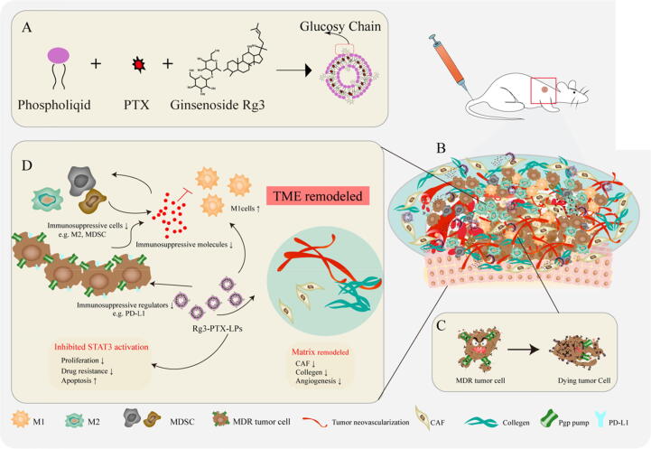
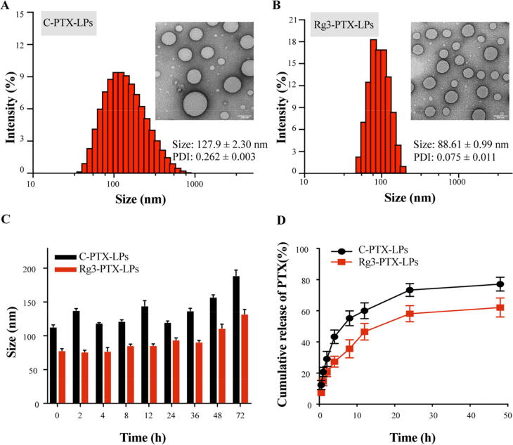
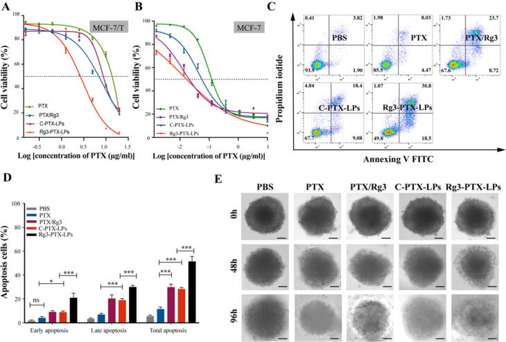
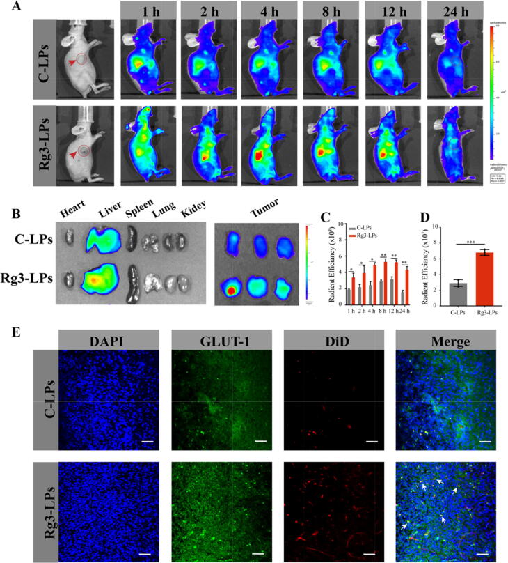
Methods: Ginsenosides Rg3 was used to formulate unique Rg3-based liposomes loaded with PTX to establish Rg3-PTX-LPs, which were prepared by the thin-film hydration method. The stability of the Rg3-PTX-LPs was evaluated by particle size analysis through dynamic light scattering. The active targeting effect of Rg3-based liposomes was examined in an MCF-7/T xenograft model by an in a vivo imaging system. To evaluate the antitumor activity and mechanism of Rg3-PTX-LP, MTT, apoptosis assays, TAM regulation, and TME remodeling were performed in MCF-7/T cells in vitro and in vivo.

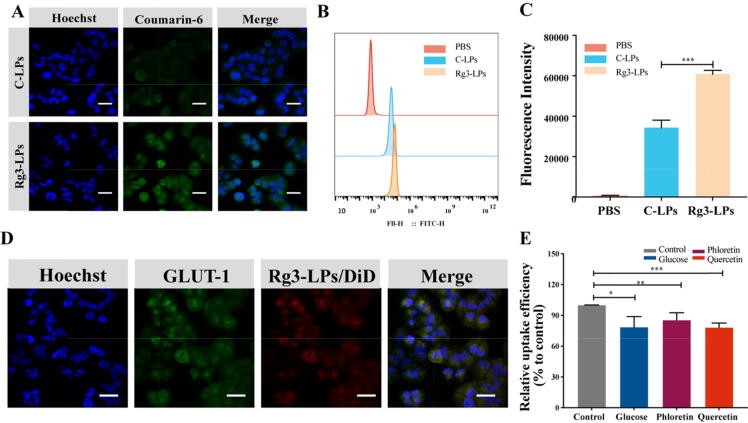
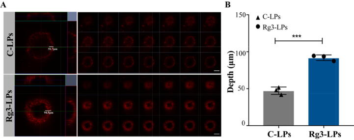
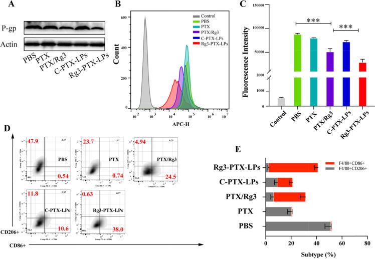
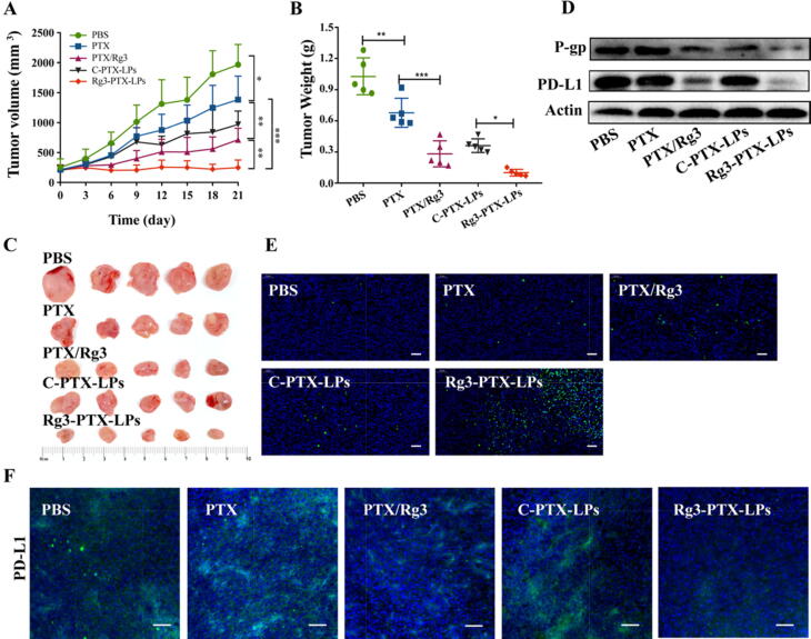
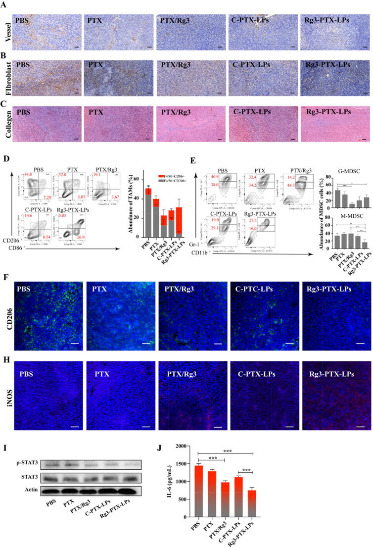
Results: Rg3-PTX-LPs could specifically distribute to MCF7/T cancer cells and TME simultaneously, mainly through the recognition of GLUT-1. The drug resistance reversing capability and in vivo antitumor effect of Rg3-PTX-LPs were significantly improved compared with conventional cholesterol liposomes. The TME remodeling mechanisms of Rg3-PTX-LPs included inhibiting IL-6/STAT3/p-STAT3 pathway activation to repolarize protumor M2 macrophages to antitumor M1 phenotype, suppressing myeloid-derived suppressor cells (MDSCs), decreasing tumor-associated fibroblasts (TAFs) and collagen fibers in TME, and promoting apoptosis of tumor cells. Hence, through the dual effects of targeting tumor cells and TME remodeling, Rg3-PTX-LPs achieved a high tumor inhibition rate of 90.3%.

Conclusion: Our multifunctional Rg3-based liposome developed in the present study offered a promising strategy for rescuing the drug resistance tumor treatment.

Keywords: Ginsenoside Rg3; Liposomes; MCF-7/T tumor; Multidrug resistance; Paclitaxel; Tumor microenvironment remodeling.


Conflict of interest statement

Declaration of Competing Interest The authors declare that they have no known competing financial interests or personal relationships that could have appeared to influence the work reported in this paper.

Figures

None
Graphical abstract
Scheme 1
Scheme 1
A schematic illustration of Rg3-PTX-LPs to reverse cancer drug resistance. (A) Ginsenoside Rg3 substitutes cholesterol as the membrane material in preparing Rg3-PTX-LPs. (B) The immunosuppressive and drug resistance tumor microenvironment. (C) Rg3-PTX-LPs downregulate the expression of Pgp and reduce the efflux of PTX from the cell. (D) Rg3-PTX-LPs remodel TME by inhibiting IL-6/STAT3/p-STAT3 pathway activation to repolarize protumor M2 macrophages to antitumor M1 phenotype, suppressing myeloid-derived suppressor cells (MDSCs), decreasing tumor-associated fibroblasts (TAFs) and collagen fibers in TME, and Inhibiting tumor angiogenesis
Fig. 1
Fig. 1
Characterization of conventional cholesterol-based liposomes loaded with PTX (C-PTX-LPs) and ginsenoside Rg3-based liposomes loaded with PTX (Rg3-PTX-LPs). (A, B) Size distribution and morphology (inset) of C-PTX-LPs and Rg3-PTX-LPs as determined by dynamic light scattering (DLS) and TEM, respectively. (C) Liposome stability of the C-PTX-LPs and the Rg3-PTX-LPs was evaluated by measuring their size after incubation in PBS containing 10% FBS at 37 °C for 72 h. (D) PTX release profiles from C-PTX-LPs and Rg3-PTX-LPs, measured in PBS containing 10% FBS.
Fig. 2
Fig. 2
Fluorescence analysis of the cellular uptake of courmarine-6-labeled C-LPs and Rg3-LPs in MCF-7/T cells. (A) Confocal fluorescence images of MCF-7/T cells in the presence of C-LPs or Rg3-LPs. (B) FACS analysis of cellular uptake of the liposomes. (C) Cellular uptake efficiency of C-LPs and Rg3-LPs. (D) In vitro binding of Rg3-LPs to MCF-7/T cells. Representative fluorescence images of Rg3-LPs and GLUT-1 localization in MCF-7/T cells. (E) Cellular uptake of Rg3-LPs in the presence of GLUT inhibitors relative to the control as determined by flow cytometry. Scale bar, 50 μm, Data are mean ± SD, n = 3. *p < 0.05, **p < 0.01, ***p < 0.001.
Fig. 3
Fig. 3
Intra-tumoral accumulation of C-LPs and Rg3-LPs in 3D MCF-7/T cell tumor spheroids in vitro. (A) Fluorescence analysis with DiD by confocal microscopy. Fluorescence intensity is a function of the accumulation of C-LPs and Rg3-LPs. A higher fluorescence intensity was observed for Rg3-LPs than C-LPs. Scale bar, 200 μm. (B) Quantitative analysis of the penetration depth of C-LPs and Rg3-LPs in MCF-7/T spheroids. The panel shows confocal z-axis, continuous top-down scanning layers that are 5 mm for each depth level. Data are mean ± SD, n = 3, *p < 0.05, **p < 0.01, ***p < 0.001.
Fig. 4
Fig. 4
In vitro anticancer activities of Rg3-PTX-LPs. (A) Cytotoxicity of different PTX formulations in MCF-7/T cells by MTT assay (n = 6). (B) Cytotoxicity of different PTX formulations in MCF-7 cells by MTT assay (n = 6). (C, D) Representative scatter plots of Annexin V/PI analysis of MCF-7/T cells treated with the various PTX formulations. The highest level of total apoptosis was observed for Rg3-PTX-LPs. (E) Representative microscopic images of MCF-7/T tumor spheroids after treatment with the different PTX formulations at 0 h, 48 h, 96 h. Scale bar, 100 μm. Data are mean ± SD, n = 3, *p < 0.05, **p < 0.01, ***p < 0.001.
Fig. 5
Fig. 5
In vitro P-gp and PD-L1 downregulation, and M2 phenotype macrophage re-education after different treatment. (A) P-gp expression in MCF-7/T cells. (B, C) FACS analysis of PD-L1 expression in MCF-7/T cells. (D, E) FACS analysis of CD206 and CD86 expression in M2 cells. Data are mean ± SD, n = 3, *p < 0.05, **p < 0.01, ***p < 0.001.
Fig. 6
Fig. 6
In vivo distribution and tumor targeting of liposomes in a breast tumor mouse model. (A) In vivo fluorescence imaging of MCF-7/T subcutaneous xenograft tumor-bearing nude mice treated with DiR-loaded liposomes at the indicated time points. (B) In vivo radiant efficiency in main organs and tumors dissected from the nude mice. (C) Fluorescence imaging of dissected major organs and tumors. (D) Ex vivo radiant efficiency at the tumors. (E) Confocal images of the liposome distribution and colocalization of liposomes with GLUT-1 in tumor sections of MCF-7/T xenograft-bearing mice after systemic administration of DiD-loaded liposomes. Red: DiD, blue: nuclei, green: GLUT-1 antibody. Bar: 50 μm. Data are mean ± SD, n = 3, *p < 0.05, **p < 0.01, ***p < 0.001.
Fig. 7
Fig. 7
Treatment efficacies in nude mice bearing MCF-7/T subcutaneous xenograft tumors. (A) Tumor growth curves. (B) Tumor weights. (C) Tumor sizes. (D) Western blotting of P-gp and PD-L1 expression in tumor tissues. (E) TUNEL assay of apoptosis in subcutaneous MCF-7/T tumors. Magnification, 10×, scale bar, 100 μm. (F) Expression of PD-L1 in MCF-7/T tumors tissues, scale bar, 50 μm. Data are mean ± SD, n = 6, *p < 0.05, **p < 0.01, ***p < 0.001.
Fig. 8
Fig. 8
Rg3-PTX-LPs remodel the TME structure of MCF-7/T tumors in an in vivo mice model. (A) At the end of the study, vessels in MCF-7/T tumors were processed for immunohistochemical staining of CD31. (B) At the end of the study, TAFs in MCF-7/T tumor tissues were processed for immunohistochemical staining for α-SMA. (C) At the end of the study, collagen fibers in MCF-7/T tumor tissues were stained with Masson’s trichrome. (Masson’s trichrome stains muscle red and collagen blue) (D) Percentages of TAM populations with specific macrophage markers (M1-type, CD11b+/F4/80+/CD86+, and M2-type, CD11b+/F4/80+/ CD206+) in tumor tissues, as detected by flow cytometry. (E) Flow-cytometry gating and quantitative analysis of CD11b+/Gr-1+ MDSCs in tumors. Double-positive cells comprise two populations, including Gr-1highCD11b+ granulocytic (G-MDSCs) and Gr-1intCD11b+ monocytic (M−MDSCs) MDSC subsets. (F) CD206 expression (M2 macrophages) in MCF-7/T tumors tissues assessed by confocal microscopy. (H) iNOS expression (M1 macrophages) in MCF-7/T tumors tissues assessed by confocal microscopy. (I) Western blotting of STAT3 and p-STAT3 expression in tumor tissues after treatments. (J) Tumor IL-6 levels after treatments assessed by ELISA. Data are mean ± SD, n = 3, *p < 0.05, **p < 0.01, ***p < 0.001.
Fig. 9
Fig. 9
Histopathological examination of major organs collected from nude mice bearing MCF-7/T cells subcutaneous xenograft tumors after the indicated treatments. Scale bar 50 μm.

References

    1. Sun Y.-S., Zhao Z., Yang Z.-N., Xu F., Lu H.-J., Zhu Z.-Y., et al. Risk factors and preventions of breast cancer. Int J Biol Sci. 2017;13(11):1387–1397. - PMC - PubMed
    1. Rutqvist L.E., Cedermark B., Glas U., Johansson H., Rotstein S., Skoog L., et al. Radiotherapy, chemotherapy, and tamoxifen as adjuncts to surgery in early breast cancer: a summary of three randomized trials. Int J Radiat Oncol Biol Phys. 1989;16(3):629–639. - PubMed
    1. Foo J., Michor F. Evolution of acquired resistance to anti-cancer therapy. J Theor Biol. 2014;355:10–20. - PMC - PubMed
    1. Huang Y., Li Y. Drug Delivery and Reversal of MDR. Mol Pharm. 2014;11(8):2493–2494. - PubMed
    1. Ling V. Multidrug resistance: molecular mechanisms and clinical relevance. Cancer Chemother Pharmacol. 1997;40(7):S3–S8. - PubMed