. 2022 Sep;10(9):e004450.
doi: 10.1136/jitc-2021-004450.

Development of GPC2-directed chimeric antigen receptors using mRNA for pediatric brain tumors

Affiliations

Development of GPC2-directed chimeric antigen receptors using mRNA for pediatric brain tumors

Jessica B Foster et al. J Immunother Cancer. 2022 Sep.

Abstract

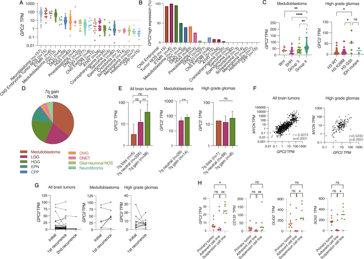
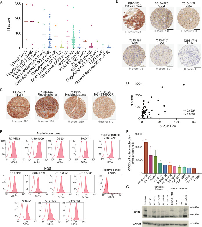
Background: Pediatric brain tumors are the leading cause of cancer death in children with an urgent need for innovative therapies. Glypican 2 (GPC2) is a cell surface oncoprotein expressed in neuroblastoma for which targeted immunotherapies have been developed. This work aimed to characterize GPC2 expression in pediatric brain tumors and develop an mRNA CAR T cell approach against this target.

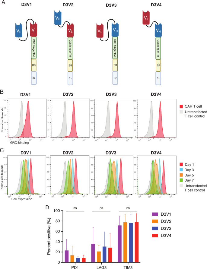
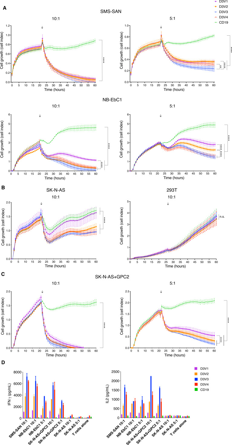
Methods: We investigated GPC2 expression across a cohort of primary pediatric brain tumor samples and cell lines using RNA sequencing, immunohistochemistry, and flow cytometry. To target GPC2 in the brain with adoptive cellular therapies and mitigate potential inflammatory neurotoxicity, we used optimized mRNA to create transient chimeric antigen receptor (CAR) T cells. We developed four mRNA CAR T cell constructs using the highly GPC2-specific fully human D3 single chain variable fragment for preclinical testing.

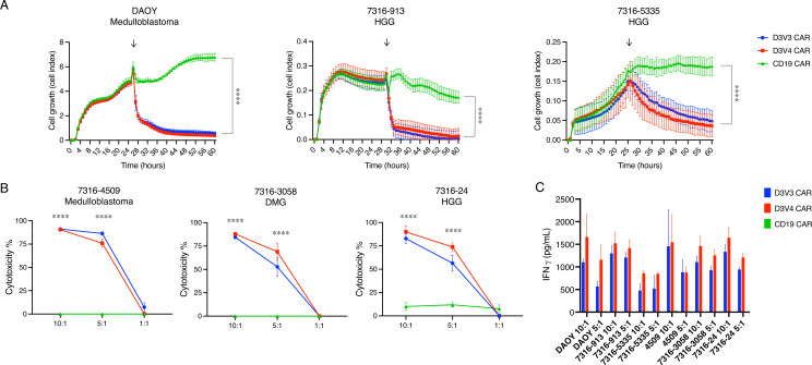
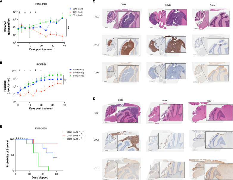
Results: We identified high GPC2 expression across multiple pediatric brain tumor types including medulloblastomas, embryonal tumors with multilayered rosettes, other central nervous system embryonal tumors, as well as definable subsets of highly malignant gliomas. We next validated and prioritized CAR configurations using in vitro cytotoxicity assays with GPC2-expressing neuroblastoma cells, where the light-to-heavy single chain variable fragment configurations proved to be superior. We expanded the testing of the two most potent GPC2-directed CAR constructs to GPC2-expressing medulloblastoma and high-grade glioma cell lines, showing significant GPC2-specific cell death in multiple models. Finally, biweekly locoregional delivery of 2-4 million GPC2-directed mRNA CAR T cells induced significant tumor regression in an orthotopic medulloblastoma model and significantly prolonged survival in an aggressive orthotopic thalamic diffuse midline glioma xenograft model. No GPC2-directed CAR T cell related neurologic or systemic toxicity was observed.

Conclusion: Taken together, these data show that GPC2 is a highly differentially expressed cell surface protein on multiple malignant pediatric brain tumors that can be targeted safely with local delivery of mRNA CAR T cells, laying the framework for the clinical translation of GPC2-directed immunotherapies for pediatric brain tumors.

Keywords: Brain Neoplasms; Cell Engineering; Central Nervous System Neoplasms; Immunotherapy; Pediatrics.


Conflict of interest statement

Competing interests: TS is currently employed by Spark Therapeutics. KK is currently employed by BioNTech and is an inventor on a patent related to use of nucleoside-modified mRNA. DMB is currently employed by Tmunity Therapeutics. JBF, DMB, JMM, and KRB hold patents for the discovery and development of immunotherapies for cancer, including patents related to glypican 2 (GPC2)-directed immunotherapies. KRB and JMM receive research funding from Tmunity for research on GPC2-directed immunotherapies and JBF, DMB, JMM, and KRB receive royalties from Tmunity for licensing of GPC2-related intellectual property. JMM is a founder of both Tantigen Bio and Hula Therapeutics, focused on cellular therapies for childhood cancers, but neither are working on GPC2-directed therapeutics. All other authors have nothing to disclose.

Figures

Figure 1
Figure 1
GPC2 is expressed in pediatric brain tumors. (A) GPC2 RNA sequencing data across OpenPBTA pediatric brain tumors cohorts. Neuroblastoma GPC2 RNA sequencing included for comparison on the left. (B) Using a cut-off value of 10 TPM, percentage of tumors in the OpenPBTA dataset with high expression of GPC2. (C) RNA sequencing of GPC2 expression in medulloblastoma and high-grade glioma subtypes. (D) Summary of tumor histotypes with chromosome 7q gain. (E) GPC2 expression stratified by chromosome 7q status. (F) Pearson correlation of MYCN and GPC2 expression across all pediatric brain tumor samples (left) and high-grade gliomas (right). (G) GPC2 expression at primary diagnosis and recurrence across all pediatric brain tumors (left), medulloblastomas (middle), and high-grade gliomas (right). (H) GPC2, CD133, SOX2, and OLIG2 expression from primary tumor-derived adherent (FBS) or suspension cell lines (serum free media). Individual cases indicated by dots, median indicated by line in A, C, and H. Data in figure part E displayed as mean with SD. ****P<0.0001; **p<0.01; *p<0.05; ns, not significant. ATRT, atypical teratoid rhabdoid tumor; CNS, central nervous system; CPC, choroid plexus carcinoma; CPP, choroid plexus papilloma; DMG, diffuse midline glioma; DNET, dysembryoplastic neuroepithelial tumor; ETMR, embryonal tumor with multilayered rosettes; GCT, germ cell tumor; HGG, high-grade glioma; LGG, low-grade glioma; NOS, not otherwise specified; TPM, transcripts per million.
Figure 2
Figure 2
GPC2 protein is expressed on the surface of pediatric brain tumors. (A) GPC2 H score from IHC of primary brain tumor TMAs. Individual samples indicated by dots, median indicated by line. (B) Representative GPC2 IHC from HGG primary tumor samples including H3G35 mutant HGG, glioblastoma multiforme (GBM), DMG, and AA. Scale bar in black is 200 µm. (C) Representative GPC2 IHC showing strongly positive staining in additional brain tumor histotypes including ETMR, pineoblastoma, medulloblastoma, and HGNET-BCOR. Scale bar in black is 200 µm. (D) Pearson correlation of GPC2 RNA expression (TPM) with GPC2 protein expression (H score) from 47 matched pediatric brain tumor samples. (E) GPC2 flow cytometry representative histograms of HGG and medulloblastoma cell lines. Red shaded curves indicate D3-GPC2-IgG1-PE staining, and non-shaded curves indicate isotype control. SMS-SAN shown as neuroblastoma positive control and T cells as negative control. (F) Summary of the relative quantification of GPC2 molecules per cell from flow cytometry of HGG and medulloblastoma cell lines (n=11). Data are represented as mean±SD of three independent experiments. (G) GPC2 western blot of a panel of medulloblastoma and HGG cell lines (n=9). SMS-SAN also presented as neuroblastoma positive control. AA, anaplastic astrocytoma; ATRT, atypical teratoid/rhabdoid tumors; BC, University of British Columbia; DMG, diffuse midline glioma; ETMR, embryonal tumor with multilayered rosettes; GPC2, glypican 2; HGG, high-grade glioma; HGNET-BCOR, high grade neuroepithelial tumor with BCOR alteration; IHC, immunohistochemistry; NOS, not otherwise specified; TMAs, tissue microarray; TPM, transcripts per million.
Figure 3
Figure 3
D3-binder based GPC2-directed mRNA CARs specifically bind GPC2 and are transiently expressed on T cells. (A) Schematic of GPC2 CAR constructs. (B) Flow cytometry representative histograms of GPC2-specific binding by GPC2 CAR T cell constructs. T cells were incubated with GPC2 protein conjugated to PE. Red line is CAR T cell, and gray line is untransfected T cell control. MFI is normalized to mode. (C) Flow cytometry representative histograms of GPC2 CAR persistence on T cells. T cells were incubated with protein L to identify CAR expression. Red line represents day 1 post-transfection, blue line represents day 3, orange line day 5, and green line day 7. Gray line is untransfected T cell control. Mean fluorescence intensity (MFI) is normalized to mode. (D) Cell surface expression of negative checkpoint regulators PD1, Lag3, and Tim3 quantified with flow cytometry at day 4 post-transfection. Data are represented as mean±SD of five independent experiments. Graphics in figure part A created with BioRender.com. 3z, CD3ζ co-stimulatory domain; BB, 41-BB costimulatory domain; CARs, chimeric antigen receptors; GPC2, glypican 2; ns, not significant; PE, phycoerythrin; TM, transmembrane domain; VH, variable heavy chain; VL, variable light chain.
Figure 4
Figure 4
GPC2-directed CAR T cells produce robust cytotoxicity in neuroblastoma cell line models. (A–C) GPC2-directed CAR T cells coincubated on RT-CES with endogenously high GPC2-expressing neuroblastoma cells lines (A), low/non-expressing cell lines (B), and cell line with forced overexpression of GPC2 (C). T cells added at approximately 24 hours as indicated by arrow. (D) Cytokine expression measured by ELISA 24 hours after coincubation of neuroblastoma and GPC2-directed CAR T cells. Data are shown as mean±SD from a representative experiment with each experiment being done two to four independent times. ****P<0.0001; **p<0.01. CAR, chimeric antigen receptor; E:T, effector-to-target ratio; GPC2, glypican 2; IFNγ, interferon-gamma; IL-2, interleukin 2; ns, not significant; RT-CES, Real-time Excelligence system.
Figure 5
Figure 5
GPC2-directed CAR T cells produce robust cytotoxicity in pediatric brain tumor cell line models. (A) GPC2-directed CAR T cells coincubated with medulloblastoma (DAOY) and HGG (7316–913, 7316–5335) cell lines. E:T ratio is 5:1, T cells added at approximately 24 hours as indicated by arrows. (B) GPC2 CAR T cells coincubated with medulloblastoma (7316–4509), HGG (7316–24), and DMG (7316–3058) cell lines with cytotoxicity measured using a luminescence assay at 48 hours. (C) Cytokine levels measured 24 hours after coincubation of GPC2 CAR T cells with each cell line. Data are shown as mean±SD from a representative experiment with each experiment being done two to four independent times. ****P<0.0001. CAR, chimeric antigen receptor; E:T ratio, effector-to-target ratio; DMG, diffuse midline glioma; GPC2, glypican 2; HGG, high-grade glioma.
Figure 6
Figure 6
GPC2-directed CAR T cells mediate antitumor responses and prolong survival in pediatric brain tumors in vivo. (A) Quantification of bioluminescence of the orthotopic group 4 medulloblastoma xenograft 7316–4509 treated with either GPC2 or CD19-directed mRNA CAR T cells. Doses indicated by arrows on graph. Data displayed as mean±SD, n=9–11 mice per arm. (B) Quantification of bioluminescence of the orthotopic group 3 medulloblastoma xenograft RCMB28 treated with either GPC2 or CD19-directed mRNA CAR T cells. Doses indicated by arrows on graph. Data displayed as mean±SD, n=10 mice per arm. (C and D) IHC of murine brains of a representative mouse per group collected at study endpoint from medulloblastoma xenograft experiments using models 7316–4509 (C) and RCMB28 (D). GPC2 staining for tumor identification and expression, CD3 stain to identify CAR T cells. Scale bar is 2000 μm at low power, 250 µm at insert. (E) Overall survival of mice implanted with thalamic DMG xenograft 7316–3058 treated with six repeated doses of 2×10⁶ CAR T cells. Doses indicated by arrows on graph. n=7 mice per arm. ****P<0.0001; **p<0.01; *p<0.05. CAR, chimeric antigen receptor; DMG, diffuse midline glioma; GPC2, glypican 2; IHC, immunohistochemistry; ns, not significant.

References

    1. Couzin-Frankel J. Cancer immunotherapy. Science 2013;342:1432–3. 10.1126/science.342.6165.1432 - DOI - PubMed
    1. Almåsbak H, Aarvak T, Vemuri MC. CAR T cell therapy: a game changer in cancer treatment. J Immunol Res 2016;2016:5474602. 10.1155/2016/5474602 - DOI - PMC - PubMed
    1. Maude SL, Frey N, Shaw PA, et al. . Chimeric antigen receptor T cells for sustained remissions in leukemia. N Engl J Med 2014;371:1507–17. 10.1056/NEJMoa1407222 - DOI - PMC - PubMed
    1. Fry TJ, Shah NN, Orentas RJ, et al. . CD22-targeted CAR T cells induce remission in B-ALL that is naive or resistant to CD19-targeted CAR immunotherapy. Nat Med 2018;24:20. 10.1038/nm.4441 - DOI - PMC - PubMed
    1. Gardner R, Finney O, Smithers H, et al. . CD19CAR T Cell Products of Defined CD4:CD8 Composition and Transgene Expression Show Prolonged Persistence and Durable MRD-Negative Remission in Pediatric and Young Adult B-Cell ALL. Blood 2016;128:219. 10.1182/blood.V128.22.219.219 - DOI

Publication types