. 2022 Sep 27;12(1):16068.
doi: 10.1038/s41598-022-20415-0.

Prophylactic administration of miR-451 inhibitor decreases osteoarthritis severity in rats

Affiliations

Prophylactic administration of miR-451 inhibitor decreases osteoarthritis severity in rats

Kayla M Scott et al. Sci Rep. .

Abstract

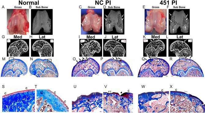
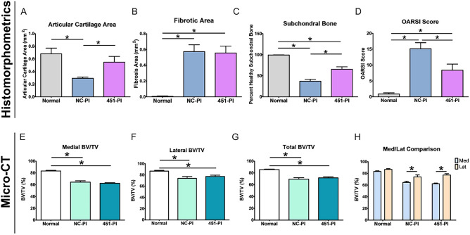
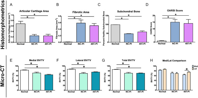
Transfection of chondrocytes with microRNA-451(miR-451), present in growth zone cartilage of the growth plate, upregulates production of enzymes association with extracellular matrix degradation. miR-451 is also present in articular cartilage and exacerbates IL-1β effects in articular chondrocytes. Moreover, when osteoarthritis (OA) was induced in Sprague Dawley rats via bilateral anterior cruciate ligament transection (ACLT), miR-451 expression was increased in OA cartilage compared to control, suggesting its inhibition might be used to prevent or treat OA. To examine the prophylactic and therapeutic potential of inhibiting miR-451, we evaluated treatment with miR-451 power inhibitor (451-PI) at the onset of joint trauma and treatment after OA had developed. The prophylactic animal cohort received twice-weekly intra-articular injections of either 451-PI or a negative control (NC-PI) beginning on post-surgical day 3. OA was allowed to develop for 24 days in the therapeutic cohort before beginning injections. All rats were killed on day 45. Micro-CT, histomorphometrics, OARSI scoring, and muscle force testing were performed on samples. 451-PI mitigated OA progression compared to NC-PI limbs in the prophylactic cohort based on histomorphometric analysis and OARSI scoring, but no differences were detected by micro-CT. 451-PI treatment beginning 24 days post-surgery was not able to reduce OA severity. Prophylactic administration of 451-PI mitigates OA progression in a post-trauma ACLT rat model supporting its potential to prevent OA development following an ACLT injury clinically.


Conflict of interest statement

Kayla M. Scott, D. Joshua Cohen, Barbara D. Boyan and Zvi Schwartz have a patent application (SCH-20–161) on the use of microRNA inhibition to prevent and treat traumatic osteoarthritis and other inflammatory diseases. The other authors declare no competing interests.

Figures

Figure 1
Figure 1
In-vivo experimental methods for miR-451-PI study. Eighteen Sprague Dawley rats underwent a bilateral ACLT surgery to induce OA. Two animals were used as controls (n = 4). The eighteen animals were split into two cohorts: 1) a prophylactic cohort that received intra-articular injections of miR-451-PI (n = 9) in left hind limbs or NC-PI (n = 9) in right hind limbs twice-weekly (red arrows) three days following surgery for a total duration of 6 weeks, and 2) a therapeutic cohort where we allowed OA to develop for 3 weeks before beginning the same injection regimen (n = 9 per treatment group, but one animal died resulting in an n = 8) for 3 weeks until animals were euthanized on day 42.
Figure 2
Figure 2
Gross images, micro-CT, and histology from the prophylactic cohort of the miR-451-PI study. Gross images (A,C,E), 3D micro-CT rendering (B,D,F), medial and lateral micro-CT images (GL). A shadow projection of the femur is included in the top corner of each micro-CT image with a red line to indicate the location each micro-CT image is taken from within the joint (GL). Masson’s trichrome histology (MR) and the respective zoomed-in images as depicted by the black box (SX) for the normal control, NC-PI, and 451-PI groups, respectively. Scale bar: 500 μm. Subchondral bone (sub bone); medial (med); lateral (lat); arrows, areas of cartilage erosion or subchondral bone remodeling; arrowheads, abnormal cartilage remodeling; C, cartilage; FT, fibrotic tissue; asterisk, microfracture.
Figure 3
Figure 3
Prophylactic administration of 451-PI decreases OA severity. Histomorphometric analysis of articular cartilage area (A), fibrotic area (B), percent healthy subchondral bone (C), and OARSI scoring (D). Micro-CT bone volume/total volume (BV/TV) analysis of medial femurs (E), lateral femurs (F), total (medial + lateral, G), and the comparison of medial and lateral femurs (H). An asterisk indicates statistical significance using an alpha equal to 0.05.
Figure 4
Figure 4
Gross images, micro-CT, and histology from the therapeutic cohort of the miR-451-PI study. Gross image (A,C,E), 3D micro-CT rendering (B,D,F), medial and lateral micro-CT images (GL). A shadow projection of the femur is included in the top corner of each micro-CT image with a red line to indicate the location each micro-CT image is taken from within the joint (GL). Masson’s trichrome histology (MR) and the respective zoomed in images as depicted by the black box (SX) for the normal control, NC-PI and 451-PI groups, respectively. Scale bar: 500 μm. Subchondral bone (sub bone); medial (med); lateral (lat); arrows, areas of cartilage erosion or subchondral bone remodeling; arrow heads, abnormal cartilage remodeling; C, cartilage; FT, fibrotic tissue; asterisk, microfracture.
Figure 5
Figure 5
Therapeutic administration of 451-PI does not decrease OA severity. Histomorphometric analysis of articular cartilage area (A), fibrotic area (B), percent healthy subchondral bone (C), and OARSI scoring (D). Micro-CT bone volume/total volume (BV/TV) analysis of medial femurs (E), lateral femurs (F), total (medial + lateral, G), and the comparison of medial and lateral femurs (H). An asterisk indicates statistical significance using an alpha equal to 0.05.

References

    1. Abramoff B, Caldera FE. Osteoarthritis: Pathology, diagnosis, and treatment options. Med. Clin. North Am. 2020;104:293–311. doi: 10.1016/j.mcna.2019.10.007. - DOI - PubMed
    1. Charlier E, et al. Insights on molecular mechanisms of chondrocytes death in osteoarthritis. Int. J. Mol. Sci. 2016;17:2146. doi: 10.3390/ijms17122146. - DOI - PMC - PubMed
    1. Nugent M. MicroRNAs: Exploring new horizons in osteoarthritis. Osteoarthr. Cartil. 2016;24:573–580. doi: 10.1016/j.joca.2015.10.018. - DOI - PubMed
    1. Lin Z, et al. MicroRNA contents in matrix vesicles produced by growth plate chondrocytes are cell maturation dependent. Sci. Rep. 2018;8:3609. doi: 10.1038/s41598-018-21517-4. - DOI - PMC - PubMed
    1. Asmussen N, Lin Z, McClure MJ, Schwartz Z, Boyan BD. Regulation of extracellular matrix vesicles via rapid responses to steroid hormones during endochondral bone formation. Steroids. 2019;142:43–47. doi: 10.1016/j.steroids.2017.12.003. - DOI - PubMed

Publication types