. 2022 Sep 27;7(1):57.
doi: 10.1038/s41536-022-00253-4.

Integrin-specific hydrogels for growth factor-free vasculogenesis

Affiliations

Integrin-specific hydrogels for growth factor-free vasculogenesis

Helena R Moreira et al. NPJ Regen Med. .

Abstract

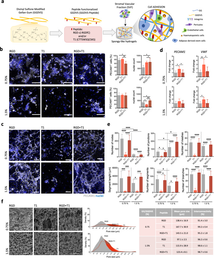
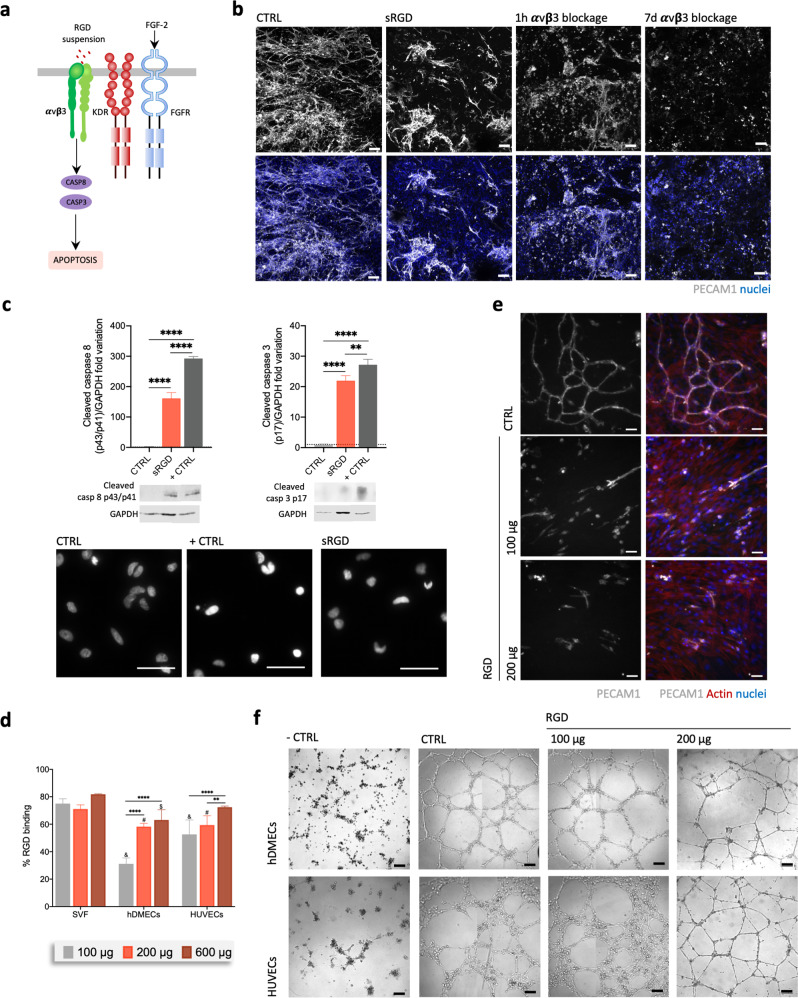
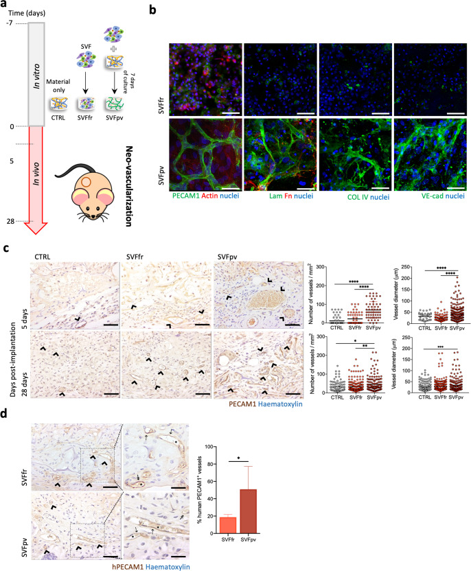
Integrin-binding biomaterials have been extensively evaluated for their capacity to enable de novo formation of capillary-like structures/vessels, ultimately supporting neovascularization in vivo. Yet, the role of integrins as vascular initiators in engineered materials is still not well understood. Here, we show that αvβ3 integrin-specific 3D matrices were able to retain PECAM1+ cells from the stromal vascular fraction (SVF) of adipose tissue, triggering vasculogenesis in vitro in the absence of extrinsic growth factors. Our results suggest that αvβ3-RGD-driven signaling in the formation of capillary-like structures prevents the activation of the caspase 8 pathway and activates the FAK/paxillin pathway, both responsible for endothelial cells (ECs) survival and migration. We also show that prevascularized αvβ3 integrin-specific constructs inosculate with the host vascular system fostering in vivo neovascularization. Overall, this work demonstrates the ability of the biomaterial to trigger vasculogenesis in an integrin-specific manner, by activating essential pathways for EC survival and migration within a self-regulatory growth factor microenvironment. This strategy represents an improvement to current vascularization routes for Tissue Engineering constructs, potentially enhancing their clinical applicability.


Conflict of interest statement

The authors declare no competing interests.

Figures

Fig. 1
Fig. 1. Integrin-specific spongy-like hydrogels modulate vasculogenesis triggering.
a Schematic representation of the rationale of using GGDVS functionalized with T1 and RGD peptides to capture cells involved in vasculogenesis from the stromal vascular fraction (SVF) of adipose tissue. b Representative immunocytochemistry images of the expression of PECAM1 in SVF cells kept in the functionalized materials after 3 days of culture and respective quantification of the percentage of PECAM1+ cells and total number of cells. Scale bar = 100 µm. c Representative immunocytochemistry images of the organization of PECAM1+ cells in the functionalized materials after 7 days of culture. Scale bar = 100 µm. d PECAM1 and VWF mRNA expression in cells kept in the functionalized materials at day 7. mRNA expression was determined by qPCR using β2M as reference gene and normalized to the respective expression at day 5 to understand the variation along time for each condition. e Quantification of the number of nodes, junctions, meshes, segments, branches, and segments length in the capillary-like structures formed after 7 days of culture. f Microstructural features (pore size and interconnectivity) of GG/GGDVS-peptides dried polymeric networks visualized by SEM and quantified by μ-CT. Scale bar = 100 µm. Quantitative results are expressed as the mean ± standard deviation where n = 5, *p < 0.05, **p < 0.01, ***p < 0.001, ****p < 0.0001, one-way or two-way ANOVA with Tukey multiple comparison post-test.
Fig. 2
Fig. 2. VEGF and FGF2 are involved in EC migration and proliferation that results in the formation of vascular structures.
a Schematic representation of the hypothesized involvement of VEGF and FGF2 pathways. KDR, VEGF, FGFR1, FGFR2, and FGF2 mRNA expression in cells cultured in the functionalized materials on day 5 and 7. mRNA expression was determined by qPCR using β2M as reference gene and normalized to the results for non-modified material on day 5. b Schematic representation of the hypothesized involvement of ANGPT1 pathway in vessel maturation. Representative immunocytochemistry images of SVF cells expressing PECAM1+ in the functionalized materials after 14 days of culture. Scale bar = 100 µm. Detail of the vascular-like network formed by PECAM1+ cells in the 0.75% GG/GGDVS-RGD spongy-like hydrogel after 14 days in culture showing several lumen (white arrows). Nuclei were counterstained with DAPI. Scale bar = 50 µm. TEK and ANGPT1 mRNA expression in cells cultured in the functionalized materials on days 5 and 7. mRNA expression was determined by qPCR using β2M as reference gene and normalized to the results for non-modified material at day 5 to allow comparison among the conditions. Quantitative results are expressed as the mean ± standard deviation where n = 5, *p < 0.05, **p < 0.01, ***p < 0.001, ****p < 0.0001, two-way ANOVA with Tukey multiple comparison post-test.
Fig. 3
Fig. 3. Integrin-specific spongy-like hydrogels prevent apoptosis of cells involved in the vasculogenic process.
a Schematic representation of the studied apoptosis signaling pathway. b Representative images of the organization of SVF PECAM1+ cells after 7 days of culture in the 0.75% GG/GGDVS-RGD when pre-incubated with RGD peptide (sRGD) or with integrin αvβ3 blocking antibody for 1 h (1 h αvβ3 blockage), or in the presence of αvβ3 blocking antibody (7d αvβ3 blockage). Control (CTRL) refers to SVF cells directly seeded in the material. Scale bar = 100 µm. c Protein level of cleaved caspase 8 and cleaved caspase 3 in SVF cells 24 h after peptide exposure or mitomycin C (+CTRL) with correspondent representative western blot bands. Plotted western blot data was determined in relation to GAPDH expression. Representative nuclei images from SVF cells labeled with Picogreen 24 h after exposure to RGD peptide or mitomycin C (+CTRL). Scale bar = 50 µm. d Percentage of RGD peptide binding to SVF cells, hDMECs, and HUVECs after 30 min incubation. e Representative images of the organization of PECAM1+ SVF cells pre-incubated with different amounts of the RGD peptide and cultured on standard 2D surfaces for 7 days. Scale bar = 50 µm. f Representative images of hDMECs and HUVECs in a Matrigel assay, pre-incubated with different amounts of the RGD peptide or in their respective basal media without any angiogenic growth factors (negative control (−CTRL)). In the positive control condition (+CTRL), cells were cultured in their optimized media without pre-incubation with RGD peptide. Scale bar = 200 µm. Quantitative results are expressed as the mean ± standard deviation where n = 3, *p < 0.05, **p < 0.01, ***p < 0.001, ****p < 0.0001, one-way ANOVA with Tukey multiple comparison post-test or Kruskal–Wallis test with Dunn’s multiple comparison post-test.
Fig. 4
Fig. 4. Involvement of VEGF and FGF2 pathways in the vasculogenesis in integrin-specific spongy-like hydrogels.
a Schematic representation of the studied signaling pathways. b Expression of P-TY397 FAK, FAK, Paxillin, Talin 1 + 2. c VEGF secretion, and KDR, P-TH202-TY204 ERK1/2, ERK1/2, P-S473 AKT1, and AKT1 expression. Expression of the proteins (b, c) was determined by western blot in SVF cells, with and without pre-incubation with RGD peptide, cultured in the 0.75% GG/GGDVS-RGD materials for 7 days. Expression of P-TH202-TY204 ERK1/2, ERK1/2 was also determined when SVF cells were pre-incubation with T1 peptides. d FGF-2 secretion and FGFR2 expression determined by western blot in SVF cells, with and without pre-incubation with RGD and T1 peptides, cultured in the 0.75% GG/GGDVS-RGD materials for 7 days. All plotted western blot data was determined in relation to GAPDH expression. e Representative images of the organization of SVF PECAM1+ cells after 7 days of culture in the 0.75% GG/GGDVS-RGD when pre-incubated with T1 peptide. Results are expressed as the mean ± standard deviation where n = 3, *p < 0.05, **p < 0.01, ***p < 0.001, ****p < 0.0001, one-way ANOVA with Tukey multiple comparison post-test or two-tailed unpaired t test.
Fig. 5
Fig. 5. Preformed human capillaries foster neovascularization in vivo.
a Schematic representation of the test conditions: CTRL (GG/GGDVS-RGD spongy-like hydrogels without cells), SVFfr (freshly isolated SVF seeded in the material), SVFpv (SVF seeded in the material and cultured for 7 days in vitro prior to implantation). b Representative immunocytochemistry images of the expression of PECAM1 (anti-human), Collagen type IV (COL4), Laminin (Lam), Fibronectin (Fn), VE-cadherin (VE-cad), and F-actin in SVF cells seeded in the 0.75% GG/GGDVS-RGD spongy-like hydrogels, without (SVFfr) and after 7 days of vitro pre-culture (SVFpv). Nuclei were counterstained with DAPI (nuclei). Scale bar = 50 µm. c Representative immunocytochemistry images showing PECAM1+ vessels (arrowhead, anti-mouse and anti-human) at the transplantation site on days 5 and 28, and respective quantification of the number of vessels and diameter. d Representative immunocytochemistry images of vessels incorporating human PECAM1+ cells (arrowhead, anti-human) at the transplantation site on day 28 and respective quantification. Higher magnification images show chimeric (⇡) and human (↟) vessels containing mouse erythrocytes (*). Scale bar = 500 µm, 100 µm, 50 µm. Quantitative results are expressed as the mean ± standard deviation, *p < 0.05, **p < 0.01, ***p < 0.001, ****p < 0.0001, Kruskal–Wallis test with Dunn’s multiple comparison post-test or two-tailed unpaired t test.

References

    1. Risau W, Flamme I. Vasculogenesis. Annu. Rev. Cell Dev. Biol. 1995;11:73–91. doi: 10.1146/annurev.cb.11.110195.000445. - DOI - PubMed
    1. Vailhé B, Vittet D, Feige JJ. In vitro models of vasculogenesis and angiogenesis. Lab. Investig. 2001;81:439–52. doi: 10.1038/labinvest.3780252. - DOI - PubMed
    1. Novosel EC, Kleinhans C, Kluger PJ. Vascularization is the key challenge in tissue engineering. Adv. Drug Deliv. Rev. 2011;63:300–311. doi: 10.1016/j.addr.2011.03.004. - DOI - PubMed
    1. Sharma D, et al. Upgrading prevascularization in tissue engineering: a review of strategies for promoting highly organized microvascular network formation. Acta Biomater. 2019;95:112–130. doi: 10.1016/j.actbio.2019.03.016. - DOI - PubMed
    1. Miklas JW, et al. QHREDGS enhances tube formation, metabolism and survival of endothelial cells in collagen-chitosan hydrogels. PLoS One. 2013;8:72956. doi: 10.1371/journal.pone.0072956. - DOI - PMC - PubMed