. 2022 Sep 27;5(1):1016.
doi: 10.1038/s42003-022-03999-1.

Phospholipid synthesis inside phospholipid membrane vesicles

Affiliations

Phospholipid synthesis inside phospholipid membrane vesicles

Sumie Eto et al. Commun Biol. .

Abstract

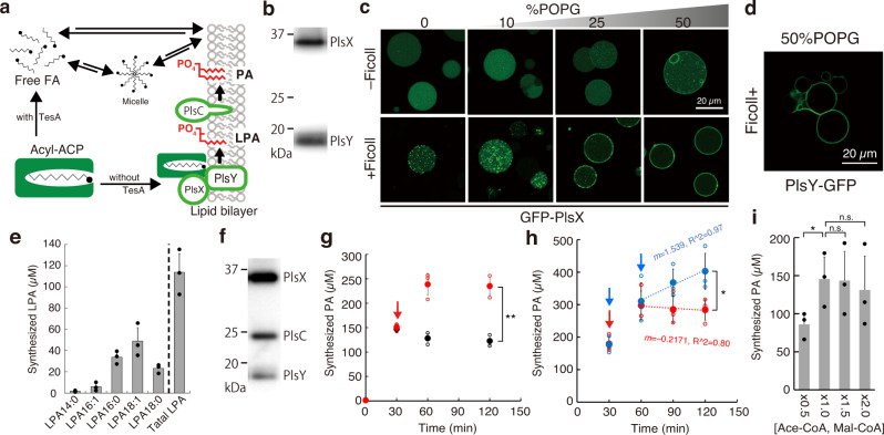
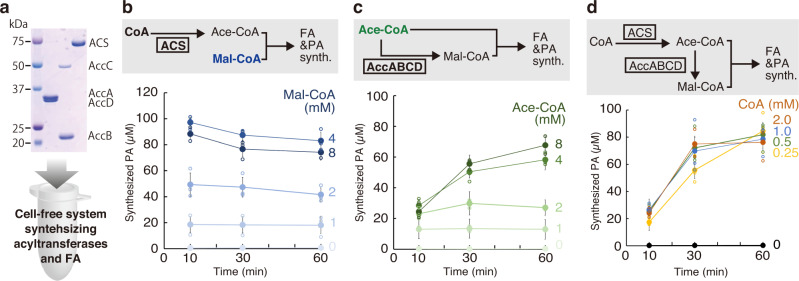
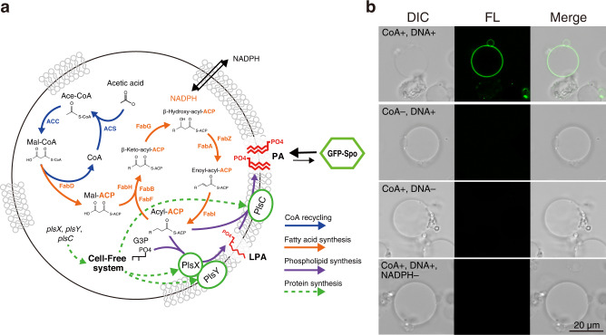
Construction of living artificial cells from genes and molecules can expand our understanding of life system and establish a new aspect of bioengineering. However, growth and division of cell membrane that are basis of cell proliferation are still difficult to reconstruct because a high-yielding phospholipid synthesis system has not been established. Here, we developed a cell-free phospholipid synthesis system that combines fatty acid synthesis and cell-free gene expression system synthesizing acyltransferases. The synthesized fatty acids were sequentially converted into phosphatidic acids by the cell-free synthesized acyltransferases. Because the system can avoid the accumulation of intermediates inhibiting lipid synthesis, sub-millimolar phospholipids could be synthesized within a single reaction mixture. We also performed phospholipid synthesis inside phospholipid membrane vesicles, which encapsulated all the components, and showed the phospholipids localized onto the mother membrane. Our approach would be a platform for the construction of self-reproducing artificial cells since the membrane can grow sustainably.


Conflict of interest statement

Y.K. is applying for a domestic patent (Japan) regarding the technology related to this work. The remaining authors declare no competing interests.

Figures

Fig. 1
Fig. 1. Fatty acid (FA) synthesis in a reconstructed cell-free system.
a Schematic overview of the cell-free lipid synthesis system. The system is composed of protein synthesis, acetyl-CoA and malonyl-CoA syntheses, FA synthesis, and phospholipid synthesis reactions. After the synthesis of acyltransferases on the liposome membranes, the FA synthesis is initiated by the addition of NAD(P)H and substrates, a mixture of acetyl-CoA and malonyl-CoA. When ACS and AccABCD are supplied, CoA, KHCO3, and acetic acid are used as substrates to synthesize acetyl-CoA and malonyl-CoA. The synthesized FAs are converted into phospholipids by the cell-free synthesized acyltransferases. b The purified Fab enzymes, ACP, and TesA. c FA synthesis in the reconstituted in vitro system containing 10 µM FabZ, 1 µM FabA, and 1 µM FabB or d 1 µM FabZ, 10 µM FabA and 10 µM FabB. The types of synthesized FAs and the total yield are shown in the inset. e Optical microscopy images of the reaction mixture after FA synthesis. +All and −ACP indicate the mixture containing all enzymes or missing ACP, respectively. f SDS-PAGE analysis of the reaction mixture in the presence or absence of ACP. P and S represent the precipitate and supernatant, respectively. g Inhibition of FA synthesis by oleic acid. h Enhanced FA synthesis by the addition of liposomes. Each dot on the graphs represents individual experimental data. Error bars indicate the standard deviation of triplicate measurements. AccABCD acetyl-CoA carboxylase ABCD, ACS acetyl-CoA synthetase, ACP acyl-carrier protein, MK molecular marker.
Fig. 2
Fig. 2. Phospholipid synthesis by cell-free synthesized acyltransferases.
a Schematic of the phospholipid synthesis pathway in a cell-free system. b SDS-PAGE image of cell-free synthesized PlsX and PlsY. Confocal microscopy images of c GFP-PlsX and d PlsY-GFP synthesized inside giant unilamellar vesicles (GUVs) in the presence (bottom) or absence (top) of Ficoll. The percentage of POPG (mol%) in the lipid composition of the GUV is described above the images. e Synthesis of lysophosphatidic acids (LPAs) by PlsX and PlsY synthesized in a bulk cell-free system. f SDS-PAGE image of cell-free synthesized PlsX, PlsY, and PlsC. g Synthesis of phosphatidic acids (PAs) by cell-free synthesized PlsX, PlsY, and PlsC. At 30 min, the resources [acetyl-CoA (Ace-CoA), malonyl-CoA (Mal-CoA), and NAD(P)H] (red) or buffer (black) were additionally supplied to the reaction mixture. h Increase of PA synthesis by supplying the resources at 30 and 60 min (blue) or only 30 min (red). The slopes indicate the transition in PA productions between 60 to 120 min. i Effect of the various concentrations of the resources supplied from the beginning of PA synthesis reaction. The concentration of 1.0× [Ace-CoA, Mal-CoA] is 2 and 4 mM, respectively. Each dot on the graphs represents individual experimental data. Double asterisk, single asterisk, and n.s. indicate that P values are less than 0.01, P values are less than 0.05, and P values are more than 0.05, respectively. Error bars indicate the standard deviation of at least triplicate measurements.
Fig. 3
Fig. 3. Phosphatidic acid synthesis by synthesizing acetyl-CoA or/and malonyl-CoA.
a An SDS-PAGE image of the purified acetyl-CoA carboxylase DA (AccDA), acetyl-CoA carboxylase BC (AccBC), and acetyl-CoA synthase (ACS). The positions of molecular size are described on the left side of the gel image. The purified enzymes were introduced into the cell-free system synthesizing acyltransferases and fatty acids. b Phosphatidic acid synthesis through the synthesis of acetyl-CoA by ACS. The reaction was initiated by the addition of CoA and various concentrations of malonyl-CoA. c Phosphatidic acid synthesis through the synthesis of malonyl-CoA by AccABCD. The reaction was initiated by the addition of various concentrations of acetyl-Co, which was converted to malonyl-CoA or directly consumed as the substrate of FA. d Phosphatidic acid synthesis through the synthesis of acetyl-CoA and malonyl-CoA by the addition of ACS and AccABCD. The reactions were initiated by the addition of various concentrations of CoA. Each dot on the graphs represents individual experimental data. Error bars indicate the standard deviation of at least triplicate measurements. FA fatty acid, PA phosphatidic acid, Ace-CoA acetyl-CoA, Mal-CoA malonyl-CoA.
Fig. 4
Fig. 4. Phospholipid synthesis inside artificial cells.
a Schematic of the phospholipid-synthesizing artificial cell. GFP-Spo was added to the exterior of the cells to visualize the synthesized PA at the membrane. b Confocal microscopy images of the artificial cells treated with GFP-Spo after the lipid synthesis reaction. The artificial cells missing CoA, DNA, or NADPH were also tested for the controls. Wide-filed images of the artificial cells are shown in (Figs. S18). CoA coenzyme A, Ace-CoA acetyl-CoA, Mal-CoA malonyl-CoA, Mal-ACP malonyl-ACP, ACP acyl-carrier protein, G3P glycerol-3-phosphate, LPA lysophosphatidic acid, PA phosphatidic acid.

References

    1. Gaut NJ, Adamala KP. Reconstituting Natural Cell Elements in Synthetic Cells. Adv. Biol. 2021;5:e2000188. doi: 10.1002/adbi.202000188. - DOI - PubMed
    1. Yeh Martín N, Valer L, Mansy SS. Toward long-lasting artificial cells that better mimic natural living cells. Emerg. Top. life Sci. 2019;3:597–607. doi: 10.1042/ETLS20190026. - DOI - PMC - PubMed
    1. Shimizu Y, et al. Cell-free translation reconstituted with purified components. Nat. Biotechnol. 2001;19:751–755. doi: 10.1038/90802. - DOI - PubMed
    1. Fujiwara K, Katayama T, Nomura SM. Cooperative working of bacterial chromosome replication proteins generated by a reconstituted protein expression system. Nucleic Acids Res. 2013;41:7176–7183. doi: 10.1093/nar/gkt489. - DOI - PMC - PubMed
    1. Su’etsugu M, Takada H, Katayama T, Tsujimoto H. Exponential propagation of large circular DNA by reconstitution of a chromosome-replication cycle. Nucleic Acids Res. 2017;45:11525–11534. doi: 10.1093/nar/gkx822. - DOI - PMC - PubMed

Publication types

MeSH terms