. 2022 Sep 27;13(1):5677.
doi: 10.1038/s41467-022-33352-3.

Fatty acids homeostasis during fasting predicts protection from chemotherapy toxicity

Affiliations

Fatty acids homeostasis during fasting predicts protection from chemotherapy toxicity

Marta Barradas et al. Nat Commun. .

Abstract

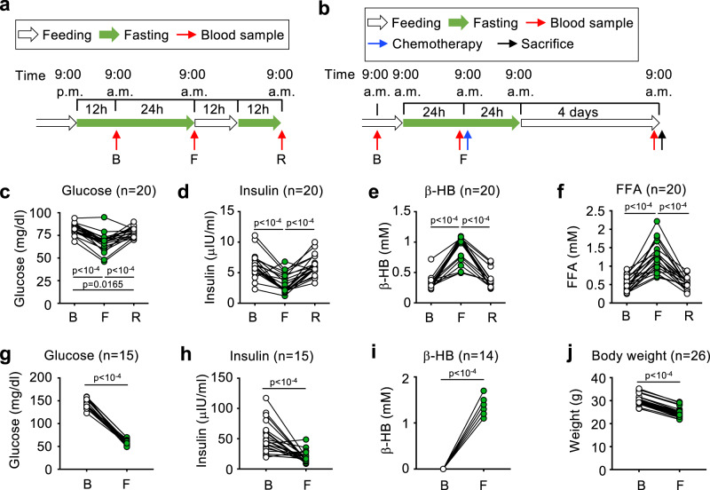
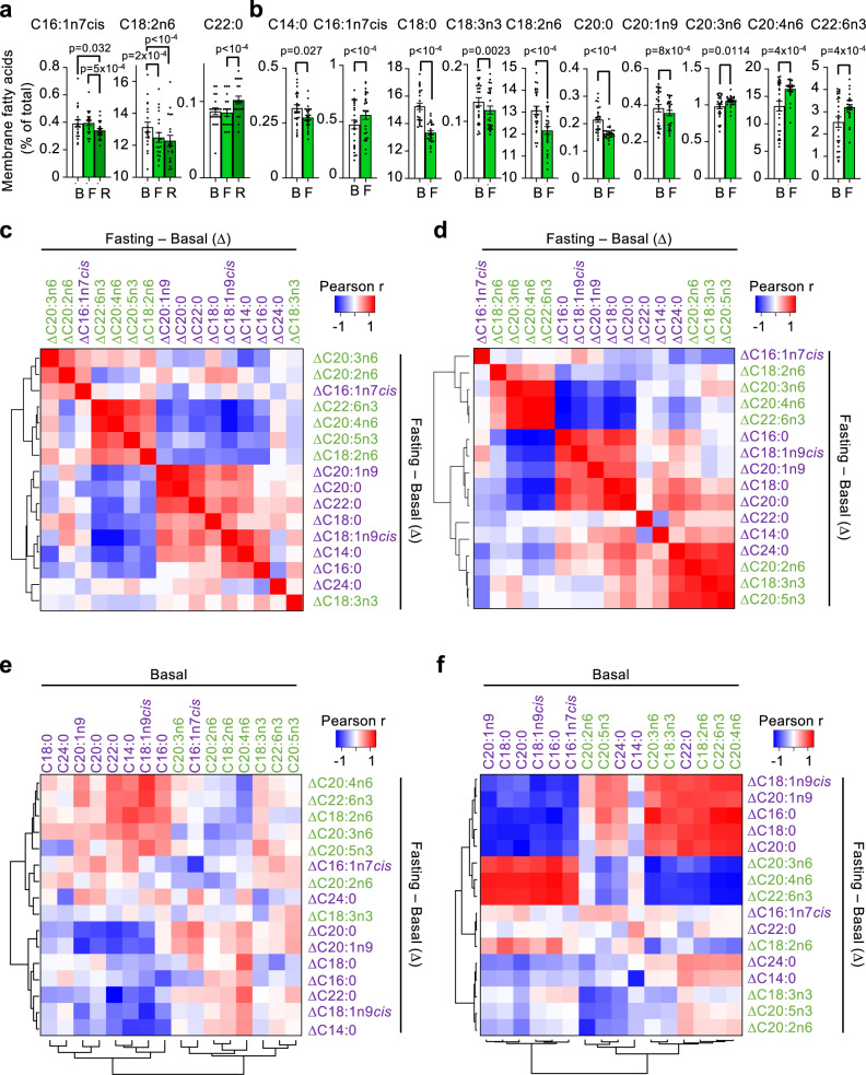
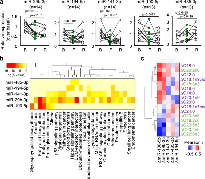
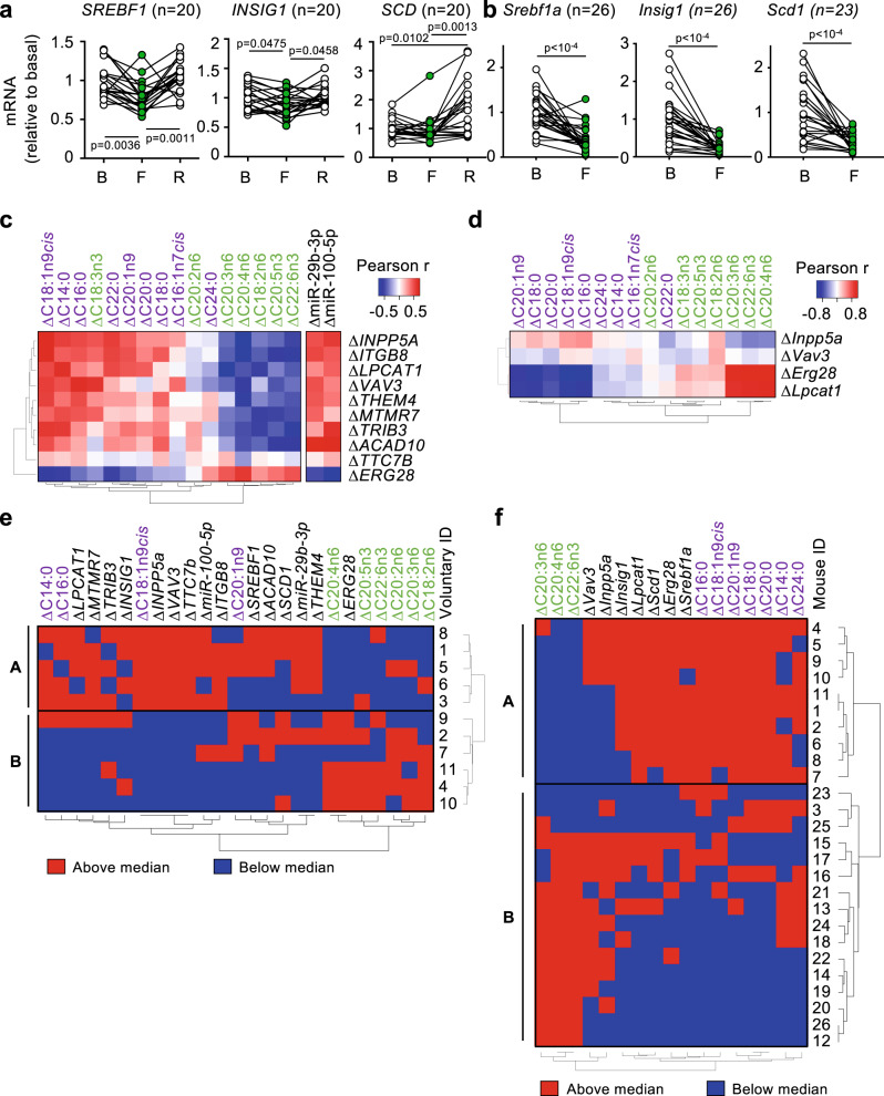
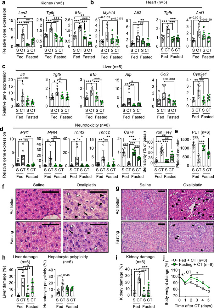
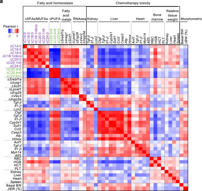
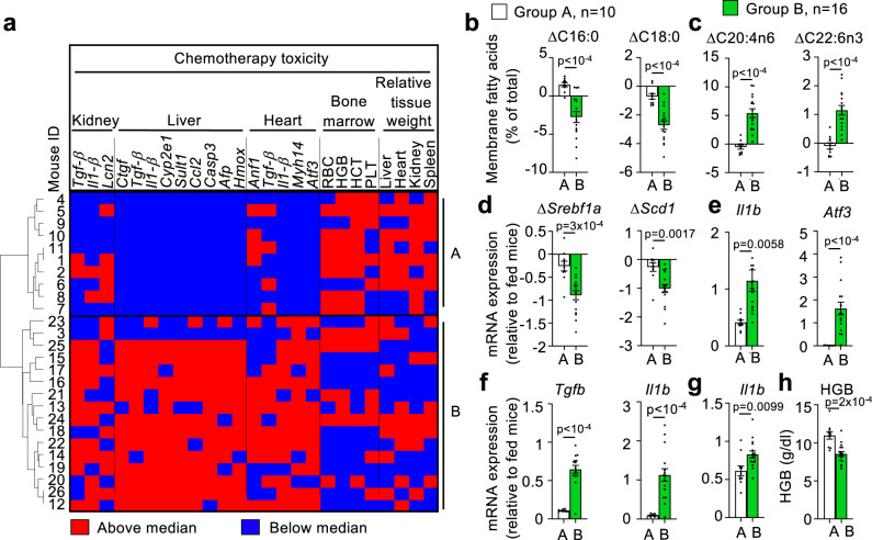
Fasting exerts beneficial effects in mice and humans, including protection from chemotherapy toxicity. To explore the involved mechanisms, we collect blood from humans and mice before and after 36 or 24 hours of fasting, respectively, and measure lipid composition of erythrocyte membranes, circulating micro RNAs (miRNAs), and RNA expression at peripheral blood mononuclear cells (PBMCs). Fasting coordinately affects the proportion of polyunsaturated versus saturated and monounsaturated fatty acids at the erythrocyte membrane; and reduces the expression of insulin signaling-related genes in PBMCs. When fasted for 24 hours before and 24 hours after administration of oxaliplatin or doxorubicin, mice show a strong protection from toxicity in several tissues. Erythrocyte membrane lipids and PBMC gene expression define two separate groups of individuals that accurately predict a differential protection from chemotherapy toxicity, with important clinical implications. Our results reveal a mechanism of fasting associated with lipid homeostasis, and provide biomarkers of fasting to predict fasting-mediated protection from chemotherapy toxicity.


Conflict of interest statement

The authors declare no competing interests.

Figures

Fig. 1
Fig. 1. Standard biomarkers of short-term fasting in healthy humans and mice.
a, b Scheme of the protocols followed for the short-term fasting experiment in humans (a) and mice (b). Green arrows indicate the times of fasting, and white arrows indicate normal feeding. Red arrows indicate blood sampling; blue arrows indicate chemotherapy administration, and black arrows indicate sacrifice. c–f Blood levels in human volunteers of glucose (c), insulin (d), β-hydroxybutyrate (β-HB, e) and free fatty acids (FFAs, f) at the basal point after 12 h of overnight fasting (B); after 36 h of fasting (F); and after 12 h of refeeding and 12 h of overnight fasting (R). g–i Blood levels of glucose (g), insulin (h) and β-HB (i) in fed conditions at first time in the morning (Basal, B) and after 24 h of fasting (F). j Mouse body weight at the indicated times. Each dot represents the data from a single individual, and is linked with a straight black line with the data of the same individual in the next point of the time course experiment. Statistical significance was performed using the one-way ANOVA test with Tukey correction for multiple comparisons (cf); or the paired two-tailed Student t test (gj). The exact p value is provided for each significant comparison. Source data are provided as a Source Data file.
Fig. 2
Fig. 2. Short-term fasting elicits changes in erythrocyte membrane fatty acids.
a, b Levels of the indicated fatty acids in erythrocyte membranes isolated at the indicated times of the fasting experiments (B: basal, after 12 h of fasting; F: after 36 h of fasting; R: after 12 h of refeeding and 12 h of fasting) in humans (a, n = 20) and mice (b, n = 26), expressed as % of total membrane fatty acids. c, d Heatmaps depicting the Pearson correlation r coefficients between changes (fasting minus basal, Δ) of all the analyzed erythrocyte membrane fatty acids from human (c) or mice (d) samples. Red color indicates direct correlations, and blue color indicates indirect correlations. Saturated and monounsaturated fatty acids are written in purple; polyunsaturated fatty acids are written in green. The precise order of each fatty acid was determined using a cluster analysis. e, f Heatmaps representing the Pearson correlation r coefficients between the changes (fasting minus basal) and the basal levels of the same erythrocyte membrane fatty acids shown in a, b. Bars represent the average of the indicated number of individuals. Error bars indicate the standard error of the mean. Statistical significance was assayed using the one-way ANOVA test with Tukey correction for multiple comparisons (a), the unpaired two-tailed Student t test (b); or the Pearson correlation test (cf). The exact p value is provided for each significant comparison. Source data are provided as a Source Data file.
Fig. 3
Fig. 3. Human circulating miRNAs change during short-term fasting.
a Expression of the indicated miRNAs measured in plasma from human volunteers at basal (B), fasting (F) and refeeding (R) points. b Heatmap showing the KEGG functions associated with genes targeted by the indicated miRNAs. c Heatmap showing the Pearson correlation r values between changes (fasting minus basal, Δ) of all the analyzed erythrocyte membrane fatty acids and changes with fasting of the indicated circulating miRNAs significantly altered with fasting. Each dot represents the data from a single individual, and is linked with a straight black line with the data of the same individual in the next point of the time course experiment. Statistical significance was performed using the one-way ANOVA test with Tukey correction for multiple comparisons (a) or with Pearson correlations (c). The exact p value is provided for each significant comparison. Source data are provided as a Source Data file.
Fig. 4
Fig. 4. Gene expression in peripheral blood mononucleated cells (PBMCs) during short-term fasting.
a, b Total RNA from PBMCs isolated from human volunteers (a) or mice (b) at the indicated time points (B basal, F fasting, R refeeding) was analyzed for the expression of the indicated lipid metabolism genes. c, d Heatmaps depicting the Pearson correlation r coefficients in humans (c) or mice (d) between the changes with fasting of erythrocyte membrane fatty acids and/or circulating miRNAs, and the changes in mRNA expression of the indicated genes identified in the PBMC RNAseq experiment. e, f Heatmaps depicting each individual, represented by a number next to each panel, and its position with respect to the median value for each of the indicated parameters: red indicates “above the median”, and blue indicates “below the median”. Both individuals and parameters were ordered according to their similarity. This unsupervised clustering defined two groups of individuals named “A” and “B” in the graphs. Line-connected dots represent the paired data for each individual (a, b). Statistical analysis was performed using the one-way ANOVA test with Tukey correction for multiple comparisons (a); the paired two-tailed Student t test (b); or Pearson correlations (c, d). The exact p value is provided for each significant comparison. Source data are provided as a Source Data file.
Fig. 5
Fig. 5. Fasting protects from oxaliplatin-induced multi-organ toxicity in mice.
a–e Expression of the indicated toxicity-reporter genes in kidney (a), heart (b) liver (c), and dorsal root ganglia (d, panels to the left) or the neurotoxicity-measuring von Frey test (d, panel to the right); and blood platelets counts (e) from mice either fed ad libitum or fasted for 24 h before and 24 h after i.p. inoculation of saline (S) or 15 mg/kg oxaliplatin (CT), and sacrificed 5 days after chemotherapy administration. f, g Representative photographs of hematoxilin and eosin staining of sections from liver (f) or kidney (g) from the same mice as in (ae), size bar = 200 μM. In liver (f), black triangles point to lipid vacuoles; white triangles point to sinusoidal dilatations; and black arrows point to polyploid hepatocytes. In kidney (g), black triangles point to tubule dilatation, and white triangles point to Bowman capsule dilatation glomerulopathy. h, i Quantification of the histological findings from liver (h) or kidney (i). j Total body weight of mice from (ai) at the indicated times after oxaliplatin (CT) administration. Bars and line-connected dots represent the average of the indicated number of mice per treatment group. Error bars represent the standard error of the mean. Statistical significance was assayed using the one-way ANOVA test (ae) or the two-way ANOVA test (j) with Tukey correction for multiple comparisons. When possible, the exact p value is provided for significant comparisons; otherwise, *p < 0.05; **p < 0.01; ***p < 0.001. Source data are provided as a Source Data file.
Fig. 6
Fig. 6. Correlation of fatty acid homeostasis biomarkers with chemotherapy toxicity in mice.
a Heatmap depicting the Pearson correlation r coefficients between the indicated fatty acid homeostasis biomarkers (membrane fatty acids, Srebf1 pathway genes in PBMCs and RNAseq-identified genes) with chemotherapy toxicity biomarkers: expression of toxicity-responding genes in kidney, liver and heart; bone marrow-associated parameters red blood cell counts (RBC), hemoglobin (HGB), hematocrit (HCT) and platelet counts (PLT); and tissue weights relative to total body weight. At the right end of the graph, the morphometric parameters of basal body weight (BW) and change in body weight with fasting in % (ΔBW (%)) are also analyzed. These correlations were performed with n = 26 mice. Source data are provided as a Source Data file.
Fig. 7
Fig. 7. Groups of individuals responding differently to fasting predict fasting-mediated protection from chemotherapy.
a Heatmap depicting the position of each individual (represented by a number next to each row) with respect to the median value for each of the indicated parameters. Red indicates “above the median”, and blue indicates “below the median”. Individuals are ordered according to the groups A and B found in Fig. 4f. RBC red blood cells. HGB hemoglobin. HCT hematocrit. PLT platelets. b–h Comparison between groups A (n = 10) and B (n = 16) of the indicated saturated (c) and polyunsaturated (c) erythrocyte membrane fatty acids; fatty acid metabolism genes (d); heart (e), liver (f) and kidney (g) toxicity-responding genes and bone marrow markers (h; hemoglobin, HGB). Bars represent the average of n = 10 (group A) and n = 16 (group B). Error bars represent the standard error of the mean. Statistical analysis was performed using the unpaired two-tailed Student t test (bh). The exact p value is provided for each significant comparison. Source data are provided as a Source Data file.

Similar articles

Cited by

References

    1. Ruderman NB. Muscle amino acid metabolism and gluconeogenesis. Annu. Rev. Med. 1975;26:245–258. - PubMed
    1. Cahill GF, Owen OE, Morgan AP. The consumption of fuels during prolonged starvation. Adv. Enzym. Regul. 1968;6:143–150. - PubMed
    1. Nuttall FQ, Almokayyad RM, Gannon MC. Comparison of a carbohydrate-free diet vs. fasting on plasma glucose, insulin and glucagon in type 2 diabetes. Metabolism. 2015;64:253–262. - PubMed
    1. Nuttall FQ, Almokayyad RM, Gannon MC. The ghrelin and leptin responses to short-term starvation vs a carbohydrate-free diet in men with type 2 diabetes; a controlled, cross-over design study. Nutr. Metab. 2016;13:47. - PMC - PubMed
    1. Merl V, et al. Serum adiponectin concentrations during a 72-hour fast in over- and normal-weight humans. Int. J. Obes. 2005;29:998–1001. - PubMed

Publication types