. 2022 Sep 27;12(1):16126.
doi: 10.1038/s41598-022-20507-x.

A critical role of calcineurin in stress responses, hyphal formation, and virulence of the pathogenic fungus Trichosporon asahii

Affiliations

A critical role of calcineurin in stress responses, hyphal formation, and virulence of the pathogenic fungus Trichosporon asahii

Yasuhiko Matsumoto et al. Sci Rep. .

Abstract

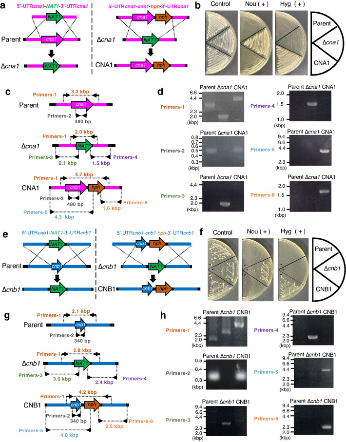
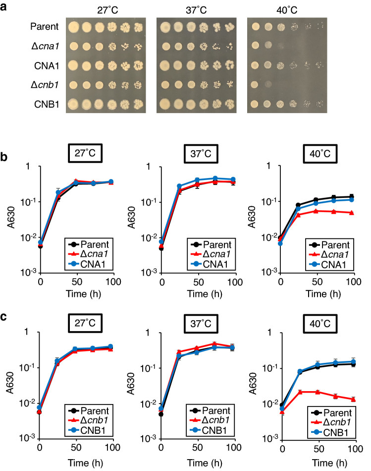
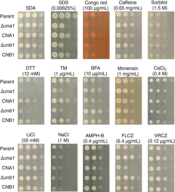
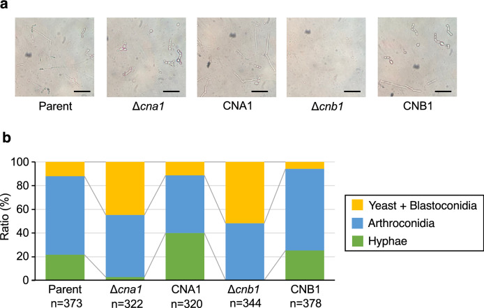
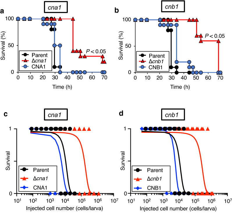
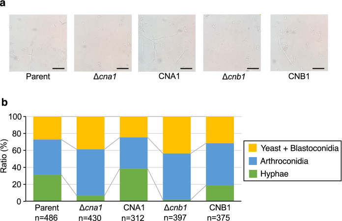
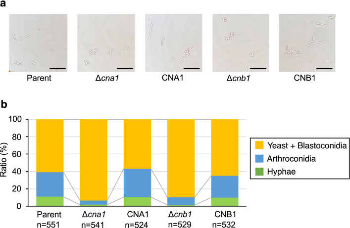
Trichosporon asahii is a conditional pathogenic fungus that causes severe and sometimes fatal infections in immunocompromised patients. While calcineurin, an essential component of a calcium-dependent signaling pathway, is known to regulate stress resistance and virulence of some pathogenic fungi, its role in T. asahii has not been investigated. Here, we demonstrated that calcineurin gene-deficient T. asahii mutants are sensitive to high temperature as well as cell-membrane and cell-wall stress, and exhibit decreased hyphal formation and virulence against silkworms. Growth of T. asahii mutants deficient in genes encoding subunits of calcineurin, cna1 and cnb1, was delayed at 40 °C. The cna1 and cnb1 gene-deficient mutants also showed sensitivity to sodium dodecyl sulfate, Congo red, dithiothreitol, and tunicamycin. On the other hand, these mutants exhibited no sensitivity to caffeine, sorbitol, monensin, CaCl2, LiCl, NaCl, amphotericin B, fluconazole, or voriconazole. The ratio of hyphal formation in the cna1 and cnb1 gene-deficient mutants was decreased. Moreover, the virulence of the cna1 and cnb1 gene-deficient mutants against silkworms was attenuated. These phenotypes were restored by re-introducing each respective gene into the gene-deficient mutants. Our findings suggest that calcineurin has a role in regulating the cellular stress response and virulence of T. asahii.


Conflict of interest statement

The authors declare no competing interests.

Figures

Figure 1
Figure 1
Generation of the cna1 and cnb1 gene-deficient T. asahii mutants and their revertants. (ad) Generation of the cna1 gene-deficient mutant and its revertant in T. asahii. (a) Strategy for generating the cna1 gene-deficient mutant (∆cna1) and its revertant (Rev.). Predicted genomes of the cna1 gene-deficient mutant and its revertant are shown. (b) The parent strain (Parent), cna1 gene-deficient mutant (∆cna1), and its revertant (Rev.) were spread on SDA with nourseothricin (Nou) (100 µg/ml) or hygromycin B (Hyg) (100 µg/ml) and incubated at 27 °C for 2 days. (c) Location of the primers for confirming the genome structure of the cna1 gene-deficient candidate by PCR using extracted genome DNA. (d) Confirmation of the genotypes of the cna1 gene-deficient mutant (∆cna1) and its revertant (Rev.) by PCR using extracted genome DNA. (eh) Generation of the cna1 gene-deficient mutant and its revertant in T. asahii. (e) Strategy for generating the cnb1 gene-deficient mutant (∆cnb1) and its revertant (Rev.). Predicted genomes of the cnb1 gene-deficient mutant and its revertant are shown. (f) The parent strain (Parent), cnb1 gene-deficient mutant (∆cnb1), and its revertant (Rev.) were spread on SDA with nourseothricin (Nou) (100 µg/ml) or hygromycin B (Hyg) (100 µg/ml) and incubated at 27 °C for 2 days. (g) Location of the primers for confirming the genome structure of the cnb1 gene-deficient candidate by PCR using extracted genome DNA. (h) Confirmation of the genotypes of the cnb1 gene-deficient mutant (∆cnb1) and its revertant (Rev.) by PCR using extracted genome DNA. Cropped blots were used. Full-length blots are presented in Supplementary Fig. S3.
Figure 2
Figure 2
Temperature sensitivity of cna1 or cnb1 gene-deficiency in T. asahii. (a) The T. asahii parent strain (Parent), the cna1 gene-deficient mutant (∆cna1), the revertant of ∆cna1 (CNA1), the cnb1 gene-deficient mutant (∆cnb1), and the revertant of ∆cnb1 (CNB1) were grown on SDA and incubated at 27 °C for 2 days. T. asahii cells was suspended in physiologic saline solution and filtered through a 40-μm cell strainer. Series of tenfold dilution of the fungal suspension were prepared using saline. Five microliters of each cell suspension was spotted on the SDA. Agar plates were incubated at 27 °C, 37 °C, or 40 °C for 24 h. (b, c) The T. asahii parent strain (Parent), the cna1 gene-deficient mutant (∆cna1), the revertant of ∆cna1 (CNA1), the cnb1 gene-deficient mutant (∆cnb1), and the revertant of ∆cnb1 (CNB1) were inoculated on Sabouraud medium and incubated at 27 °C, 37 °C, or 40 °C. Absorbance of the culture at 630 nm was monitored. Data are shown as means ± standard error of the mean (SEM).
Figure 3
Figure 3
Sensitivity of the cna1 and cnb1 gene-deficient mutants against stress inducers. The T. asahii parent strain (Parent), the cna1 gene-deficient mutant (∆cna1), the revertant of ∆cna1 (CNA1), the cnb1 gene-deficient mutant (∆cnb1), and the revertant of ∆cnb1 (CNB1) were grown on SDA and incubated at 27 °C for 2 days. T. asahii cells was suspended in physiologic saline solution and filtered through a 40-μm cell strainer. Series of tenfold dilution of the fungal suspension were prepared using saline. Five microliters of each cell suspension was spotted on the SDA containing SDS (0.00625%), Congo red (100 µg/ml), caffeine (0.65 mg/ml), sorbitol (1.5 M), DTT (12 mM), TM (1 µg/ml), BFA (10 µg/ml), monensin (1 mg/ml), CaCl2 (0.4 M), LiCl (55 mM), NaCl (1 M), amphotericin-B (0.4 µg/ml), fluconazole (6.4 µg/ml), or voriconazole (0.12 µg/ml). Each agar plate was incubated at 37 °C for 24 h.
Figure 4
Figure 4
Effect of cna1 or the cnb1 gene-deficiency on T. asahii morphology in Sabouraud dextrose medium. The T. asahii parent strain (Parent), the cna1 gene-deficient mutant (∆cna1), the revertant of ∆cna1 (CNA1), the cnb1 gene-deficient mutant (∆cnb1), and the revertant of ∆cnb1 (CNB1) were grown on SDA and incubated at 27 °C for 2 days. T. asahii cells was suspended in physiologic saline solution and filtered through a 40-μm cell strainer. Absorbance at 630 nm of the T. asahii cell suspension was adjusted to 1–1.5. The cell suspension (10 µl) was added to Sabouraud dextrose medium (100 µl). The solutions were incubated at 37 °C for 48 h. The incubated solution was placed on glass slides and covered by a glass coverslip. (a) Samples were examined with bright light under a microscope. The scale bar indicated 50 µm. (b) The pictures were randomly obtained. The numbers of three cell types: yeast + blastoconidia, arthroconidia, and hyphae, were counted. The representative cells to determine the cell types are shown in Supplementary Fig. S2.
Figure 5
Figure 5
Attenuated pathogenicity in the cna1 or the cnb1 gene-deficient T. asahii mutants against silkworms. (a,b) The T. asahii parent strain (Parent; 2.9 × 105 cells/larva), the cna1 gene-deficient mutant (∆cna1; 7.4 × 105 cells/larva), the revertant from ∆cna1 (CNA1; 4.2 × 105 cells/larva), the cnb1 gene-deficient mutant (∆cnb1; 6.1 × 105 cells/larva), or the revertant from ∆cnb1 (CNB1; 7.1 × 105 cells/larva) were injected into the silkworm hemolymph and the silkworms were incubated at 37 °C. Silkworm survival was monitored for 72 h. The significance of differences between the parent strain group and the cnb1 gene-deficient mutant groups was calculated by the log-rank test based on the curves by the Kaplan–Meier method. P < 0.05 was considered significant. n = 10/group. (c,d) Number of surviving silkworms at conditions under 37 °C was determined at 48 h after administration of the fungal cells (50 to 1.2 × 106 cells/larva) into the silkworm hemolymph. Surviving and dead silkworms are indicated as 1 and 0, respectively. n = 4/group. Curves were drawn from combined data of 2–3 independent experiments by a simple logistic regression model.
Figure 6
Figure 6
Low hyphal growth of cna1 or the cnb1 gene-deficient T. asahii mutants in harvested silkworm hemolymph. The T. asahii parent strain (Parent), the cna1 gene-deficient mutant (∆cna1), the revertant of ∆cna1 (CNA1), the cnb1 gene-deficient mutant (∆cnb1), and the revertant of ∆cnb1 (CNB1) were grown on SDA and incubated at 27 °C for 2 days. T. asahii cells was suspended in physiologic saline solution and filtered through a 40-μm cell strainer. Absorbance at 630 nm of the T. asahii cell suspension was adjusted to 1–1.5. The cell suspension (10 µl) was added to harvested silkworm hemolymph (100 µl) and the solutions were incubated at 37 °C for 48 h. The incubated solution was placed on glass slides and covered by a glass coverslip. (a) Samples were examined with bright light under a microscope. The scale bar indicated 50 µm. (b) The pictures were randomly obtained. The numbers of three cell types: yeast + blastoconidia, arthroconidia, and hyphae, were counted.
Figure 7
Figure 7
Effect of cna1 or the cnb1 gene-deficiency on T. asahii morphology in human serum. The T. asahii parent strain (Parent), the cna1 gene-deficient mutant (∆cna1), the revertant of ∆cna1 (CNA1), the cnb1 gene-deficient mutant (∆cnb1), and the revertant of ∆cnb1 (CNB1) were grown on SDA and incubated at 27 °C for 2 days. T. asahii cells was suspended in physiologic saline solution and filtered through a 40-μm cell strainer. Absorbance at 630 nm of the T. asahii cell suspension was adjusted to 1–1.5. The cell suspension (10 µl) was added to human serum (100 µl) and the solutions were incubated at 37 °C for 48 h. The incubated solution was placed on glass slides and covered by a glass coverslip. (a) Samples were examined with bright light under a microscope. The scale bar indicated 50 µm. (b) The pictures were randomly obtained. The numbers of three cell types: yeast + blastoconidia, arthroconidia, and hyphae, were counted.

Similar articles

Cited by

References

    1. Sugita T, Nishikawa A, Ichikawa T, Ikeda R, Shinoda T. Isolation of Trichosporon asahii from environmental materials. Med. Mycol. 2000;38:27–30. doi: 10.1080/mmy.38.1.27.30. - DOI - PubMed
    1. Sugita T, et al. Genetic diversity and biochemical characteristics of Trichosporon asahii isolated from clinical specimens, houses of patients with summer-type-hypersensitivity pneumonitis, and environmental materials. J. Clin. Microbiol. 2001;39:2405–2411. doi: 10.1128/JCM.39.7.2405-2411.2001. - DOI - PMC - PubMed
    1. Zhang E, Sugita T, Tsuboi R, Yamazaki T, Makimura K. The opportunistic yeast pathogen Trichosporon asahii colonizes the skin of healthy individuals: Analysis of 380 healthy individuals by age and gender using a nested polymerase chain reaction assay. Microbiol. Immunol. 2011;55:483–488. doi: 10.1111/j.1348-0421.2011.00341.x. - DOI - PubMed
    1. Colombo AL, Padovan ACB, Chaves GM. Current knowledge of Trichosporon spp. and Trichosporonosis. Clin. Microbiol. Rev. 2011;24:682–700. doi: 10.1128/CMR.00003-11. - DOI - PMC - PubMed
    1. Gouba N, Raoult D, Drancourt M. Eukaryote culturomics of the gut reveals new species. PLoS ONE. 2014;9:e106994. doi: 10.1371/journal.pone.0106994. - DOI - PMC - PubMed

Publication types

MeSH terms

Supplementary concepts