

Severe Congenital Diaphragmatic Hernia With Trisomy 9: A Case Report and Review of the Literature
- PMID: 36168364
- PMCID: PMC9506681
- DOI: 10.7759/cureus.28395
Severe Congenital Diaphragmatic Hernia With Trisomy 9: A Case Report and Review of the Literature
Abstract
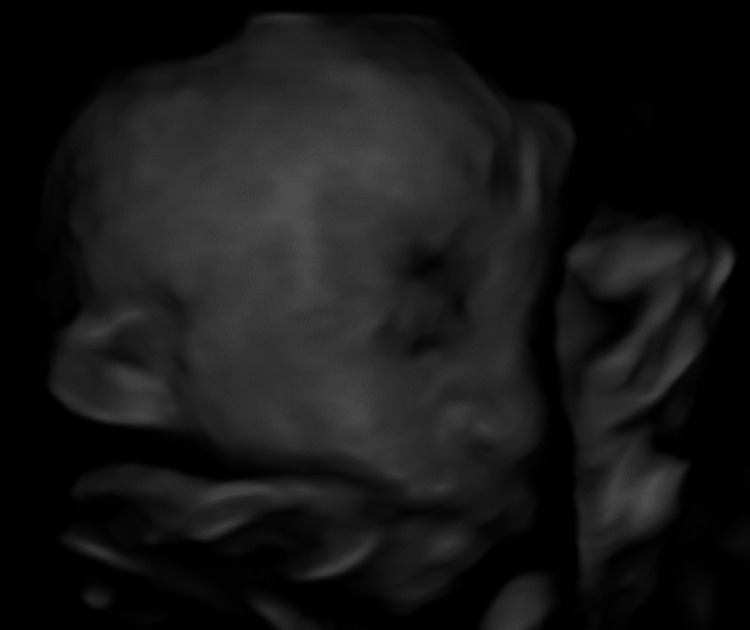
Congenital diaphragmatic hernia (CDH) is known to be complicated with various chromosomal abnormalities. However, the grade of pulmonary hypoplasia of CDH complicated by trisomy 9 is not known. This information is essential to the mother who has had a fetus with the same complication. We report a case of severe CDH with trisomy 9. The fetus had fetal growth restriction and multiple anomalies, including severe left CDH (observed/expected lung-to-head ratio 13.7%, liver-up, stomach grade 3 in Kitano classification), mild ventriculomegaly, low-set ear, rocker bottom, and single umbilical artery. Chromosomal test by amniocentesis showed a karyotype of 47,XX,+9. The neonate was born alive at 34 weeks but died 49 minutes after birth. In the literature review, this case and seven cases of complete trisomy 9 had CDH, and four of them were explained as "large" or "severe" CDH. In conclusion, trisomy 9 might be occasionally complicated by severe CDH.
Keywords: amniocentesis; congenital diaphragmatic hernia; fetal ultrasonography; prenatal genetic testing; trisomy 9.
Copyright © 2022, Fuma et al.
Conflict of interest statement
The authors have declared that no competing interests exist.
Figures


Similar articles
-
Prenatal diagnosis and array comparative genomic hybridization characterization of trisomy 21 in a fetus associated with right congenital diaphragmatic hernia and a review of the literature of chromosomal abnormalities associated with congenital diaphragmatic hernia.Taiwan J Obstet Gynecol. 2015 Feb;54(1):66-70. doi: 10.1016/j.tjog.2014.12.001. Taiwan J Obstet Gynecol. 2015. PMID: 25675923 Review.
-
Outcomes of congenital diaphragmatic hernia: a population-based study in Western Australia.Pediatrics. 2005 Sep;116(3):e356-63. doi: 10.1542/peds.2004-2845. Pediatrics. 2005. PMID: 16140678
-
Decreased neonatal morbidity in 'stomach-down' left congenital diaphragmatic hernia: implications of prenatal ultrasound diagnosis for counseling and postnatal management.Ultrasound Obstet Gynecol. 2021 Nov;58(5):744-749. doi: 10.1002/uog.23630. Ultrasound Obstet Gynecol. 2021. PMID: 33724570
-
Second-trimester diagnosis of complete trisomy 9 associated with abnormal maternal serum screen results, open sacral spina bifida and congenital diaphragmatic hernia, and review of the literature.Prenat Diagn. 2004 Jun;24(6):455-62. doi: 10.1002/pd.900. Prenat Diagn. 2004. PMID: 15229846 Review.
-
Severe left diaphragmatic hernia limits size of fetal left heart more than does right diaphragmatic hernia.Ultrasound Obstet Gynecol. 2015 Dec;46(6):688-94. doi: 10.1002/uog.14790. Epub 2015 Nov 9. Ultrasound Obstet Gynecol. 2015. PMID: 25597867
Cited by
-
Case report: A case report and literature review of complete trisomy 9.Front Genet. 2023 Aug 31;14:1241245. doi: 10.3389/fgene.2023.1241245. eCollection 2023. Front Genet. 2023. PMID: 37719705 Free PMC article.
References
-
- Re-evaluation of stomach position as a simple prognostic factor in fetal left congenital diaphragmatic hernia: a multicenter survey in Japan. Kitano Y, Okuyama H, Saito M, et al. Ultrasound Obstet Gynecol. 2011;37:277–282. - PubMed
-
- Trends, correlates, and survival of infants with congenital diaphragmatic hernia and its subtypes. Ramakrishnan R, Salemi JL, Stuart AL, Chen H, O'Rourke K, Obican S, Kirby RS. Birth Defects Res. 2018;110:1107–1117. - PubMed
-
- Pathological findings in the complete trisomy 9 syndrome: three case reports and review of the literature. Ferreres JC, Planas S, Martínez-Sáez EA, et al. Pediatr Dev Pathol. 2008;11:23–29. - PubMed
Publication types
LinkOut - more resources
Full Text Sources