. 2022 Sep 28;289(1983):20221398.
doi: 10.1098/rspb.2022.1398. Epub 2022 Sep 28.

Fossil basicranium clarifies the origin of the avian central nervous system and inner ear

Affiliations

Fossil basicranium clarifies the origin of the avian central nervous system and inner ear

Guillermo Navalón et al. Proc Biol Sci. .

Abstract

Among terrestrial vertebrates, only crown birds (Neornithes) rival mammals in terms of relative brain size and behavioural complexity. Relatedly, the anatomy of the avian central nervous system and associated sensory structures, such as the vestibular system of the inner ear, are highly modified with respect to those of other extant reptile lineages. However, a dearth of three-dimensional Mesozoic fossils has limited our knowledge of the origins of the distinctive endocranial structures of crown birds. Traits such as an expanded, flexed brain, a ventral connection between the brain and spinal column, and a modified vestibular system have been regarded as exclusive to Neornithes. Here, we demonstrate all of these 'advanced' traits in an undistorted braincase from an Upper Cretaceous enantiornithine bonebed in southeastern Brazil. Our discovery suggests that these crown bird-like endocranial traits may have originated prior to the split between Enantiornithes and the more crownward portion of avian phylogeny over 140 Ma, while coexisting with a remarkably plesiomorphic cranial base and posterior palate region. Altogether, our results support the interpretation that the distinctive endocranial morphologies of crown birds and their Mesozoic relatives are affected by complex trade-offs between spatial constraints during development.

Keywords: birds; brains; dinosaurs; ear; endocranium; labyrinth.


Conflict of interest statement

We declare we have no competing interests.

Figures
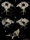
Figure 1.
Figure 1.
Enantiornithine braincase MPM-334-1 from the Late Cretaceous of southeastern Brazil. Three-dimensional meshes digitally rendered using Blender. (a) Ventral view, (b) dorsal view, (c) caudal view, (d) anterior view, and (e) left lateral view. Light brown arrow in (e) indicates orientation of the foramen magnum. Cn: exits of cranial nerves.
Figure 2.
Figure 2.
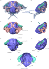
Main endocranial cavities of MPM-334-1. (a,c,e) caudal view, (b,d,f) left lateral view, and (g) ventral view. Teal arrow in (d) indicates the caudoventral direction in which the optic tectum pushes the anterior semicircular canal, arguably generating the characteristic crown bird-like ventral deflection in this structure. Cn: exits of cranial nerves.
Figure 3.
Figure 3.
Anatomy of the endosseous labyrinth of the vestibular system and other inner ear structures of MPM-334-1. Teal arrow in (c) indicates the caudoventral deflection of the anterior semicircular canal of the endosseous labyrinth (caused by the displacement of the optic tectum, figure 2). (a) Inset displays the same view of the endocranial structures of MPM-334-1 displayed in figure 2a. (bd) show different detailed views of the left endosseous labyrinth, oriented with its lateral semicircular canal completely horizontal (neutral Lsc). (b) Caudal (oriented to neutral Lsc), (c) lateral (oriented to neutral Lsc), and (d) dorsal (oriented to neutral Lsc). Note that the cochlear duct is not preserved on the left endosseous labyrinth but is visible on the right labyrinth (figure 2).
Figure 4.
Figure 4.
Comparative anatomy of the endosseous labyrinth and cochlear duct across selected birds (extinct and extant) and closely related non-avian dinosaurs. Labyrinths of extant birds sourced from [34]. Byronosaurus, Archaeopteryx and Hesperornis from [19]. Cerebavis from [32]. Enaliornis from [33]. Labyrinths are positioned following a neutral-cranial-base orientation for the whole crania (completely horizontal parasphenoid rostrum). Stem-based and node-based clade names are explicitly illustrated. Illustrations are not to scale.
Figure 5.
Figure 5.
Main structures of central nervous system (including optic tectum) and inner ear (vestibular system and cochlear duct) across selected birds (extinct and extant) and closely related non-avian dinosaurs. While the orientation of the foramen magnum varies across phylogeny, the degree of ventralization of the foramen magnum in MPM-334-1 is significantly greater than that of other stem taxa, and comparable to that of certain crown birds (e.g. Accipiter and Selasphorus). Stem-based and node-based clade names are explicitly illustrated. Illustrations are not scaled.
Figure 6.
Figure 6.
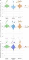
Orientation of the foramen magnum across birds and other saurischians. Violin plots show the distribution (contour) of the species values (large dot = mean value; line = interquartile ranges) for the angle between the foramen magnum and the parasphenoid rostrum (top left inset figure). Dashed line represents the angular value for MPM-334-1; n equals the number of specimens/species used for each taxon. Stem-based and node-based clade names are explicitly illustrated.
Figure 7.
Figure 7.
The relationship between orientation of the foramen magnum and body mass in crown birds. Dashed line indicates the angle for MPM-334-1. The convex hulls indicate the region encompassed by each major clade of crown birds (e.g. Palaeognathae, Galloanserae and Neoaves).
Figure 8.
Figure 8.
Orientation of the foramen magnum in Neoaves. Violin plots show the distribution (contour) of the species values (large dot = mean value; line = interquartile ranges) for the angle between the foramen magnum and the parasphenoid rostrum; dashed line indicates the angle for MPM-334-1. Three increasingly inclusive groups of neoavians with mostly aquatic ecologies are figured: (a) Ardeae; (b) Aequorlithornithes and an expanded grouping of water-linked clades; and (c) Aequorlithornithes + Gruiformes.
Figure 9.
Figure 9.
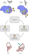
Conceptual diagram showing the non-flexed and flexed brain morphologies exhibited by extant birds, and the interplay of key hypothesized factors influencing the morphology of various systems within the avian cranium. Teal arrow pointing towards the labyrinth of Accipiter indicates the caudoventral deflection of the anterior semicircular canal of the endosseous labyrinth caused by the displacement of the optic tectum; figure 2.

References

    1. Romer AS. 1959. Osteology of the Reptiles. Krieger Publishing Company. Malabar, Florida, USA.
    1. Hanken J, Hall BK. 1993. The skull, volume 2: patterns of structural and systematic diversity. Chicago, IL: University of Chicago Press.
    1. Bhullar BAS, Marugán-Lobón J, Racimo F, Bever GS, Rowe TB, Norell MA, Abzhanov A. 2012. Birds have paedomorphic dinosaur skulls. Nature 487, 223-226. (10.1038/nature11146) - DOI - PubMed
    1. Bhullar BAS, Hanson M, Fabbri M, Pritchard A, Bever GS, Hoffman E. 2016. How to make a bird skull: major transitions in the evolution of the avian cranium, paedomorphosis, and the beak as a surrogate hand. Integr. Comp. Biol. 56, 389-403. (10.1093/icb/icw069) - DOI - PubMed
    1. O'Connor JK, Chiappe LM. 2011. A revision of enantiornithine (Aves: Ornithothoraces) skull morphology. J. Syst. Paleontol. 9, 135-157. (10.1080/14772019.2010.526639) - DOI

Publication types

LinkOut - more resources