. 2022 Dec;11(23):e2201955.
doi: 10.1002/adhm.202201955. Epub 2022 Oct 17.

3D-Printed Radiopaque Bioresorbable Stents to Improve Device Visualization

Affiliations

3D-Printed Radiopaque Bioresorbable Stents to Improve Device Visualization

Yonghui Ding et al. Adv Healthc Mater. 2022 Dec.

Abstract

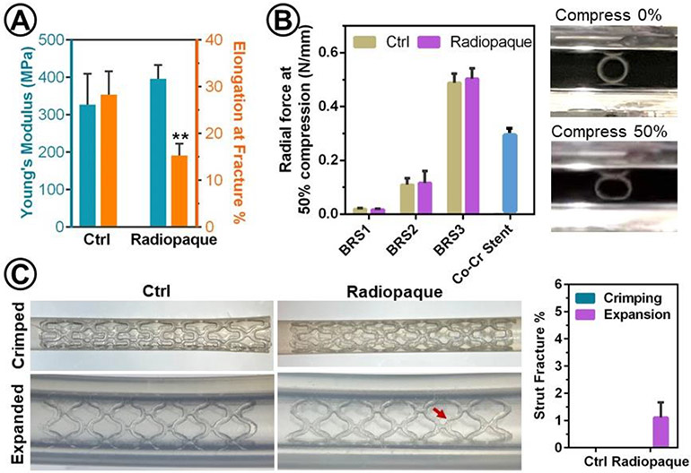
Bioresorbable stents (BRS) hold great promise for the treatment of many life-threatening luminal diseases. Tracking and monitoring of stents in vivo is critical for avoiding their malposition and inadequate expansion, which often leads to complications and stent failure. However, obtaining high X-ray visibility of polymeric BRS has been challenging because of their intrinsic radiolucency. This study demonstrates the use of photopolymerization-based 3D printing technique to fabricate radiopaque BRS by incorporating iodixanol, a clinical contrast agent, into a bioresorbable citrate-based polymer ink. The successful volumetric dispersion of the iodixanol through the 3D-printing process confers strong X-ray visibility of the produced BRS. Following in vitro degradation, the 3D-printed BRS embedded in chicken muscle maintains high X-ray visibility for at least 4 weeks. Importantly, the 3D-printed radiopaque BRS demonstrates good cytocompatibility and strong mechanical competence in crimping and expansion, which is essential for minimally invasive stent deployment. In addition, it is found that higher loading concentrations of iodixanol, e.g. 10 wt.%, results in more strut fractures in stent crimping and expansion. To conclude, this study introduces a facile strategy to fabricate radiopaque BRS through the incorporation of iodixanol in the 3D printing process, which could potentially increase the clinical success of BRS.

Keywords: X-ray visibility; bioresorbable stents; iodixanol; radiopacity.


Conflict of interest statement

Conflict of Interest

The authors declare no conflict of interest.

Figures

Figure 1.
Figure 1.
3D-printing of radiopaque BRS. (A) Schematic of preparation and 3D-printing of radiopaque ink. (B) CAD models and photos of 3D-printed bioresorbable vascular stent and esophageal stent. (C) Representative cross-sectional scanning electron microscope (SEM) images of 3D-printed BRS1, BRS2, and BRS3 containing 5 wt% iodixanol with increased strut thickness as well as their measured dimensions, including strut thickness and inner diameter (n = 4). The designed values of strut thickness were 137 μm for BRS1, 255 μm for BRS2, and 510 μm for BRS3.
Figure 2.
Figure 2.
Energy-dispersive X-ray spectrometry (EDS) elemental analysis validates the presence of iodixanol within BRS. (A) Cross-sectional electron microscope (EM) images and EDS mapping data. The magenta signal indicates the localization of elemental iodine. (B) Typical EDS spectrum of BRS that are prepared from mPDC and mPDC/iodixanol composite with 5 wt% iodixanol and molecular structure of iodixanol with highlighted iodine. The Au and Pd peaks are resulted from the conductive coating layer to avoid charging problem in SEM.
Figure 3.
Figure 3.
Micro-CT and fluoroscopy images of BRS reveal their radiopacity in air and chicken thigh. (A) Representative radiographic images of BRS, including Ctrl and radiopaque BRS with 5 wt% iodixanol, were acquired by placing them in air or inside a chicken thigh in the presence of bone and micro-CT scanning. (B) The radiopacity of BRS was quantified by mean Hounsfield unit (relative to their control), which was increased with strut thickness (n = 3). (C) The fluoroscopic images of BRS were acquired in vitro. All the radiopaque BRS here contain 5 wt% iodixanol.
Figure 4.
Figure 4.
The impacts of iodixanol on mechanical properties of BRS. (A) Young’s moduli and elongation at fracture were measured by tensile test using 3D-printed dog-bone samples. Significant difference is marked as ** for p < 0.01 (n=4) as compared to Ctrl. (B) Radial forces of BRS and a Co-Cr stent (XIENCE Sierra metal stent for coronary artery disease from Abbott Vascular) as measured by two-parallel plate compression test and representative photographs of BRS1 before and after compression by 50% of its initial diameter (n=4). (C) Representative photographs of BRS and quantification of their strut facture (%, n = 3) after crimping (crimped) and deployment (expanded) into an artificial lumen, i.e. silicone tube with inner dimeter of 2.5 mm using balloon dilation catheter. Arrows indicate struct fractures. Control (Ctrl): mPDC; Radiopaque: mPDC/iodixanol composite. All the radiopaque BRS here contain 5 wt% iodixanol.
Figure 5.
Figure 5.
Iodixanol release and BRS degradation in their radiopacity, weight, and radial strength over time. (A) The cumulative release of iodixanol from BRS were evaluated by incubating BRS in PBS at 37°C under agitation and measuring absorbance at 244 nm (n = 6). (B) BRS3 radiographs and (C) the quantified mean Hounsfield unit (n = 3) were accessed by micro-CT scanning of BRS embedded in chicken thigh muscle after releasing for 2, 4, and 12 weeks. (D) Mass loss and (E) decrease of radial force at 50% compression of BRS over 12-week degradation (n = 4).
Figure 6.
Figure 6.
Cytocompatibility of Radiopaque BRS. HUVECs were seeded on tissue culture polystyrene and incubated in extracts from the radiopaque BRS and growth medium as control. BRS extracts showed high cell viability (MTT assay) and healthy cell morphology (light microscope), which were comparable to cells incubated in growth medium. n = 4. All the radiopaque BRS here contains 5 wt% iodixanol.

References

    1. Bauersachs R, Zeymer U, Brière J-B, Marre C, Bowrin K, Huelsebeck M, Cardiovasc. Ther 2019, 2019. - PMC - PubMed
    1. Desai JP, Moustarah F, StatPearls [Internet] 2021.
    1. Hoare D, Bussooa A, Neale S, Mirzai N, Mercer J, Advanced Science 2019, 6, 1900856; - PMC - PubMed
    2. Jinnouchi H, Torii S, Sakamoto A, Kolodgie FD, Virmani R, Finn AV, Nature Reviews Cardiology 2019, 16, 286. - PubMed
    1. Zhu Y, Yang K, Cheng R, Xiang Y, Yuan T, Cheng Y, Sarmento B, Cui W, Mater. Today 2017, 20, 516.
    1. Van Lith R, Baker E, Ware H, Yang J, Farsheed AC, Sun C, Ameer G, Advanced Materials Technologies 2016, 1;
    2. Ware HOT, Farsheed AC, Akar B, Duan C, Chen X, Ameer G, Sun C, Materials Today Chemistry 2018, 7, 25.

Publication types