. 2022 Sep;12(9):e1028.
doi: 10.1002/ctm2.1028.

Positive epigenetic regulation loop between AR and NSUN2 promotes prostate cancer progression

Affiliations

Positive epigenetic regulation loop between AR and NSUN2 promotes prostate cancer progression

Wenkai Zhu et al. Clin Transl Med. 2022 Sep.

Abstract

Background: Prostate cancer (PCa) is a major type of cancer in man worldwide. Androgen deprivation therapy (ADT) and the next-generation androgen receptor (AR) pathway inhibitors have acquired great success in treating PCa. However, patients treated with ADT or AR targeted therapy are inevitably developing into castration-resistant prostate cancer (CRPC) or becoming drug resistance. The role of mRNA 5-methylcytosine (m5C) modification in cancers is largely unknown. This study aimed to explore the role of the m5C methyltransferase NSUN2 in Prostate cancer (PCa).

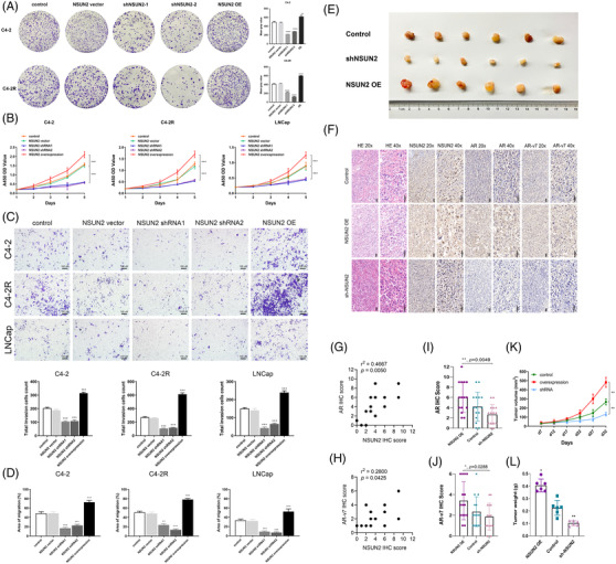
Methods: The expression of NSUN2 and its clinicopathological impact were evaluated in PCa cohorts. The effect of NSUN2 on the biological characteristics of PCa cells was investigated on the basis of gain-offunction and loss-of-function analyses. Subcutaneous models further uncovered the role of NSUN2 in tumor growth. Epi-transcriptome assays with RNA bisulfite sequencing (RNA-BisSeq) analysis and in vitro enzyme reaction assays were performed to validate the targeted effect of NSUN2 on AR. AR-binding sites in the NSUN2 promoter were investigated by ChIP and luciferase assays to uncover the interplay between NSUN2 and AR signaling. RIP-qPCR and EMSA methods were performed to confirm that YBX1 binds to AR m5 C sites.

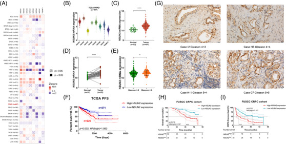
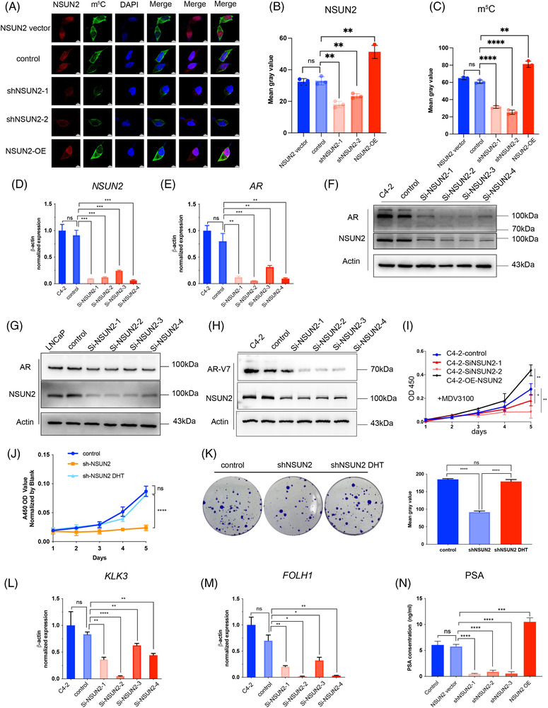
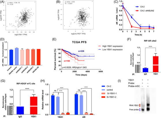
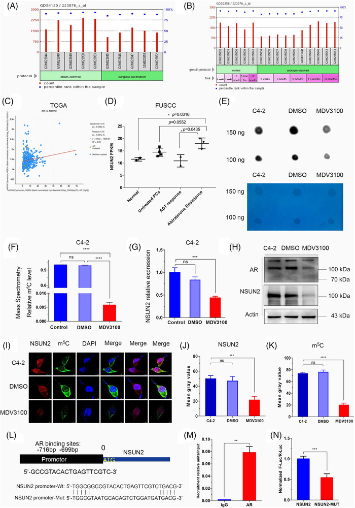
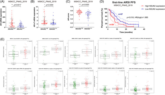
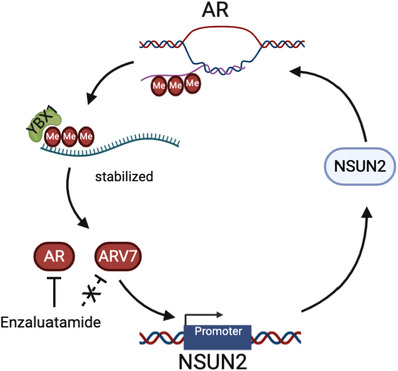
Results: NSUN2 is highly expressed in PCa and predicts poor outcome. NSUN2 plays roles as a PCa oncogene both in vitro and in vivo. Depletion of NSUN2 results in decreased expression and activities of AR, including AR-V7. Mechanistically, NSUN2 posttranscriptionally stabilized AR by cluster m5 C modification in a m5CYBX1-dependent manner. Strikingly, treatment with enzalutamide, an effective AR inhibitor, reduces NSUN2 expression and decreases the m5C modification level in prostate cancer cells. Finally, we found that AR transcriptionally regulates NSUN2.

Conclusion: NSUN2 stabilizes AR mRNA through cluster 5-methylcytosine modification and activates a positive feedback loop to promote prostate cancer.

Keywords: AR-V7; NSUN2; androgen receptor; castration-resistant prostate cancer; cluster; cytosine-5 methylation; prostate cancer.


Conflict of interest statement

The authors declare no conflict of interest.

Figures

FIGURE 1
FIGURE 1
NSUN2 upregulation in PCa indicates worse prognosis. (A) Pan‐cancer overall survival outcome analysis of cytosine‐5‐methyltransferases in TCGA cohorts using Cox proportional hazard model. (B) Box and whisker dot plot of cytosine‐5‐methyltransferases expression levels in PCa tissues from the TCGA cohort. (C) Scatter plot of NSUN2 mRNA expression in normal tissues and PCa tissues from the TCGA cohort. (D) Scatter diagram of NSUN2 mRNA expression in 52 paired PCa and adjacent normal tissues. (E) Scatter plot of NSUN2 mRNA expression in tissues from patients with high Gleason scores (Gleason > = 9). (F) K‐M curves of the PFS of 497 patients from the TCGA cohort. (G) Representative images (×40 magnification) of NSUN2 expression in prostate cancer tissues examined by IHC. (H) Kaplan–Meier analysis shows that high expression of NSUN2 predicts poor OS for PCa patients. The bottom panel shows the number of patients at risk of the corresponding node. (FUSCC cohort; n = 88 cases). (I) Kaplan–Meier analysis shows that high expression of NSUN2 predicts poor CRPC‐free survival for PCa patients. The bottom panel shows the number of patients at risk of the corresponding node. (FUSCC cohort; n = 88 cases). PCa, prostate cancer
FIGURE 2
FIGURE 2
NSUN2 regulates the proliferation, invasion and migration of PCa cells both in vitro and in vivo. (A) Colony formation assay of C4‐2 and C4‐2R cells with NSUN2 knockdown or OE (n = 3 independent experiments). The p values were determined using a two‐sided unpaired student's t‐test. (B) CCK‐8 assay of C4‐2, C4‐2R and LNCaP cells with NSUN2 knockdown or OE (n = 3 independent experiments). The p values were determined using a two‐sided unpaired student's t‐test. (C) Transwell assay of C4‐2, C4‐2R and LNCaP cells with NSUN2 knockdown or OE (n = 3 independent experiments). The p values were determined using a two‐sided unpaired student's t‐test. (D) Wound healing assay of C4‐2, C4‐2R and LNCaP cells with NSUN2 knockdown or OE (n = 3 independent experiments). The p values were determined using a two‐sided unpaired student's t‐test. (E) A xenograft mouse model was constructed by subcutaneously injecting 6‐week‐old BALB/cA nude mice with C4‐2 cells. Decreased tumour volume and weight were observed in the NSUN2 shRNA‐2 group, and increased tumour size was observed in the NSUN2 OE group compared with the control group. (F) HE staining and IHC were implemented to detect the expression levels of NSUN2, AR and AR‐V7 using tumour tissues harvested from xenograft model mice. IHC of tumour tissues from the negative control group showing (G) NSUN2, AR and (H) AR‐V7 expression. Plots showing the IHC scores of (I) AR and (J) AR‐V7. Plots of tumour (K) volume and (L) weight
FIGURE 3
FIGURE 3
Knockdown of NSUN2 reduces the m5C level and decreases AR expression and activity. (A–C) Immunocytochemical staining and cell imaging were performed to detect the NSUN2 and m5C levels in C4‐2 cells with or without NSUN2 knockdown or OE. (n = 3 independent experiments). The p values were determined using a two‐sided unpaired student's t‐test. D‐E qPCR analysis of the mRNA levels of AR (D) and NSUN2 (E) in C4‐2 cells with or without NSUN2 knockdown. (F,G) Immunoblot analysis of AR, NSUN2 and β‐actin expression in C4‐2 (F) and LNCaP (G) cells with or without NSUN2 knockdown. (H) Immunoblot of AR‐V7 and NSUN2 in C4‐2 cells with or without NSUN2 knockdown. (I) CCK‐8 assay of MDV3100 in C4‐2 cells with or without NSUN2 knockdown or NSUN2 OE. (n = 3 independent experiments). The p values were determined using a two‐sided unpaired student's t‐test. (J) CCK‐8 assay of DHT in LNCaP cells with or without NSUN2 knockdown (shNSUN2‐2 was used). (n = 3 independent experiments). The p values were determined using a two‐sided unpaired student's t‐test. (K) Colony formation assay of LNCaP cells with or without NSUN2 knockdown (shNSUN2‐2 was used) treated with DHT (n = 3 independent experiments). The p values were determined using a two‐sided unpaired student's t‐test. L‐M qPCR analysis of the mRNA levels of KLK3 (L) and FOLH1 (M) in C4‐2 cells with or without NSUN2 knockdown or OE. (N) Detection of PSA levels in C4‐2 cells with or without NSUN2 knockdown or OE by immunofluorescence (n = 3 independent experiments). The p values were determined using a two‐sided unpaired student's t‐test
FIGURE 4
FIGURE 4
NSUN2 mediates the m5C modification of AR mRNA. (A) Diagram of the two predicted m5C sites in AR mRNA. (B) Modification site 2 identified by RNA‐BisSeq of mRNA from C4‐2 cells. (C) Synthetic RNA probes of AR m5C modification regions pulled down the NSUN2 protein. (D) Assessment of the m5C modification of AR mRNA by m5C‐RIP qPCR (n = 3 independent experiments). The p values were determined using a two‐sided unpaired student's t‐test. (E) Assessment of the m5C modification of AR mRNA by m5C‐RIP qPCR in C4‐2 cells with or without NSUN2 knockdown (siNSUN2‐2 was used) (n = 3 independent experiments). The p values were determined using a two‐sided unpaired student's t‐test. (F) Identification of binding sites between the NSUN2 protein and AR mRNA by NSUN2‐RIP qPCR (n = 3 independent experiments). The p values were determined using a two‐sided unpaired student's t‐test. (G,H) Identification of the specific m5C modification site through Sanger sequencing of bisulphite conversion probes modified by an in vitro RNA m5C transferase reaction. (I) Dot blot detection of AR m5C modification site 2 by in vitro RNA m5C transferase reaction. (J) Dot blot detection of the m5C modification level of the wild‐type or mutated AR probes modified by in vitro RNA m5C transferase reaction. Methylene blue was used as a loading control. (K) Pull‐down assay with wild‐type or mutated probes to confirm the NSUN2 binding site for AR mRNA. (L) Relative luciferase activity of the luciferase reporter gene, with or without the AR modification region in C4‐2 cells. Each well (∼106 cells) was transfected with 2 µg luciferase reporter plasmid and 1 µg pRL‐TK plasmid (Renilla luciferase reporter). (M) Relative luciferase activity of the luciferase reporter gene containing the AR modification region in C4‐2 cells with or without NSUN2 knockdown (siNSUN2‐2 was used). Each well (∼106 cells) was transfected with 2 µg luciferase reporter plasmid and 1 µg pRL‐TK plasmid (Renilla luciferase reporter) (n = 3 independent experiments). The p values were determined using a two‐sided unpaired student's t‐test. Immunoblot detection of NSUN2 and β‐actin expression in C4‐2 cells with or without NSUN2 knockdown (siNSUN2‐2 was used)
FIGURE 5
FIGURE 5
YBX1 is the reader of AR mRNA m5C sites. (A) Scatterplot of YBX1 expression and AR expression in the TCGA cohort. (B) Scatterplot of ALYREF expression and AR expression in the TCGA cohort. (C) qPCR analysis of the mRNA stability of AR, shNSUN2‐2 was used in the shNSUN2 group. (D) Nuclear and cytoplasmic mRNA were extracted, and AR mRNA was detected by qRT‐PCR in C4‐2 cells with or without NSUN2 knockdown or OE. (E) K‐M plot of the disease‐free survival of PCa patients with high or low YBX1 expression in the TCGA cohort. (F) Assessment of the binding abilities of YBX1 with AR mRNA in C4‐2 cells by YBX1‐RIP qPCR (n = 3 independent experiments). The p values were determined using a two‐sided unpaired student's t‐test. (G) Assessment of the HDGF m5C modification sites in C4‐2 cells by YBX1‐RIP qPCR (n = 3 independent experiments). The p values were determined using a two‐sided unpaired student's t‐test. (H) Silencing YBX1 in C4‐2 cells decreased AR mRNA expression (n = 3 independent experiments). The p values were determined using a two‐sided unpaired student's t‐test. (I) Assessment of the ability of YBX1 protein (200 ng) to bind with modified (the right line) or unmodified (the left line) AR site 2 probes (20 ng) by EMSA (n = 3 independent experiments)
FIGURE 6
FIGURE 6
AR inhibition decreases the mRNA m5C level and downregulates NSUN2 expression. (A) GEO database (GDS4120) analyses the NSUN2 expression after surgical castration. (B) GEO database (GDS3358) analyses NSUN2 expression after androgen deprivation. (C) Scatterplot of NSUN2 expression and AR expression in the TCGA cohort. (D) NSUN2 expression was detected by RNA‐Seq in normal prostate (radical cystectomy sample), untreated PCa, ADT‐responsive (3‐month ADT‐treated), and abiraterone‐resistant PCa (PSA progression after abiraterone) samples from the FUSCC cohort. (E) m5C dot blot assay using poly(A)+ mRNA of C4‐2 cells treated with or without enzalutamide. The experimental group was treated with 50 nM enzalutamide for 72 h. Methylene blue was used as a loading control (n = 3 independent experiments). (F) RNA m5C quantification by LC/MS/MS using poly(A)+ mRNA of C4‐2 cells treated with or without enzalutamide; the experimental group was treated with 50 nM enzalutamide for 72 h (n = 3 independent experiments). The p values were determined using a two‐sided unpaired student's t‐test. (G) qPCR analysis of the mRNA levels of NSUN2 in C4‐2 cells; the experimental group was treated with 50 nM enzalutamide for 72 h. (H) Immunoblot analysis of AR, NSUN2 and β‐actin expression in C4‐2 cells; the experimental group was treated with 50 nM enzalutamide for 72 h. (I–K) Immunocytochemical staining and cell imaging were performed to detect the NSUN2 and m5C levels in C4‐2 cells; the experimental group was treated with 50 nM enzalutamide for 72 h (n = 3 independent experiments). The p values were determined using a two‐sided unpaired student's t‐test. (L) Diagram of the NSUN2 promoter and the AR binding region. (M) ChIP assay confirmed the 19‐bp AR binding site in the NSUN2 promoter (n = 3 independent experiments). The enrichment abundance of each group was normalized by the input value. The p values were determined using a two‐sided unpaired student's t‐test. (N) Mutation of the AR binding site decreased the promoter activity in the luciferase reporter assay (n = 3 independent experiments). Each well (∼106 cells) was transfected with 2 µg luciferase reporter plasmid and 1 µg pRL‐TK plasmid (Renilla luciferase reporter). The p values were determined using a two‐sided unpaired student's t‐test. ADT, androgen deprivation therapy; PCa, prostate cancer
FIGURE 7
FIGURE 7
NSUN2 expression is positively correlated with the expression of AR signalling‐related genes and predicts PFS in multiple cohorts. (A–C) AR (A) and AR‐V7 (B) mRNA expression and AR score (C) were plotted for 91 PCa patients from the MSKCC cohort stratified according to NSUN2 expression. (D) The Kaplan–Meier (K–M) curve of first‐line ARSI PFS was plotted for the MSKCC cohort. (E) Scatter diagrams of NSUN2 mRNA expression in high or low AR signaling from MSKCC, SMMU, SU2C‐PCF cohorts. PCa, prostate cancer
FIGURE 8
FIGURE 8
Model of the AR and NSUN2 positive feedback loop. AR pre‐mRNA is modified by NSUN2 and recognized by YBX1 and maintains its stability. At the same time, AR can act as a transcription factor to regulate the expression of NSUN2. In this way, a positive feedback loop is formed between NSUN2 and AR to promote the progression of prostate cancer. AR inhibitors can inhibit the transcriptional capacity of AR, but not AR alternative spliceosomes, such as AR‐V7. NSUN2 can regulate the stability of AR, and inhibition of NSUN2 in combination with AR inhibitors may better inhibit the progression of prostate cancer

Similar articles

Cited by

References

    1. Chen W, Zheng R, Baade PD, et al. Cancer statistics in China, 2015. CA Cancer J Clin. 2016;66(2):115‐132. - PubMed
    1. Culp MB, Soerjomataram I, Efstathiou JA, Bray F, Jemal A. Recent global patterns in prostate cancer incidence and mortality rates. Eur Urol. 2020;77(1):38‐52. - PubMed
    1. De Laere B, Rajan P, Gronberg H, et al. Androgen receptor burden and poor response to abiraterone or enzalutamide in TP53 wild‐type metastatic castration‐resistant prostate cancer. JAMA Oncol. 2019;5(7):1060‐1062. - PMC - PubMed
    1. Gravis G, Fizazi K, Joly F, et al. Androgen‐deprivation therapy alone or with docetaxel in non‐castrate metastatic prostate cancer (GETUG‐AFU 15): a randomised, open‐label, phase 3 trial. Lancet Oncol. 2013;14(2):149‐158. - PubMed
    1. Fizazi K, Tran N, Fein L, et al. Abiraterone plus prednisone in metastatic, castration‐sensitive prostate cancer. N Engl J Med. 2017;377(4):352‐360. - PubMed

Publication types

MeSH terms