. 2022 Sep 28;79(10):531.
doi: 10.1007/s00018-022-04564-z.

Single cell analysis of PANoptosome cell death complexes through an expansion microscopy method

Affiliations

Single cell analysis of PANoptosome cell death complexes through an expansion microscopy method

Yaqiu Wang et al. Cell Mol Life Sci. .

Abstract

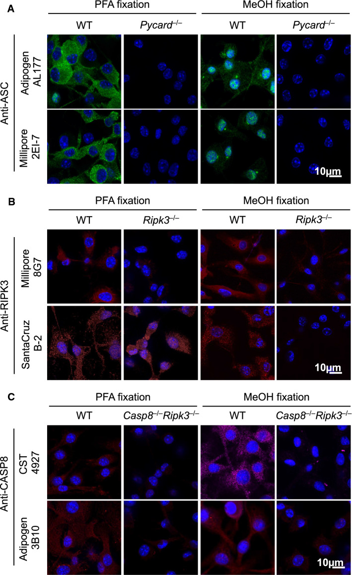
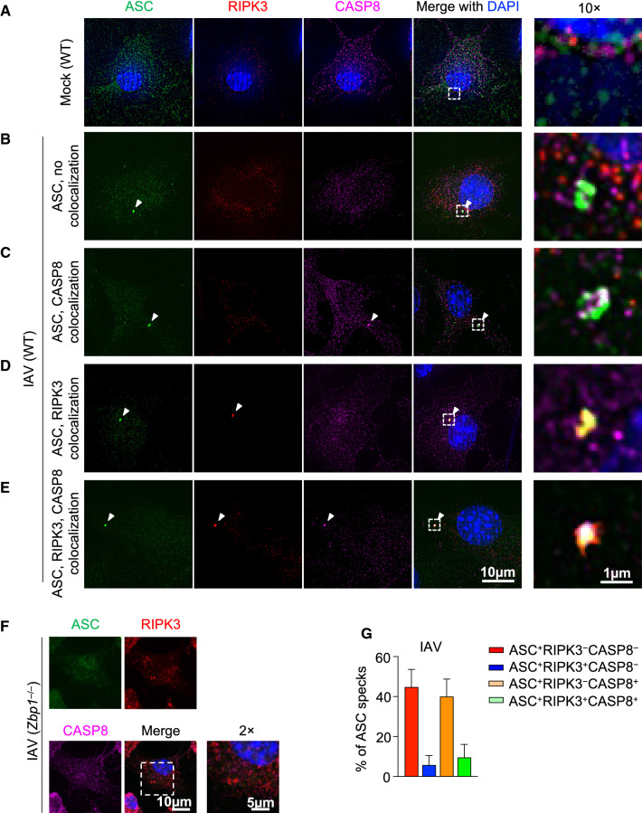
In response to infection or sterile insults, inflammatory programmed cell death is an essential component of the innate immune response to remove infected or damaged cells. PANoptosis is a unique innate immune inflammatory cell death pathway regulated by multifaceted macromolecular complexes called PANoptosomes, which integrate components from other cell death pathways. Growing evidence shows that PANoptosis can be triggered in many physiological conditions, including viral and bacterial infections, cytokine storms, and cancers. However, PANoptosomes at the single cell level have not yet been fully characterized. Initial investigations have suggested that key pyroptotic, apoptotic, and necroptotic molecules including the inflammasome adaptor protein ASC, apoptotic caspase-8 (CASP8), and necroptotic RIPK3 are conserved components of PANoptosomes. Here, we optimized an immunofluorescence procedure to probe the highly dynamic multiprotein PANoptosome complexes across various innate immune cell death-inducing conditions. We first identified and validated antibodies to stain endogenous mouse ASC, CASP8, and RIPK3, without residual staining in the respective knockout cells. We then assessed the formation of PANoptosomes across innate immune cell death-inducing conditions by monitoring the colocalization of ASC with CASP8 and/or RIPK3. Finally, we established an expansion microscopy procedure using these validated antibodies to image the organization of ASC, CASP8, and RIPK3 within the PANoptosome. This optimized protocol, which can be easily adapted to study other multiprotein complexes and other cell death triggers, provides confirmation of PANoptosome assembly in individual cells and forms the foundation for a deeper molecular understanding of the PANoptosome complex and PANoptosis to facilitate therapeutic targeting.

Keywords: AIM2; ASC; Apoptosis; Caspase-1; Caspase-8; Cell death; HSV-1; IFN; Infection; Inflammasome; Inflammation; Influenza; Innate immunity; KPT-330; Method; Microscopy; NLRP3; Necroptosis; PANoptosis; PANoptosome; Protocol; Pyroptosis; RIPK3; ZBP1.


Conflict of interest statement

T.-D.K. is a consultant for Pfizer.

Figures

Fig. 1
Fig. 1
Optimizing conditions for staining mouse ASC, CASP8, and RIPK3. AC Bone marrow-derived macrophages (BMDMs) from the indicated genotypes were fixed by 4% paraformaldehyde (PFA) or methanol (MeOH) and stained with indicated primary antibodies to evaluate staining specificity. Images are representative of at least three independent experiments
Fig. 2
Fig. 2
ASC specks are heterogenous in response to IAV infection. AE Wild-type (WT) bone marrow-derived macrophages (BMDMs) were mock treated (A) or infected with influenza A virus (IAV) (B–E) and stained for ASC, RIPK3, and CASP8. Different compositions of ASC specks are shown. F ASC, RIPK3, and CASP8 staining of IAV-infected Zbp1–/– BMDMs. G Compositional analysis of ASC specks (n ≥ 100) in IAV-infected WT BMDMs at 12 h post-infection. Mean ± standard error is shown. Images are representative of at least three independent experiments
Fig. 3
Fig. 3
Inflammasome stimulation by LPS + ATP induces formation of ASC specks containing RIPK3 and CASP8. A Representative images of ASC specks from bone marrow-derived macrophages (BMDMs). Cells of the indicated genotypes were primed with 100 ng/μl LPS for 4 h, then stimulated with 5 mM ATP (i.e., LPS + ATP) for 10 min. B Expansion microscopy views of a RIPK3/CASP8-containing ring-shaped ASC speck induced by LPS + ATP. C Fluorescence intensity of ASC, RIPK3, and CASP8 in the ring section along the white arrow. X axis indicates the distance along the white arrow traveling from the base of the arrow to the arrowhead. D 3D view of the ASC speck in (B). Images are representative of at least three independent experiments

References

    1. Gullett JM, Tweedell RE, Kanneganti TD (2022) It's All in the PAN: crosstalk, plasticity, redundancies, switches, and interconnectedness encompassed by panoptosis underlying the totality of cell death-associated biological effects. Cells 11(9):1495 - PMC - PubMed
    1. Kuriakose T, Man SM, Malireddi RK, Karki R, Kesavardhana S, Place DE, et al (2016) ZBP1/DAI is an innate sensor of influenza virus triggering the NLRP3 inflammasome and programmed cell death pathways. Sci Immunol 1(2):aag2045 - PMC - PubMed
    1. Malireddi RKS, Karki R, Sundaram B, Kancharana B, Lee S, Samir P et al (2021) Inflammatory cell death, PANoptosis, mediated by cytokines in diverse cancer lineages inhibits tumor growth. Immunohorizons 5(7):568–580 - PMC - PubMed
    1. Kesavardhana S, Malireddi RKS, Burton AR, Porter SN, Vogel P, Pruett-Miller SM et al (2020) The Zα2 domain of ZBP1 is a molecular switch regulating influenza-induced PANoptosis and perinatal lethality during development. J Biol Chem 295(24):8325–8330 - PMC - PubMed
    1. Banoth B, Tuladhar S, Karki R, Sharma BR, Briard B, Kesavardhana S, et al (2020) ZBP1 promotes fungi-induced inflammasome activation and pyroptosis, apoptosis, and necroptosis (PANoptosis). J Biol Chem 295(52):18276–18283 - PMC - PubMed

LinkOut - more resources